Prediction of risk factors of bronchial mucus plugs in children with Mycoplasma pneumoniae pneumonia
←
→
Page content transcription
If your browser does not render page correctly, please read the page content below
Zhang et al. BMC Infectious Diseases    (2021) 21:67
https://doi.org/10.1186/s12879-021-05765-w

 RESEARCH ARTICLE                                                                                                                                  Open Access

Prediction of risk factors of bronchial
mucus plugs in children with Mycoplasma
pneumoniae pneumonia
Jiahui Zhang†, Ting Wang†, Rongrong Li†, Wei Ji, Yongdong Yan, Zhichao Sun, Jiahong Tan, Jinfeng Wu,
Li Huang* and Zhengrong Chen*

  Abstract
  Background: Recently, many cases of pneumonia in children with Mycoplasma pneumoniae infection have been
  shown to have varying degrees of intrabronchial mucus plug formation. The clinical, laboratory, radiological
  characteristics, and treatment of patients with Mycoplasma infection are analyzed in this study. The risk factors for
  M. pneumoniae pneumonia (MPP) mucus plug formation in children are explored, and a risk factor scoring system is
  established.
  Methods: MPP patients treated with bronchoscopy were retrospectively enrolled in the study from February 2015
  to December 2019. The children were divided into a mucus plug group and a control group according to the
  presence or absence of mucus plug formation. The clinical, laboratory, radiological characteristics, and treatment of
  the two groups of children were compared. Univariate and multivariate logistic regression models were used to
  identify the risk factors for MPP mucus plug formation. The receiver operating characteristic (ROC) curve was drawn
  to evaluate the regression model and establish the MPP mucous plug risk factor scoring system.
  Results: A univariate analysis showed that the children in the mucous group were older and had a longer fever
  duration, longer hospital stay, higher fever peak, more cases of wheezing symptoms and allergies, and
  azithromycin or corticosteroids were administered later. In addition, neutrophil, C-reactive protein (CRP), lactate
  dehydrogenase (LDH), D-dimer (DD), sputum MP-DNA copy number, and total immunoglobulin A (IgA) levels were
  higher, while prealbumin (PA) levels were lower. The ROC curve analysis showed that children with MPP had PA
  ≤144.5 mg/L, had used corticosteroids during the course of the illness of ≥4.5 days, CRP ≥12.27 mg/L, an LDH ≥
  462.65 U/L, and there was a possibility of intra-airway mucus formation. The independent risk factors were scored
  according to their odds ratio (OR) value. Among the 255 children with MPP, the high-risk group had 44 (83.02%)
  mucus plugs out of 53; the middle-risk group had 35 (34.3%) mucus plugs out of 102; and the low-risk group had
  11 (11%) mucus plugs out of 100.
  Conclusions: PA levels, timing of corticosteroid use (use in the first few days), CRP levels, and LDH levels were
  independent risk factors for MPP mucus plug formation. This provides a basis for the early identification of MPP in
  children combined with mucus plug formation.
  Keywords: Mycoplasma pneumoniae pneumonia, Children, Mucus plug, Risk factors

* Correspondence: szdalv@163.com; chen_zheng_rong@163.com
†
 Jiahui Zhang, Ting Wang and Rongrong Li contributed equally to this work.
Department of Respiratory Medicine, Children’s Hospital of Soochow
University, Suzhou 215003, China

                                       © The Author(s). 2021 Open Access This article is licensed under a Creative Commons Attribution 4.0 International License,
                                       which permits use, sharing, adaptation, distribution and reproduction in any medium or format, as long as you give
                                       appropriate credit to the original author(s) and the source, provide a link to the Creative Commons licence, and indicate if
                                       changes were made. The images or other third party material in this article are included in the article's Creative Commons
                                       licence, unless indicated otherwise in a credit line to the material. If material is not included in the article's Creative Commons
                                       licence and your intended use is not permitted by statutory regulation or exceeds the permitted use, you will need to obtain
                                       permission directly from the copyright holder. To view a copy of this licence, visit http://creativecommons.org/licenses/by/4.0/.
                                       The Creative Commons Public Domain Dedication waiver (http://creativecommons.org/publicdomain/zero/1.0/) applies to the
                                       data made available in this article, unless otherwise stated in a credit line to the data.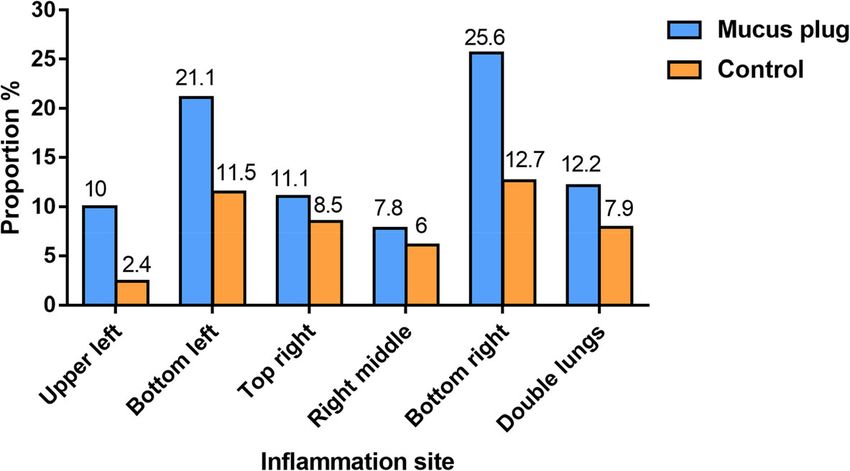
Zhang et al. BMC Infectious Diseases   (2021) 21:67                                                          Page 2 of 8

Background                                                   aspirates (NPA) MP-DNA > 1.0 × 10⁵ copies/L. The ex-
M. pneumoniae is an important and common pathogen            clusion criteria included those patients with chronic lung
in respiratory infections in children [1, 2]. M. pneumo-     disease, recurrent respiratory tract infections, recurrent
niae pneumonia (MPP) accounted for 34.75% of                 wheezing or a medical history of asthma, bronchopul-
community-acquired pneumonia (CAP) in hospitalized           monary dysplasia, immunosuppression or defective dis-
children from the Children’s Hospital of Soochow Uni-        ease, severe heart, liver, kidney disease, malignant
versity from January 2011 to December 2015, with chil-       tumors, and incomplete case information.
dren over 5 years of age being more commonly affected          Laboratory tests were completed within 24 h after the
than younger children [3–6]. The acute phase of MPP          children were admitted to the hospital, including neutro-
can present with varying degrees of damage to the air-       phil, CRP, LDH, DD, MP-IgM, PA levels, and NPAs.
way mucosa, which in severe cases can lead to mucosal        Demographic and clinical data of 255 children with
embolization of the orifice and inflammatory stenosis or     MPP, including epidemiological, clinical, laboratory, and
even occlusion. In addition, more than 30% of refractory     radiological characteristics, and bronchoscopy results, as
MPP have been found to form bronchial mucus plugs            well as treatments and outcomes, were collected and
(BMPs) [7].                                                  recorded.
  BMPs are endogenous bronchial foreign bodies that
are caused by inflammation, bleeding, necrosis, abnor-       Fiberoptic bronchoscopy
mal secretion of bronchial mucus in the bronchus,            The children were fasted for 4–6 h prior to surgery.
mucus elimination obstacles, and then mucus accumula-        Atropine (0.01–0.02 mg/kg) and midazolam (0.1–0.3
tion and agglomeration in the bronchus, forming bron-        mg/kg, the maximum amount was 4 mg/time) were
chial mucus plugging [8, 9]. If not cleared in time, they    injected intramuscularly 30 min prior to surgery. A
can lead to bronchodilatation, pulmonary arrhythmia,         solution of 2% lidocaine was swallowed nasally and
occlusive bronchitis, and even acute respiratory failure     orally three to four times, and 1% rosemary nasal
and the blockage can be seriously life-threatening.          drops were administered to the right nose. The
  A study by Xu Q et al. showed that in patients aged 5      choice of bronchoscopy model depended on patient
years and older, higher IL-10 levels and higher IFN-γ        age. The bronchoscope reached the openings of the
levels had an important predictive value for mucus plug      trachea and the left and right bronchi through the
formation [10]. In the predicted nomogram, atelectasis       nose and epiglottis. The front end of the broncho-
and pleural effusion had the highest score and the high-     scope reached the lesion and was then embedded in
est weight, which is a powerful indicator for bronchos-      the lumen. Bronchial alveolar lavage was performed
copy intervention [11]. The aim of this study was to         using 0.9% saline at 37 °C. For local lavage, where it
analyze the risk factors for the formation of BMP in chil-   was difficult to remove the mucus tie, it was re-
dren with MPP. This evaluation of risk factors can assist    moved with a brush or biopsy forceps and slowly
clinicians to judge whether there is a possibility of BMP    pulled out of the fiberoptic bronchoscope.
formation and ascertain the opportunity for reasonable
treatment. Thus, it has important significance for redu-     Definitions
cing the occurrence of irreversible damage.                  According to the performance of bronchoscopy, pa-
                                                             tients were divided into a mucous plug group and a
Materials and methods                                        control group. In the mucus group, the sputum plugs
Patients and data collection                                 could be seen in the bronchial cavity of the lungs
This retrospective study was conducted in the Children’s     that blocked the lumen, and some plastic sputum
Hospital of Soochow University. A total of 255 children      plugs had formed. These are not easily removed and
who met the diagnosis of MPP were selected and treated       require the use of a brush. Some even require the use
with bronchoscopy during hospitalization from February       of foreign body pliers. In the control group, there
2015 to December 2019. The age range of the selected         were no mucus plugs in the lumen of the bronchus
255 children with MPP ranged from 2 months to 16             under the bronchoscope, but a few flocculent or thin
years. They all met the diagnostic criteria of the MPP       secretions in the lumen could be seen.
diagnosis and treatment expert consensus (2015 version)
for children. The clinical manifestations were fever,        Statistical analysis
cough, and dyspnea, and with or without other systemic       SPSS25.0 statistical software was used for the data
manifestations, such as dry lungs and wet rales, with        analysis. Measurement data conforming to a normal
signs of pulmonary consolidation and changes in lung         distribution are expressed as means ± standard devia-
imaging. In addition, the conditions that needed to be       tions (x ± s). The comparison between the two groups
met were a serum MP-IgM > 1.1 or nasopharyngeal              used an independent sample t test. Non-normal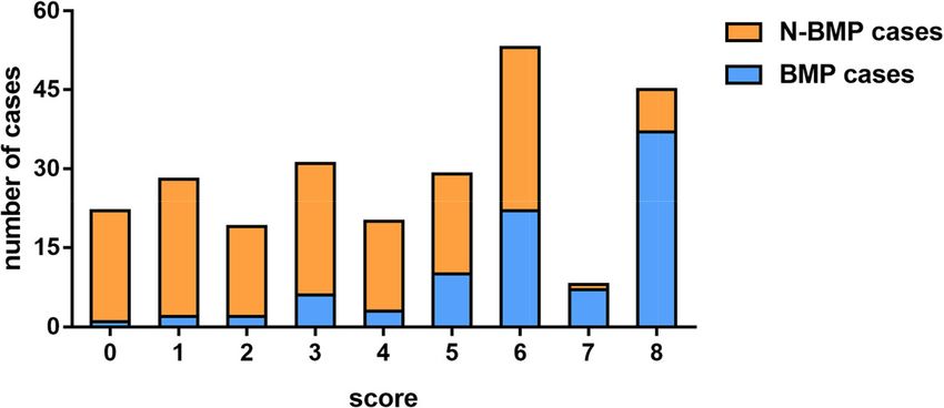
Zhang et al. BMC Infectious Diseases       (2021) 21:67                                                         Page 3 of 8

Table 1 Clinical characteristics of patients in the mucus plug and control group
Variables                                                 Mucus plug group (n = 90)   Control group (n = 165)      P value
Characteristics
  Gender (male/female, n)                                 49/41                       89/76                        0.938
       −
  Age ( x ± s)/year                                       6.10 ± 2.85                 4.50 ± 2.93                  < 0.01
  Length of hospitalization (−x ± s) /d                   10.72 ± 3.47                8.81 ± 2.72                  < 0.01
  Course before admission [M(P25–P75)]/d                  8 (6, 10)                   7 (5, 11)                    0.869
  Allergic constitution [n (%)]                           40 (44.4)                   33 (21.2)                    < 0.01
Signs and symptoms
  Fever [n (%)]                                           86 (95.6)                   135 (81.8)                   0.002
               −
  Heat range ( x ± s) /d                                  9.72 ± 3.80                 5.00 ± 3.98                  < 0.01
  Hot peak [M(P25–P75), °C]                               39.6 (39, 40)               39 (38.5, 39.7)              < 0.01
  Shortness of breath [n (%)]                             6 (6.7)                     8 (4.8)                      0.542
  Breather [n (%)]                                        7 (7.8)                     41 (24.8)                    0.01
  Lung rales [n (%)]                                      40 (44.4)                   68 (41.2)                    0.618
  Lung wheezing [n (%)]                                   6 (6.6)                     27 (16.3)                    0.27
  Reduced breath sounds [n (%)]                           14 (15.6)                   21 (1.3)                     0.531
Laboratory characteristics
  WBC [M(P25–P75)]/ × 109 L−1                             7.63 (5.97, 11.35)          7.97 (6.12, 10.64)           0.844
  Neutrophil (−x ± s)/%                                   67.34 ± 12.78               57.53 ± 15.20                < 0.01
  Eos [M(P25–P75)]/×109 L− 1                              0.4 (0.045, 1.325)          0.3 (0.07, 1.5)              0.866
  CRP [M(P25–P75)]/mg·L−1                                 25.72 (12.62, 51.23)        7.58 (2.58, 18.23)           < 0.01
  PLT [M(P25–P75)]/×109 L−1                               275.5 (231.75, 356)         321 (232, 394)               0.109
  LDH [M(P25–P75)]/U·L−1                                  545.9 (397.3, 655.5)        389.3 (340.0, 389.3)         < 0.01
  PA [M(P25–P75)]/mg·L−1                                  114 (92.75, 134)            142 (115, 195.5)             < 0.01
  DD [M(P25–P75)]/μg·L−1                                  1207 (484.5, 3677.75)       367 (187, 367)               < 0.01
                                  −1
  CK-MB [M(P25–P75)]/ng·mL                                0.9 (0.5, 1.5)              1.1 (0.6, 1.75)              0.295
  ALT [M(P25–P75)]/U·L−1                                  17.6 (12.55, 30.725)        12.9 (10.1, 17.8)            < 0.01
  AST [M(P25–P75)]/U·L−1                                  34.6 (27.425, 43.725)       30.2 (24.95, 37.95)          0.003
  Serum MP-IgM                                            3.46 (1.36, 5.49)           3.2 (1.15, 5.14)             0.154
Sputum MP-DNA copy number [n (%)]
  Low load group                                          3 (3.3)                     20 (12.1)                    0.019
  Medium load group                                       13 (14.4)                   42 (25.5)                    0.041
  High load group                                         74 (82.2)                   103 (62.4)                   0.001
Humoral immunity
  IgG (−x ± s)/g·L−1                                      9.79 ± 2.86                 9.43 ± 3.08                  0.357
                        −1
  IgA [M(P25–P75)]/g·L                                    1.27 (0.96, 1.76)           1.04 (0.6, 1.53)             0.007
  IgM [M(P25–P75)]/g·L−1                                  1.49 (1.09, 2.20)           1.37 (0.95, 1.92)            0.061
Cellular immunity [M(P25–P75), %]
  CD3+                                                    66.96 (61.2, 73.05)         65.5 (60.05, 72.15)          0.634
  CD3 + CD4+                                              34.14 (28.3, 40.32)         34.2 (30.25, 39.2)           0.524
  CD3 + CD8+                                              27.25 (21.5, 27.25)         25.8 (21.5, 30.2)            0.24
  CD4+/CD8+                                               1.30 (1.0, 1.3)             1.3 (1.1, 1.7)               0.117
  CD3-CD (15 + 56) +                                      10.35 (5.7, 15.7)           10.2 (7.1, 14.8)             0.715
  CD3-CD19+                                               19.2 (14.02, 25.92)         18.7 (12.95, 26.85)          0.983
  CD19 + CD23+                                            7.9 (5.4, 12.72)            9.4 (6.55, 13.6)             0.023
Zhang et al. BMC Infectious Diseases (2021) 21:67 Page 4 of 8
Table 1 Clinical characteristics of patients in the mucus plug and control group (Continued)
Variables Mucus plug group (n = 90) Control group (n = 165) P value
Radiological characteristics [n (%)]
Lung consolidation 84 (93.3) 126 (76.36) 0.001
Atelectasis 9 (10) 8 (4.8) 0.123
Pleural effusion 19 (21.1) 18 (10.9) 0.040
Pleural effusion site [n (%)]
Left side 9 (10) 10 (6.1) 0.331
Right side 10 (11.1) 11 (6.7) 0.238
Treatment
Medication time [M(P25–P75)]/Day n of course
Azithromycin 5.5 (4, 7) 4 (3, 6) 0.012
corticosteroids 7 (5, 9) 5 (3, 8) 0.003
Fog time before tracheoscopy[M(P25–P75)]/d 3 (2, 4) 3 (2, 5) 0.525
Sputum MP-DNA copy number: low load group, < 1 × 10⁴ L−1; medium load group, 1 × 10⁴ L−1 –106 L−1; high load group, > 1 × 10⁶ L−1
ALT alanine aminotransferase; AST aspartate aminotransferase; CD cluster of differentiation; CK-MB creatine kinase-MB; CRP C reactive protein; DD D-dimer;
IgA immunoglobulin A; IgG immunoglobulin G; LDH lactate dehydrogenase; MP Mycoplasma pneumonia; PA Prealbumin; PLT Platelets; WBC white blood cells
distribution data is expressed as the median, and the com- (64.7%) cases. The mean ages of the mucus plug and
parison between the two groups used the Wilcoxon rank control group patients were 6.10 ± 2.85 years and 4.50 ±
sum test. A P < 0.05 was considered statistically significant. 2.93 years, respectively, with a significant difference (P <
The count data is expressed as a percentage (%), and a 0.05). The males in the mucus plug group and the con-
comparison between the groups was performed using a χ2 trol group were 49 (54.4%) of 90 and 89 (53.9%) of 165,
test. A logistic regression analysis of the risk factors re- respectively, and the difference was not statistically sig-
lated to intratracheal mucus plug formation in children nificant (P > 0.05; Table 1). In the mucus plug group,
with MPP was also performed (variable selection criteria children < 2 years of age were significantly less than 2–6
were P < 0.05 and elimination criteria were P > 0.1; test years old and > 6 years old, with children > 6 years old
level was bilateral α = 0.05). The receiver operating charac- being the majority, and the difference was statistically
teristic (ROC) curve was drawn, and the area under curve significant (P < 0.017; Fig. 1). The univariate analysis
(AUC) was used to evaluate the predictive value of each showed that the children in the mucus plug group were
independent risk factor in the formation of mucus plugs. older, had a longer fever duration, longer hospital stay,
A P < 0.05 was considered statistically significant. higher fever peak, more cases of fever, wheezing symp-
toms and allergies, azithromycin (intravenous or oral) or
Results corticosteroids (intravenous or oral) were administered
Clinical characteristics later, and the neutrophil, C-reactive protein (CRP), lac-
There were 255 children with MPP who were treated tate dehydrogenase (LDH), D-dimer (DD), sputum MP-
with bronchoscopy. The mucus plug group accounted DNA copy number, total IgA, and the CD19 + CD23+
for 90 (35.3%) cases and the control group for 165 levels were higher, while the PA levels were lower. In
Fig. 1 Age distribution at onsetZhang et al. BMC Infectious Diseases (2021) 21:67 Page 5 of 8
Fig. 2 Inflammation site distribution
addition, lung consolidation and pleural effusion cases and 48.5%, 78.9 and 40%, 76.7 and 63.6%, and 65.6 and
were greater (Table 1). The chest radiograph or com- 72.1%, respectively (Fig. 4).
puted tomography (CT) showed that the lung inflamma-
tion in the mucous group was primarily in the bottom Logistic regression analysis after assigning risk factors for
right lung (23 [25.6%] of 90) and the bottom left lung BMP formation
(19 [21.1%]; Fig. 2). Figure 3 is a bronchoscopic perform- The critical values of the independent factors were
ance and imaging features of a 10-year-old MPP patient. assigned to evaluate the risk of BMP formation. The lo-
gistic regression analysis showed a statistical significance
(P < 0.05, Table 3). Based on the logistic regression ana-
Analysis of risk factors for BMP formation in children with lysis, a lower PA lever (> 144.5 vs ≤144.5 mg/L; OR,
MPP 6.514; 95% CI, 3.410–12.443), later corticosteroid ther-
A univariate logistic regression showed that the PA level apy (≥4.5 vs < 4.5 d; OR, 2.333; 95% CI, 1.298–4.195), a
(odds ratio [OR], 1.015; 95% confidence interval [CI], higher CRP level (< 12.27 vs ≥12.27 mg/L; OR, 5.409;
1.001–1.030), timing of corticosteroids use (use in the 95% CI, 3.041–9.622), and a higher LDH level (< 462.65
first few days; OR, 0.802; 95% CI, 0.663–0.970), CRP vs ≥462.65 U/L; OR, 4.377; 95% CI, 2.533–7.565) were
level (OR, 0.986; 95% CI, 0.944–0.992), and LDH level each independently associated with BMP formation
(OR, 0.996; 95% CI, 0.992–0.999) were independent risk (Table 3).
factors for MPP mucus plug formation (P < 0.05,
Table 2). The ROC curve analysis showed that when the Percentage of mucous plug patients in the MPP scoring
optimal thresholds for PA, use of corticosteroids during groups
the course, CRP, and LDH were ≤ 144.5 mg/L, ≥4.5 days, The independent risk factors were scored according to
≥12.27 mg/L, and ≥ 462.65 U/L, respectively, their sensi- their OR value. The time of corticosteroids application
tivity and specificity to predict BMP formation were 87.8 ≥4.5 days was one point; a CRP ≥12.27 mg/L and an
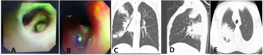
Fig. 3 This is a bronchoscopic performance and imaging features of a MPP patient. Bronchoscopy performance (A and B): A is the plastic sputum
plug of the upper right and the posterior branch. The wall of the tube is smooth after the brush removes it; B is the shaped sputum plug on the
left side of B8 and B9, and the lumen is smooth after lavage; C and D are the normal and lateral pictures of this child’s chest radiograph; and E is
a CT slice of this child. CT = computed tomography; MPP = M. pneumoniae pneumoniaZhang et al. BMC Infectious Diseases (2021) 21:67 Page 6 of 8
Table 2 Logistic regression analysis of risk factors related to BMP formation in MPP
Variable Partial regression coefficient (β) SE Wald χ2 value P value OR (95% CI)
CRP (mg/L) −0.033 0.013 6.604 0.01 0.986 (0.944–0.992)
LDH (U/L) −0.004 0.002 5.419 0.02 0.996 (0.992–0.999)
PA (mg/L) 0.015 0.007 4.655 0.031 1.015 (1.001–1.030)
Corticosteroids (d) −0.220 0.097 5.151 0.023 0.802 (0.663–0.970)
Constant 14.063 6.451 4.753 0.029 –
Corticosteroids, use corticosteroids for the first few days
BMP bronchial mucus plug; CI confidence interval; CRP C reactive protein; LDH lactate dehydrogenase; MPP . pneumoniae pneumonia; OR odds ratio;
PA prealbumin; SE standard error
LDH ≥462.65 U/L was two points each; and a PA a higher LDH level (≥462.65 U/L) were significantly as-
≤144.5 mg/L was three points. According to the scores, sociated with presence of BMP in children with MPP.
the MPP patients were divided into a high-risk group Xu Q et al. found that CRP, LDH, age, and fever dur-
(7–8 points), a middle-risk group (4–6 points), and a ation were associated with the formation of BMPs in
low-risk group (0–3 points). Among them, 53 cases were children with refractory Mycoplasma pneumoniae pneu-
in the high-risk group and 44 cases (83.02%) were monia (RMPP) [10]. Xu X et al. did not find a significant
caused by mucus plugs. There were 102 cases in the difference in serum LDH and CRP levels between the
middle-risk group, 35 cases (34.3%) with mucus plugs, children with and without BMPs [13]. It was found in
100 cases in the low-risk group, and 11 cases (11%) with this study that a CRP ≥12.27 mg/L and an LDH ≥462.65
mucus plugs (Fig. 5). were associated with BMP formation in children with
MPP. This is related to the body’s excessive immune in-
flammatory response that leads to a numerous inflam-
Discussion matory factors. These inflammation factors further lead
In recent years, with the increase in the incidence of to serious airway mucosal damage, ciliary clearance dys-
MPP and the resistance to macrolide antibiotics, the in- function, and epithelial cell shedding, eventually forming
cidence of refractory and severe MPP has increased. a mucus plug to block the airway.
Studies have shown that BMP may become an important In contrast to previous studies, this study showed that
factor in the difficulty of MPP treatment [12]. The re- the PA level had a higher predictive value. A PA ≤144.5
sults of this study suggest that clinical variables, includ- mg/L was associated with BMP formation in children
ing a lower PA level (≤144.5 mg/L), later corticosteroid with MPP. PA is a negative acute phase protein synthe-
therapy (≥4.5 d), a higher CRP level (≥12.27 mg/L), and sized by the liver and is a non-specific host defense
Fig. 4 a is the ROC curve analysis of the CRP (blue line), LDH (red line), and the timing of corticosteroids application (green line), which predicts
the sensitivity and specificity of MPP BMP formation at 76.7 and 63.6%, 65.6 and 72.1%, and 78.9 and 40%, respectively. b is the ROC curve
analysis of the PA, which predicts that the sensitivity and specificity of MPP BMP formation at 87.8 and 48.5%, respectively. BMP = bronchial
mucus plugs; CRP = C reactive protein; LDH = lactate dehydrogenase; MPP = Mycoplasma pneumoniae pneumonia; PA = prealbumin; ROC =
receiver operating characteristicZhang et al. BMC Infectious Diseases (2021) 21:67 Page 7 of 8 Table 3 Logistic regression analysis after assigning risk factors for BMP formation Variable (assignment) Wald χ2 P OR (95% CI) CRP (< 12.27 mg/L = 0 ≥ 12.27 mg/L = 1) 33.000 0.000 5.409 (3.041–9.622) LDH (< 462.65 U/L = 0 ≥ 462.65 U/L = 1) 27.983 0.000 4.377 (2.533–7.565) PA (> 144.5 mg/L = 0 ≤ 144.5 mg/L = 1) 32.210 0.000 6.514 (3.410–12.443) corticosteroids (< 4.5 d = 0 ≥ 4.5 d = 1) 8.018 0.005 2.333 (1.298–4.195) Corticosteroids, use corticosteroids for the first few days BMP bronchial mucus plug; CI confidence interval; CRP C reactive protein; LDH lactate dehydrogenase; OR odds ratio; PA prealbumin substance. Therefore, in an acute infection, the PA OR value. Among them, a PA ≤144.5 mg/L was three serum level can be rapidly reduced. Previous studies points; a CRP ≥12.27 mg/L and an LDH ≥462.65 U/L have found that PA is associated with the severity and was two points each; and a time of CS therapy applica- prognosis of many diseases [13]. Shen et al. conducted a tion of ≥4.5 d was one point. Therefore, children who retrospective analysis of 174 children with community- met the above indicators obtained the highest score of 8 acquired pneumonia (CAP) and found that the sensitiv- points. According to the scores, MPP patients with 7–8 ity of PA to diagnose CAP was higher than that of other points belonged to the high-risk group for BMP forma- inflammation indicators, which is an independent pro- tion. For example, an MPP child with a PA level of tective factor for children with CAP. Combined with ≤144.5 mg/L, CS therapy after 4.5 d, a CRP level of CRP, PA can effectively improve the diagnostic efficiency ≥12.27 mg/L, and an LDH level of ≥462.65 U/L strongly of children’s CAP and assess the severity of pneumonia indicated the presence of a BMP. [14]. Research by Wang et al. showed that the PA level This study had some limitations. First, laboratory sam- can reflect the severity of severe MPP, suggesting that ples were not collected for the same period as the pres- the PA level may become an objective indicator for pre- ence of disease, which produced a bias. Second, a dicting the progress of severe MPP [15]. prospective study is required to further confirm the reli- In addition, it was found that earlier use of corticoster- ability for this retrospective study. Third, with the lim- oid (CS) therapy can reduce the formation of BMP in ited number of cases, it was difficult to make the children with MPP. The optimal threshold was less than number of cases in the two groups similar. 4.5 days. CS therapy has a direct inhibitory effect on many inflammatory cells, which can inhibit neutrophil Conclusions apoptosis, promote eosinophil apoptosis, and reduce the PA level, timing of CS therapy use (use in the first few number of mast cells in the airway [16–18]. CS therapy days), CRP level, and LDH level were independent risk can also inhibit inflammatory factors, improve clinical factors for MPP mucus plug formation. According to the symptoms, reduce airway microvascular leakage, and re- scoring system used in this study, the higher the score of duce BMP production [19, 20]. children with MPP, the higher the risk of forming BMP. In this study, the predictive values of the various risk The scoring system does have the potential to be used factors for mucus plug formation were different, and for the identification of BMP in children with MPP, they were assigned a predictive value according to their thereby contributing to a rational therapeutic choice. Fig. 5 Correspondence between the scoring model and the formation of mucus plugs. According to the score, 255 children with Mycoplasma pneumoniae pneumonia (MPP) were divided into a high-risk group (7–8 points), a middle-risk group (4–6 points), and a low-risk group (0–3 points). Among them, the high-risk group had 44 [83.02%] mucus plugs out of 53; the middle-risk group had 35 [34.3%] mucus plugs out of 102; and the low-risk group had 11[11%] mucus plugs out of 100.
Zhang et al. BMC Infectious Diseases              (2021) 21:67                                                                                                  Page 8 of 8

Abbreviations                                                                          6.    Sondergaard MJ, Friis MB, Hansen DS, Jorgensen IM. Clinical manifestations
ALT: Alanine aminotransferase; AST: Aspartate aminotransferase; AUC: Area                    in infants and children with Mycoplasma pneumoniae infection. PLoS One.
under curve; BMP: Bronchial mucus plugs; CAP: Community-acquired                             2018;13(4):e195288.
pneumonia; CD: Cluster of differentiation; CI: Confidence interval; CK-                7.    Huang L, Huang X, Jiang W, Zhang R, Yan Y, Huang L. Independent
MB: Creatine kinase isoenzyme; CRP: C reactive protein; CS: Corticosteroid;                  predictors for longer radiographic resolution in patients with refractory
CT: Computed tomography; DD: D-dimer; EOS: Eosinophil count;                                 Mycoplasma pneumoniae pneumonia: a prospective cohort study. BMJ
EPO: Eosinophil peroxidase; IgA: Immunoglobulin A; IgG: Immunoglobulin G;                    Open. 2018;8(12):e23719.
LDH: Lactate dehydrogenase; MPP: Mycoplasma pneumoniae pneumonia;                      8.    Brogan TV, Finn LS, Pyskaty DJ Jr, Redding GJ, Ricker D, Inglis A, et al. Plastic
NPA: Nasopharyngeal aspirates; OR: Odds ratio; PA: Prealbumin;                               bronchitis in children: a case series and review of the medical literature.
ROC: Receiver operating characteristic; WBC: White blood cells                               Pediatr Pulmonol. 2002;34(6):482–7.
                                                                                       9.    Noizet O, Leclerc F, Leteurtre S, Brichet A, Pouessel G, Dorkenoo A, et al.
Acknowledgements                                                                             Plastic bronchitis mimicking foreign body aspiration that needs a specific
We thank LetPub (www.letpub.com) for its linguistic assistance during the                    diagnostic procedure. Intensive Care Med. 2003;29(2):329–31.
preparation.                                                                           10.   Xu Q, Zhang L, Hao C, Jiang W, Tao H, Sun H, et al. Prediction of bronchial
                                                                                             mucus plugs formation in patients with refractory Mycoplasma pneumoniae
Authors’ contributions                                                                       pneumonia. J Trop Pediatr. 2017;63(2):148–54.
All authors have read and approved the manuscript. JZ and ZC conceived                 11.   Xu X, Li H, Sheng Y, Wu L, Wang D, Liu L, et al. Nomogram for prediction of
and designed the study. LH, TW and RL made contributions to the analysis                     bronchial mucus plugs in children with Mycoplasma pneumoniae
and interpretation of data. WJ and YY collected the clinical data. JT, JW and                pneumonia. Sci Rep. 2020;10(1):4579.
ZS contributed to the statistical analysis.                                            12.   Lee KY, Lee HS, Hong JH, Lee MH, Lee JS, Burgner D, et al. Role of
                                                                                             prednisolone treatment in severe Mycoplasma pneumoniae pneumonia in
Funding                                                                                      children. Pediatr Pulmonol. 2006;41(3):263–8.
This work was supported by grants from the following projects: Social                  13.   Wang W, Wang CS, Ren D, Li T, Yao HC, Ma SJ. Low serum prealbumin
Development Projects of Jiangsu Province (grant NO. BE2019671); Science                      levels on admission can independently predict in-hospital adverse cardiac
and Technology Program of Suzhou (grant NO. SS201869); the National                          events in patients with acute coronary syndrome. Medicine. 2018;97(30):
Natural Science Foundation of China (grant NO. 81970027; 81870006;                           e11740.
81771676; 81971490); Jiangsu Provincial Medical Youth Talent (grant NO.                14.   Shen Y, Zhou J, Shao X. Value of prealbumin in diagnosis of children with
QNRC2016766); Suzhou Medical Youth Talent (grant NO. GSWS2019047); Key                       community-acquired pneumonia in China and assessment of severity. Chin
Lab of Respiratory Disease of Suzhou (grant NO. SZS201714); Suzhou Medical                   J Nosocomiol. 2018;28(14):2173–7.
Technology Projects of Clinical Key Diseases (grant NO. LCZX201809); The               15.   Wang C, Liu G, Wang S, Zhang J. Clinical significance of prealbumin in
Postgraduate Research & Practice Innovation Program of Jiangsu Province                      children with severe Mycoplasma pneumoniae pneumonia. Chin J Appl Clin
(grant NO. KYCX20 2727). The funders had no role in study design, data                       Pediatr. 2020;35(4):302–4.
collection and analysis, decision to publish, or preparation of the manuscript.        16.   Belvisi MG. Regulation of inflammatory cell function by corticosteroids. Proc
This work was supported by the following projects.                                           Am Thorac Soc. 2004;1(3):207–14.
                                                                                       17.   Kankaanranta H, Lindsay MA, Giembycz MA, Zhang X, Moilanen E, Barnes PJ.
Availability of data and materials                                                           Delayed eosinophil apoptosis in asthma. J Allergy Clin Immunol. 2000;106(1
The datasets used and/or analysed during the current study are available                     Pt 1):77–83.
from the corresponding author on reasonable request.                                   18.   Saffar AS, Ashdown H, Gounni AS. The molecular mechanisms of
                                                                                             glucocorticoids-mediated neutrophil survival. Curr Drug Targets. 2011;12(4):
Ethics approval and consent to participate                                                   556–62.
This study protocol was approved by the Ethical Review Committee of                    19.   Boschetto P, Rogers DF, Fabbri LM, Barnes PJ. Corticosteroid inhibition of
Children’s Hospital of Soochow University with judgment’s reference number                   airway microvascular leakage. Am Rev Respir Dis. 1991;143(3):605–9.
2020CS078. Written consent was obtained from the parents or guardians of               20.   Hauber HP, Goldmann T, Vollmer E, Wollenberg B, Zabel P. Effect of
all participants before data collection.                                                     dexamethasone and ACC on bacteria-induced mucin expression in human
                                                                                             airway mucosa. Am J Resp Cell Mol. 2007;37(5):606–16.
Consent for publication
Not applicable.                                                                        Publisher’s Note
                                                                                       Springer Nature remains neutral with regard to jurisdictional claims in
Competing interests                                                                    published maps and institutional affiliations.
The authors declare that they have no competing interests.

Received: 6 August 2020 Accepted: 4 January 2021

References
1. Cao B, Ren LL, Zhao F, Gonzalez R, Song SF, Bai L, et al. Viral and
    Mycoplasma pneumoniae community-acquired pneumonia and novel
    clinical outcome evaluation in ambulatory adult patients in China. Eur J Clin
    Microbiol Infect Dis. 2010;29(11):1443–8.
2. Meyer Sauteur PM, van Rossum AM, Vink C. Mycoplasma pneumoniae in
    children: carriage, pathogenesis, and antibiotic resistance. Curr Opin Infect
    Dis. 2014;27(3):220–7.
3. Jain S, Williams DJ, Arnold SR, Ampofo K, Bramley AM, Reed C, et al.
    Community-acquired pneumonia requiring hospitalization among U.S.
    children. N Engl J Med. 2015;372(9):835–45.
4. Defilippi A, Silvestri M, Tacchella A, Giacchino R, Melioli G, Di Marco E, et al.
    Epidemiology and clinical features of Mycoplasma pneumoniae infection in
    children. Respir Med. 2008;102(12):1762–8.
5. Xinxing Z, Wenjing G, Zhengrong C, et al. Epidemiological analysis of
    refractory mycoplasma Pneumoniae pneumonia in children in Suzhou from
    2011 to 2015. J Pediatr Pharm. 2019;25(08):7–10.
You can also read