Case Report Thyroid tuberculosis: two cases report and review of the literature
←
→
Page content transcription
If your browser does not render page correctly, please read the page content below
Int J Clin Exp Med 2016;9(11):22477-22485
www.ijcem.com /ISSN:1940-5901/IJCEM0024338

Case Report
Thyroid tuberculosis: two cases
report and review of the literature
You-Ding Jiang1, Jian-Qi Gao1, Sui Chen1, Xiao-Yan Deng2
1
 Department I of Thoracic Surgery, 2Department of Ultrasonography, Guangzhou Chest Hospital, Tuberculous
Institute of State Key Laboratory of Respiratory Disease, No. 62, Hengzhigang Road, Yuexiu District, Guangzhou
510095, Guangdong, People’s Republic of China
Received January 18, 2016; Accepted April 10, 2016; Epub November 15, 2016; Published November 30, 2016

Abstract: Thyroid tuberculosis (TTB) is a rare form of extrapulmonary tuberculosis. We report two cases of young
women with this disease. One woman presented with a solid, painless swelling in the left side of the neck, the other
woman had a similar swelling in the right side of the neck and some palpable cervical lymph nodes in both sides
of the neck. They did not complain dysphagia, dyspnea or any tuberculosis specific symptoms. Neither signs nor
symptoms of hypothyroidism or hyperthyroidism were present. Patients had no other tuberculosis focus or history.
Chest X-ray was normal. Sputum for acid fast bacilli (AFB) was negative, but tuberculin skin tests (TST) were positive
and strongly positive respectively. Ultrasonography revealed a 45 mm × 44 mm × 33 mm hypoechoic mass and
might mimic thyroid adenoma on the first woman. On the second woman, Ultrasonography, computed tomography
(CT) and positron emission tomography-computed tomography (PET-CT) examinations showed a 18 mm × 15 mm ×
15 mm hypoechoic mass and malignant cervical lymphadenectasis. She was made thyroid malignancy in overstage
the primary diagnosis. Hemithyroidectomy on the first woman and total thyroidectomy on the second woman were
performed. Both histopathologic examinations of the thyroid biopsy material revealed caseating granulomas with
epithelioid histiocytes and giant cells. Cultures and stains for AFB in the specimens were either negative. The final
diagnosis were TTB, TTB and tuberculosis lymphadenitis respectively. Antituberculous therapies were started after
surgery and there was no recurrence in the thyroid lesion during the subsequent regular follow-up in outpatient
clinic.

Keywords: Thyroid, thyroid gland, tuberculosis

Introduction                                                   ed. There have been isolated case reports and
                                                               few case series of TTB in the literature so far.
Tuberculosis is still highly prevalent in some                 Here, we report two cases of TTB with the con-
areas of the world. Tuberculosis affects almost                sent obtained from the patients and have a
all organs of the human body, the lung is the                  review of the literature to discuss the clinical
main target in primary infection. Extrapulmonary               characteristics and treatment for an attempt to
tuberculosis was reported to constitute 15.33%                 improve the understanding of TTB.
of all the tuberculosis cases in the world
and 0.76% in China [1]. However, TTB is still                  Cases report
extremely uncommon, compared to another
prevalent extrapulmonary tuberculosis, such                    Case 1
as tuberculosis pleural effusion, tuberculous
cervical lymphadenitis, etc, TTB may be asymp-                 A 49-year-old female farmer presented to our
tomatic or present with an occasionally elusiv-                hospital on January 27, 2007 with a solid, pain-
en spectrum of manifestations. Its clinical                    less swelling that had gradually increased in
manifestations and radiological features are                   size over two months in the left and anterior
similar findings to those of thyroid malignancy                neck and radiologic findings of a focal thyroid
or other thyroid masses. Consequently, TTB is                  lesion. There was no constitutional symptoms
often overlooked, mis-diagnosed and mis-treat-                 such as dysphagia, dyspnea, irritability, fever,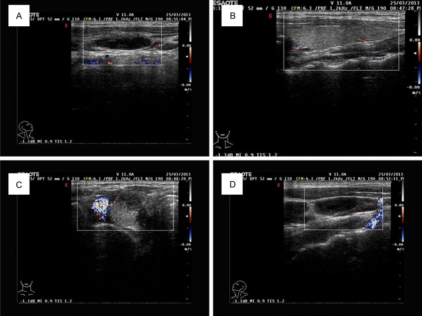
Thyroid and tuberculosis

                                                                                 in the lesion. Hemithyroide-
                                                                                 ctomy was performed with
                                                                                 the diagnose of goiter with
                                                                                 adenomatous preoperatively.
                                                                                 Cut section showed the lesion
                                                                                 filled with colloid alonge with
                                                                                 a focus of caseous necrosis.
                                                                                 Microscopically histopatho-
                                                                                 logic examination revealed
                                                                                 the characteristic features of
Figure 1. Histological images revealed epitheloid cell, Langerhans giant cell,   caseating necrosis, epitheli-
necrotic caseation along with surrounding thyroid follicles (H&E, 100x).         oid cell granulomas and
                                                                                 Langhan’s giant cells (Figure
                                                                                 1). Gram stain and tissue cul-
night sweats, weight loss, fatigue, or anorexia.               ture, stain and culture for AFB in the thyroid
She denied past or family tuberculosis history                 material did not reveal bacteria or mycobacte-
except BCG vaccination history against tuber-                  rium. The final diagnosis was TTB. The patient
culosis. On examination, a solid, non-tender,                  was started on standard antituberculous
painless, 40 mm × 30 mm in size, poor-defined                  therapy postoperatively ie, Rifampicin (R),
margins swelling was noted in the left and                     Isoniazid (H), Ethambutol (E) and Pyrazinamide
anterior neck. The swelling moved with de-                     (Z) for initial phase of 3 months followed by
glutition and its overlying skin was normal,                   continue phase with H, R, E for next 6 months.
suggesting that it originated from the thyroid.                The patient was followed up for 9 months
No enlargement of the regional lymph nodes                     without recurrence and her general wellbeing
was found. Trachea was slightly pushed to the                  was remarkable.
right side. White blood cell (WBC) count in the
                                                               Case 2
peripheral blood was 6.9 × 10⁹/L (normal
range, 3.5-9.5 × 10⁹/L), neutrophil level was                  A 34-year-old female farmer was transferred to
45% and erythrocyte sedimentation rate (ESR)                   our hospital on July 8, 2013 because of the
was 9 mm/h (normal range, 0-26 mm/h).                          pharynx nasalis tuberculous diagnosed histo-
Thyroid profile revealed normal level of the                   pathologically by the specimens obtained from
thyroid hormones [T3 1.37 nmol/L (normal                       nasopharyngoscope in the other hospital. She
range, 1.3-3.1 nmol/L), T4 92.25 nmol/L (nor-                  was presented with a solid, painless swellings
mal range, 66-181 nmol/L), free T3 4.23                        that had slowly increased in size over two
pmol/L (normal range, 3.7-6.8 pmol/L), free                    months in the right side of neck. There was no
T4 13.83 pmol/L (normal range, 12-22                           tuberculosis specific symptoms. She denied
pmol/L), thyroid stimulating hormone (TSH)                     past or family history of tuberculosis except
2.08 mU/L (normal range, 0.27-4.2 mU/L)].                      BCG vaccination history against tuberculosis.
Human immunodeficiency virus testing was                       No signs or symptoms of hypo- or hyperthyroid-
negative. Tumor markers including CA19-9,                      ism were present. A solid, non-tender, painless,
CEA and Calcitonin were within normal range.                   15 mm × 15 mm in size, infiltrative margins
Coagulation profile, liver and renal functions                 swelling was examed in the anterior neck more
were with normal limits. Chest X-ray revealed                  toward to the right side. It had a smooth sur-
no pulmonary abnormalities. TST (1:10,000)                     face with firm consistency, but moved with
was positive (14.5 mm). Ziehl Neelsen stain-                   swallowing. The enlargement of the regional
ings for AFB using sputum in the continuous                    lymph nodes on both sides of the neck was pal-
three mornings were negative. Ultrasonogra-                    pable. Coagulation profile, liver and renal data,
phy of the neck revealed a nodular lesion in-                  tumor markers revealed normal. ESR was 15
volving most of the left lobe of thyroid, the left             mm/h. Thyroid hormones revealed normal (TT3
lobe of thyroid was enlarged, measuring 44                     1.85 nmol/L, TT4 121.40 nmol/L, FT3 4.0
mm × 45 mm × 33 mm in size. The lesion was                     pmol/L, FT4 14.94 pmol/L, TSH 1.65 mU/L).
hypoechoic and heterogeneous in echogeni-                      Human immunodeficiency virus testing was
city. The hypoechoic areas did not extend into                 negative. Chest X-ray revealed no pulmonary
the isthmus. Poor blood flow signal was found                  abnormalities. TST was strongly positive (20

22478                                                          Int J Clin Exp Med 2016;9(11):22477-22485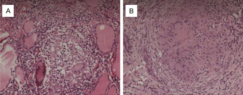
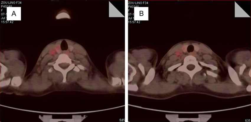
Thyroid and tuberculosis

Figure 2. Thyroid ultrasound revealed an enlarged cervical lymph node on the right lobe of the thyroid (A) and a het-
erogenous hypoechoic lesion with 18 mm of maximal dimension (C). On the left lobe of the thyroid, a similar lesion
with 3 mm of maximal dimension was identified in the enlarged cervical lymph node (B) and the other left lobe was
seen to be normal (D).

mm and a water bulb in skin). Sputum for AFB                   roidectomy was performed with the thyroid
was negative. Ultrasonography revealed a nod-                  malignancy in overstage primary diagnosis pre-
ular lesion involving most of the right lobe of                operatively. Cut section did not show caseous
thyroid. The right lobe of thyroid was enlarged,               necrosis, but histopathological examination
measuring 18 mm × 15 mm × 15 mm in size.                       revealed that the lesions on both lobes of thy-
The lesion was hypoechoic and heterogeneous                    roid and the extracted lymph nodes consisted
and did not extend into the isthmus. Poor blood                of granuloma with caseous necrosis, Langhan’s
flow signal was found in the lesion (Figure 2). A              giant cells and epithelioid cells (Figure 4). Gram
similar swelling which was 3 mm × 3 mm × 2                     stain and tissue cultures, staining and culture
mm in size, heterogeneous hypoechoic lesion,                   for AFB in the specimen showed negative. The
was identified in the left lobe of thyroid by US.              final diagnosis was TTB and tuberculosis lymph-
CT of the neck in the other hospital revealed a                adenitis. There was no postoperative complica-
low density mass in the right lobe of thyroid                  tions. The patient was treated postoperatively
which micked malignancy alonge with some                       by the same anti-tuberculous option prior to a
enlarged cervical lymph nodes. PET-CT exami-                   permanent substitution with L-thyroxin (LT)
nation on July 15, 2007 suggested the right                    (100 ug per day). Thyroid hormones were nor-
lobe lesion of thyroid might be malignancy with                mal (T3 1.95 nmol/L, T4 135.10 nmol/L, FT3
metastatic cervical lymphadenectasis due to                    5.22 pmol/L, FT4 20.36 pmol/L, TSH 0.459
the maximal standard uptake value (SUV) of                     mU/L) at the hospital discharge. Patient was
the lesion was 2.9, its avenue SUV was 2.3, and                asymptomatic and euthyroid during the 8
its value of CT was 65 HU (Figure 3). A total thy-             months follow-up.

22479                                                             Int J Clin Exp Med 2016;9(11):22477-22485
Thyroid and tuberculosis
the symptoms and insuffi-
cient knowledge of TTB, but
in 2-7% of cases during autop-
sy [7]. The reasons for so low
incidence may be [4, 8-10]:
(1) some antagonistic actions
and immunity between myco-
bacterium tuberculosis and
thyroid hormone or iodine; (2)
the affluent blood flow and
oxygenation in thyroid were
not suitable for mycobacteri-
Figure 3. PET-CT revealed areas of increased 18F-fluorodeoxyglucose (18F- um tuberculosis growth. Hen-
FDG) uptake in the right lobe lesion of thyroid with cervical lymphadenecta- ce, unhealthy thyroid may
sis. lose or reduce such protec-
tion against tuberculosis and
develop TTB.
Some investigators [7, 11, 12]
considered that the spread
ways for TTB might include
military spread, direct spread
from an adjacent focus, such
as cervical lymph nodes tu-
berculosis, laryngeal tubercu-
losis, trachea tuberculosis,
mediastinal lymph node tu-
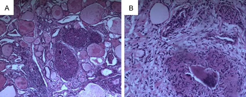
Figure 4. Histological images showed epitheloid cell, Langerhans giant cell, berculosis, and lymphatic
necrotic caseating granuloma along with surrounding thyroid follicles (H&E,
spread. The more common
100x).
way might be miliary spread
as one part of generalized dis-
Discussion semination [4]. Occasionally, military spread
may occur in preexisting thyroid enlargement
Thyroid was regarded in the 19th century as [13]. The lymphatic spread route is controver-
never been infected tuberculosis [3]. However, sial so far based on the literature [9, 13]. Our
TTB was firstly reported by Lebert [2] in 1862 in later patient presented with tuberculosis
a disseminated miliary tuberculosis woman pharynx nasalis which was not contiguous,
whose autopsy showed TTB. Then in 1878, hence the spread was likely to be direct
Chiari [3] described 7 TTB cases in 100 autop- spread or lymphatic spread. However, that
sies of patients died from disseminated tuber- whether the TTB was infected by the tuberculo-
culosis. Bruns [3] in 1893 described the first sis lymphadenitis or the tuberculosis lymphad-
TTB case diagnosed in a woman with a rapidly enitis was by TTB, was not clear yet. The first
enlarging goitre and cervical lymphadenopathy, patient did not have extrathyroid focus or
but no evidence of pulmonary tuberculosis. The disseminated tuberculosis. Hence it was likely
successful drainage of tuberculosis thyroid to be primary TTB in which there was not known
abscess was firstly reported in 1894 [3]. Rankin tuberculosis focus outside of the thyroid.
and Graham [4] identified 21 TTB cases from
20,758 thyroidectomy specimens, an incidence The clinical presentation of TTB is asymptom-
of 0.1%. El Malki HO, et al [5], diagnosed 8 TTB atic and variable. TTB may be primary or associ-
cases in 2,426 partial thyroidectomy speci- ated with disseminated tuberculosis and there
mens, an incidence of 0.3%. In Turkey, the inci- is no consensus on which is the most frequent
dence of TTB was 0.6-1.15% [6]. Physicians [8, 9]. BulbulogluE et al [8] reviewed 76 TTB
diagnosed TTB in 0.1-1% of cases dued to the cases reported from 1905 to 2004 and found
difficulties of differential diagnosis according to that 49 cases were presented most commonly
22480 Int J Clin Exp Med 2016;9(11):22477-22485Thyroid and tuberculosis
with solitary nodules or multinodular goiter, 10 ic. The features of CT, MRI and PET-CT were
cases were abscess, 2 cases were cyst, most less described in the 21 cases reports. Other
cases were primary TTB, and only 10 cases co- investigators [20] reported that CT might show:
existent pulmonary tuberculosis. Anirban (1) an enlarged thyroid and any other infection
Ghosh et al [9] reviewed about 200 TTB cases focus in the neck; (2) the parenchymal lesions
reported before 2006 and found that almost all appear hypodense against the enhancing
cases had primary foci elsewhere in the body, normal thyroid; (3) abscesses, either within
and isolated TTB was extremely rare. We per- the gland or in the subcutaneous plane, show
formed a PubMed and library search using the peripheral rim enhancement; (4) along with or
key words “thyroid tuberculosis” “tuberculosis without regional lymphadenopathy. MRI was
and thyroid” and retrieved detailed articles sporadically reported [21]: (1) the normal thy-
from the English-language literatures after roid gland was homogeneously hyperintense
2006 (Table 1). relative to the neck muscles on both T1- and
T2-weighted images; (2) the tuberculous lesion
The 21 cases in Table 1 revealed a slight mid- showed an intermediate signal on both T1- and
dle-aged female preponderance. There were T2-weighted images, the signal intensity was
12 cases presented with solitary nodule, 5 higher than normal glandular parenchyma
cases with cystic mass or recurrent cyst, 3 due to the presence of densely cellular inflam-
cases with multinodular goiter, 2 cases alonged matory granulation tissue, with tuberculous
with swollen regional lymph nodes, 1 case with granulomas with or without minimal necrosis;
cold abscess, and no case with chronic skin (3) the abscess appeared hypointense on T1-
sinust. Thyroid swelling or neck swelling was and hyperintense on T2-weighted images, and
often the main initial symptom in 21 cases, peripheral rim enhancement on contrast-
alonge occasionally with some specific tubercu- enhanced MRI; (4) regional lymph nodes were
losis symptoms such as intermittent cough, low better seen on T1-weighted images. There
grade fever, night sweating, weight loss, fatigue, was a paucity of reports highlighting the role of
anorexia, emaciation, hemoptysis, and some PET-CT to assess TTB presently. The lesions
hyper- or hypothyroidism symptoms such as were seen as areas of increased 18F-
exophthalmos, hoarseness of voice, palpita- fluorodeoxyglucose uptake in active regions of
tion, dysphagia, dyspnea and irritability. There granulomatous inflammation with cold abscess
were 4 cases with extrathyroid tubercular focus representing necrosed tissue [22]. In our latter
such as pulmonary tuberculosis, cervical spine patient, PET-CT showed the increased
tuberculosis and tuberculosis lymphadenitis. 18F-fluorodeoxyglucose uptake and increased
Like some reports [14-16], tuberculous thyroid- CT value in the lesion.
itis can often be mistaken for carcinoma.
Among the 21 lesions, 11 lesions located in the TTB is difficult to diagnose because the initial
right lobe, 7 lesions in the left lobe and 3 symptom, laboratory examination and the
lesions in both the lobes. All of the lesions appearance of imaging may not provide a sub-
moved with deglutition. PPD was positive or stantial clue or specific information for an accu-
strongly positive. Thyroid function was pre- rate and timely diagnosis. The diagnosis of TTB
served in most of the 21 cases and reports of is frequently delayed and may represent an
thyroid function abnormalities were extremely incidental finding at pathological examination.
infrequent [17, 18]. There were 4 cases with Neither Sometimes PPD was positive or strong-
hyper- or hypothyroidism symptoms in Table 1. ly positive. Some cases co-exsist tuberculosis
These observations might be associated with or tuberculosis history. These important clues
the short disease history of TTB and the small may raise the suspicion of TTB. Most investiga-
number of literature. tors [2, 6, 12, 23] accept the definitive histo-
logical and bacteriological evidences as the
The features of US in the 21 cases, like the diagnostic essential criterion. Histopathological
report [19], may show a solitary nodule, multi- features include caseating necrosis, epithelioid
ple nodules, diffuse goiter, and regional lymph- cell granulomas and Langhan’s giant cells [2, 6,
adenopathy occasionally, but rarely cold 12, 23]. However, many diseases may cause
abscess. The lesion is usually heterogeneous epithelioid granulomatous in thyroid, like granu-
hypoechoic except that the abscess is anecho- lomatous thyroiditis, palpation thyroiditis, fun-
22481 Int J Clin Exp Med 2016;9(11):22477-22485Thyroid and tuberculosis
Table 1. Summary of cases of TTB from English-language literatures in recent 10 years
Case No/Sex/ Co-exsist- TST Thyroid
Presentations Neck USG Examination and diagnosis Treatment and follow-up Ref No.
Age (years) ing illness (mm) function
1/M/65 recurrent thyroid cyst, intermit- active PTB - normal LLT, cystic lesion AFBS (+), TTB diagnosis by FNAC FNAC+local incision, abscess Chuang TJ, et
tent cough drainage, medications al. (1), 2015
2/F/20 Thyromegaly no - normal LLT, 2.2 x 2 x 2-cm, caseating necrosis, epithelioid cell partial thyroidectomy+ De-Tao Yin,
heterogenous granulomas and Langhan’s giant medications/asymptomatic et al. (2012),
hypoechogenicity lesion cells/TTB diagnosis 9-year 2012
3/F/34 neck mass, low fever, night no - normal RLT, 2 x 2 x 2-cm, AFBC (+)/TTB diagnosis partial thyroidectomy+ De-Tao Yin,
sweating cystic lesion medications/asymptomatic et al. (2012),
7-year 2012
4/F/68 aggravate in neck mass, low no - normal RLT, 3.5 x 3 x 3-cm, caseating necrosis, epithelioid cell subtotal thyroidectomy+ De-Tao Yin,
fever, weight loss hypoechogenicity, and granulomas and Langhan’s giant medications/asymptomatic et al. (2012),
swollen lymph nodes cells/RLT-TTB, LLT-goiter 6-year 2012
5/F/28 neck mass, exophthalmos, old PTB - normal RLT, 1.5 x 1.5 x 1-cm, caseating necrosis, epithelioid cell subtotal thyroidectomy, no De-Tao Yin,
emaciated, fatigue hypoechogenicity granulomas and Langhan’s giant medications/asymptomatic et al. (2012),
cells/RLT-TTB+goiter 5-year 2012
6/M/45 rapid neck swelling, cough, ctive PTB 22.00 hypothyroidism RLT, 4.7 cm, AFBC (+) AFBS (-), necrosis, FNAC, 3HREZ/?HR/well Luiz HV, et al.
hemoptysis, ysphagia, evening and AFBC (+) heterogenous epithelioid cell granulomas and 6-months (2), 2013
fever, night sweats, weight loss for sputum hypoechogenicity multinucleated giant cells without
neoplastic cells/TTB
7/F/56 thyroid nodule, multinodular no strong hypothyroidism BLT, 0.5 x 1.1-cm, caseating necrosis, epithelioid total thyroidectomy+ Liwei Meng,
goiter positive hypoechogenicity cell granulomas and Langhan’s selective neck et al. (7), 2014
in both lobes giant cells, BLT-TTB+tuberculosis dissection+6HRE+LL
lymphadenitis+palilary thyroid
cancer
8/F/45 neck swelling, low fever, weight no 18x18 normal RLT, 6 x 5-cm AFBC (+), caseating necrosis, FNAC, 2HREZ/4HR/well Sunita Sanehi,
loss epithelioid cell granulomas and follow-up et al. (59),
Langhan’s giant cells/TTB 2007
9/F/47 neck swelling no negative normal RLT, many hypoechogen- caseating necrosis, epithelioid cell partial thyroidectomy+ Ines Zendah,
ic nodules in both lobes, granulomas and Langhan’s giant 2HREZ/6HR et al. (4),
the maximal ones (3 x cells/TTB+LNTB 2008
1-cm) in the right lobe
10/F/35 neck swelling, hoarseness of no - normal RLT, 4.5 x 3-cm, heterog- AFBS(+), caseating necrosis, partial thyroidectomy, no Sant Parkash
voice enous hypoechogenicity epithelioid cell granulomas and medications Kataria, et al.
Langhan’s giant cells/TTB (10), 2012
11/M/45 neck swelling no strong normal LLT, 3.4 x 2.7 x 2.4-cm, AFBS (-), multifocal granulomatous FNAC+3(HR+ Gao-Yi Yang,
positive inhomogeneously caseous necrosis/TTB ciprofloxacine)/well-3 et al. 2015
hypoechogenicity lesion months
12/M/49 neck swelling, sweating, no - normal RLT, 5.5-cm, heterog- caseating necrosis, epithelioid cell FNAC+thyroidectomy+no Kon Stantinos
evening fever, palpitation, enous hypoechogenicity granulomas and Langhan’s giant medications Terzidis, et al.
weight loss and cystic cells/TTB (1), 2007
13/F/21 neck swelling, fever, weight TB contact positive normal - AFBC (+) AFBS (-), caseation FNAC+3HREZ/6HRE/well Uzma Majid,
loss history necrosis/TTB 6-months et al. 2011
14/F/51 neck swelling no - normal hypoechoic nodule (left), possibility granulomatous (RLT), total thyroidectomy+ Uzma Majid,
small solid and cystic inflammation (LLT) 3HREZ/6HRE+LL et al. 2011
nodules
22482 Int J Clin Exp Med 2016;9(11):22477-22485Thyroid and tuberculosis
15/M/32 solitary nodule in right neck no - normal RLT, 4 x 4-cm, caseation necrosis/TTB FNAC+3HREZ/6HRE Uzma Majid,
multinodular goister et al. 2011
16/M/21 neck swelling, malaise, weak- cervical - normal LLT, hypoechogenicity AFBS (+), caseating necrosis, epithe- FNAC+medications Madhusudhan
ness spine TB nodule lioid cell granulomas and Langhan’s KS, et al. (7),
giant cells/TTB 2009
17/F/26 neck lump no 34.00 normal RLT, 3.5 x 1.8-cm, AFBC (+) FNAC+HREZ Prince Cheri-
cystic mass yan Modayil,
et al. 2009
18/F/11 neck swelling no - normal LLT, 1.8 x 1.6-cm, AFBS (+), caseating necrosis, epithe- FNAC+medications Anita Bodh, et
multi-lymphadenopathy lioid cell granulomas and Langhan’s al. (1), 2014
giant cells/TTB
19/F/34 neck swelling, multi-thyroid hypothyroid - hypothyroid BLT, 2 x 1.5-cm, heterog- AFBS (+), caseating necrosis, epithe- FNAC+subtotal thyroidec- Chaudhary A,
enlargement 10 years enously hypoechogenici- lioid cell granulomas and Langhan’s tomy, no medications et al. 2010
ty in both lobes giant cells, adenomatous goiter/TTB
20/F/49 neck swelling no - normal LLT, 4.5 x 4.4 x 3.3-cm, AFBS (-) AFBC (-) caseating necrosis, partial thyroidectomy+ our cases
heterogenous epithelioid cell granulomas and 3HREZ/6HRE/asymptomatic
hypoechogenicity lesion Langhan’s giant cells/TTB 9-months
21/F/34 neck swelling no - normal RLT, 1.8 x 1.5 x 1.5-cm, AFBS (-) AFBC (-) caseating necrosis, total thyroidectomy+ our cases
hypoechogenicity/LLT, epithelioid cell granulomas and 3HREZ/6HRE+LL/
0.3 x 0.3 x 0.2-cm, Langhan’s giant cells, adenomatous asymptomatic 6-months
hypoechogenicity/ goiter/TTB+LNTB
regional lymph node on
both sides
Abbreviations: – = no detail about TST; F = female; M = male; LT = levothyroxine; AFBS/AFBC = staining/culture for AFB; AFBS (-)/AFBC (-) = staining/culture for AFB was negtive; AFBS (+)/AFBC (+) = staining/culture for AFB was positive; PTB
= pulmonary tuberculosis; LLT/RLT = left/right lobe of thyroid.
22483 Int J Clin Exp Med 2016;9(11):22477-22485Thyroid and tuberculosis
gal infection, tuberculosis, sarcoidosis, goitrous berculosis drugs injection is available [8]. Some
autoimmune thyroiditis, granulomatous vascu- practices in Table 1 had affirmed FNAC more
litis, thyroglossal duct cyst, lipomas or thyroid micro-invasive and simpler than surgery,
neoplasia and foreign body reaction [23, 24]. because FNAC can avoid some unnecessary
Caseating necrosis and the demonstration of thyroidectomy in some events.
AFB in the specimens are the distinguishing
feature to TTB. The smear of AFB in the speci- However, there were still some defects for FNAC
men is not always positive, but the diagnosis is in our opinions, such as subcutaneous haema-
still not be precluded because the simultane- toma, accidental tracheal injury, local infection,
ous culture of AFB reveal positive more fre- subcutaneous tracheal abscess formation and
quently than staining. We should wait patiently the small number of tissue which was not
the culture result for 4-6 weeks. Among the 21 always convenient for histological and bacterio-
cases, there are 10 cases with positive demon- logical examinations. What’s more, with the
stration of AFB in the specimens, 5 cases with development of mini-invasive and fast-recovery
staining positive and 5 cases with culture posi- surgery, and to avoid progression to a thyroid
tive. The small number of case can not explain abscess formation for failed drug therapy, and
the whole incidence of staining positive and with the advantages of the whole removal of
culture positive. Importantly, TTB may coexist some simultaneous lesions such as goiter,
with thyroid carcinoma [14-16]. Some authors hypothyroidism, hyperthyroidism and carcino-
highly recommended FNAC and US-guided ma, surgery is still necessary. Surgical removal
FNAC biopsy for the timely and accurate diag- or thyroidectomy should occur in the large
nosis. The tissue obtained by FNAC was exam- lesion, infected cysts, TTB case coexisting with
ined pathologically and stained, cultured for thyroid carcinoma, mimicking malignancy
AFB. We summarized some diagnosis points lesions [24], and the cases whose diagnoses
and clues as following: (1) young adults, espe- are not confirmed by FNAC. Surgical drainage
cially women; (2) people with tuberculosis his- should be performed in abscess and subcuta-
tory or close contact with tuberculosis history, neous abscess formed after FNAC. Sole che-
or the existence of other parts of tuberculosis motherapy or FNAC may be performed in the
lesions, or there were some tuberculosis symp- small nodule and non-abscess case. Thyroid
toms; (3) painless neck mass; (4) short-term function should be monitored during TTB treat-
diagnostic antituberculosis treatment affirm ment [8]. In the 21 cases and all other previous
significant improvement; (5) the lesions with reports, no death and recurrence were found
caseous necrosis and (or) the demonstration of and TTB responded well to the medications and
AFB were found after surgery or FNAC. some surgeries. Its overall treatment effect
Treatment for TTB is mainly based on antituber- was satisfactory.
culosis medications and additional surgical
Summary
removal or drainage. Drug regimen and dura-
tion vary among authors and different tubercu- Thyroid tuberculosis is rare and difficult to dis-
losis diseases. We chose H, R, E and Z for initial cern clinically and on examinations. A greater
phase of 3 months followed by continuing awareness of the differential diagnosis of thy-
phase with H, R and E for next 6 months
roid masses is necessary. Its final diagnosis is
(3HREZ/6HRE). Some patients in the litera-
made on the definitive histological or bacterio-
tures [2, 10, 15, 17, 18, 24] performed H, R, E
logical evidence by surgery or FNAC. The overall
and Z for initial phase of 2 to 3 months followed
response to antituberculous therapy with or
by H, R and with or without E for next 4 to 6
without surgery or FNAC is good.
months [2~3HREZ/4~6HR(E)]. Among the 21
cases, 2 cases did not accept any medications Acknowledgements
after surgery, but responded well to the sole
surgery. Some authors did not recommend a This work was partly supported by Department
total thyroidectomy due to consequent hypothy- of pathology from Guangzhou Chest Hospital.
roidism. A total thyroidectomy was performed
in our latter patient and no findings of hypothy- Disclosure of conflict of interest
roidism by the permant therapy of LL in the fol-
low-up. The methods of percutaneous antitu- None.
22484 Int J Clin Exp Med 2016;9(11):22477-22485Thyroid and tuberculosis
Address correspondence to: You-Ding Jiang, [12] Chandanwale SS, Buch AC, Vimal SS, Sachdeva
Department I of Thoracic Surgery, Guangzhou Chest P. Thyroid tuberculosis: presenting symptom of
Hospital, Tuberculous Institute of State Key La- mediastinal tuberculous lymphadenitis-an un-
boratory of Respiratory Disease, No. 62, Heng- usual case. Indian J Tuberc 2014; 61: 84-7.
zhigang Road, Yuexiu District, Guangzhou 510095, [13] Coller FA, Huggins CB. Tuberculosis of the thy-
roid gland: a review of the literature and report
Guangdong, People’s Republic of China. E-mail: jyd-
of five new cases. Ann Surg 1926; 84: 804-20.
mgkyc@sina.com
[14] Al-Mulhim AA, Zakaria HM, Abdel Hadi MS, Al-
Mulhim FA, Al-Tamimi DM, Wosornu L. Thyroid
References
tuberculosis mimicking carcinoma: report of
two cases. Surg Today 2002; 32: 1064-7.
[1] World Health Orgnization. Global tuberculosis
[15] Meng L, Hu S, Huang L, Xu C. Papillary thyroid
report 2012. WHO, Geneva,Switzerland,2012.
cancer coexisting with thyroid tuberculosis: a
www.who.int/entity/tb/publications/global_
case report. Oncol Lett 2014; 7: 1563-1565.
report/en/.
[16] Ishinaga H, Hamaguchi N, Suzuki H, Miyamura
[2] Sanehi S, Dravid C, Chaudhary N, Rai
T, Nakamura S, Otsu K, Takeuchi K. Case of
AK. Primary thyroid tuberculosis. Indian J
papillary cancinoma of the thyroid gland with
Otolaryngol Head Neck Surg 2007; 59: 154-
concurrent tuberculos lymphadenitis. Nihon
156.
Jibiinkoka Gakkai Kaiho 2013; 116: 1315-9.
[3] Barnes P, Weatherstone R. Tuberculosis of the
[17] Majid U, Islam N. Thyroid tuberculosis:a case
thyroid: two case reports. Br J Dis Chest 1979;
series and a review of the literature. J Thyroid
73: 187-91.
Res 2011; 2011: 359864.
[4] Fred W, Rankin A. Stephens Graham.
[18] Raman L, Murray J, Banka R. Primary tubercu-
Tuberculosis of the thyroid gland. Ann Surg
losis of the thyroid gland:an unexpected cause
1932; 96: 625-648.
of thyrotoxicosis. BMJ Case Rep 2014; 2014.
[5] El Malki HO, Mohsine R, Benkhraba K,
[19] Yang GY, Zhao D, Zhang WZ, Meng J, Li J, Li XH,
Amahzoune M, Benkabbou A, El Absi M, Ifrine
Wan HF. Role of ultrasound evaluation for the
L, Belkouchi A, Balafrej S. Thyroid tuberculosis:
diagnosis and monitoring of thyroid tuberculo-
diagnosis and treatment. Chemotherapy
sis: A case report and review of the literature.
2006; 52: 46-9.
Oncol Lett 2015; 9: 227-230.
[6] Akbulut S, Sogutcu N, Arikanoglu Z, Bakir S,
[20] Kang BC, Lee SW, Shim SS, Choi HY, Baek SY,
Ulku A, Yagmur Y. Thyroid tuberculosis in south-
Cheon YJ. US and CT findings of tuberculosis of
eastern Turkey: is this the resurgence of a
the thyroid gland:three case reports. Clin
stubborn disease? World J Surg 2011; 35:
Imaging 2000; 24: 283-6.
1847-52.
[21] Madhusudhan KS, Seith A, Khadgawat R, Das
[7] Simkus A. Thyroid tuberculosis. Medicina
P, Mathur S. Tuberculosis of the thyroid gland:
(Kaunas) 2004; 40: 201-4.
magnetic resonance imaging appearances.
[8] Bulbuloglu E, Ciralik H, Okur E, Ozdemir G,
Singapore Med J 2009; 50: e235-8.
Ezberci F, Cetinkaya A. Tuberculosis of the thy-
[22] S Harkirat, SS Anana, LK Indrajit, AK Dash.
roid gland: review of the literature. World J Surg
Pictorial essay: PET/CT in tuberculosis. Indian
2006; 30: 149-55.
J Radiol Imaging 2008; 18: 141-147.
[9] Ghosh A, Saha S, Bhattacharya B,
[23] Kataria SP, Tanwar P, Singh S, Kumar S.
Chattopadhay S. Primary tuberculosis of thy-
Primary tuberculosis of the thyroid gland: a
roid gland: a rare case report. Am J Otolaryngol
case report. Asian Pac J Trop Biomed 2012; 2:
2007; 28: 267-70.
839-40.
[10] Luiz HV, Pereira BD, Silva TN, Veloza A, Matos
[24] Zendah I, Daghfous H, Ben Mrad S, Tritar F.
C, Manita I, Cordeiro MC, Raimundo L, Portugal
Primary tuberculosis of the thyroid gland.
J. Thyroid tuberculosis with abnormal thyroid
Hormones (Athens) 2008; 7: 330-3.
function-case report and review of the litera-
ture. Endocr Pract 2013; 19: e44-9.
[11] Das SK, Bairagya TD, Bhattacharya S, Barman
DC. Tuberculosis of the thyroid gland. Indian J
Lepr 2012; 84: 151-4.
22485 Int J Clin Exp Med 2016;9(11):22477-22485You can also read