Wound Infections From Taiwan Cobra Bites: Determining Bac-teriology, Antibiotic Susceptibility, and the Use of Antibiotics- A Cobra BITE Study
←
→
Page content transcription
If your browser does not render page correctly, please read the page content below
Preprints (www.preprints.org) | NOT PEER-REVIEWED | Posted: 27 January 2021

     Article

     Wound Infections From Taiwan Cobra Bites: Determining Bac-
     teriology, Antibiotic Susceptibility, and the Use of Antibiotics-
     A Cobra BITE Study
     Heng Yeh 1,2, Shi-Ying Gao 1 and Chih-Chuan Lin 1,2*

                                       1 Department of Emergency Medicine, Lin-Kou Medical Center, Chang Gung Memorial Hospital, Taoyuan
                                         33305, Taiwan; bearuncle@yahoo.com (C.-C.L.) ; tsubame800329@gmail.com(H.Y.)
                                       2 College of Medicine, Chang Gung University, Taoyuan 33302, Taiwan

                                       * Correspondence: bearuncle@yahoo.com

                                       Abstract: Taiwan cobra (Naja atra) bites account for approximately 20% of all venomous snakebites
                                       in Taiwan. In Taiwan, the rates of wound necrosis and secondary infection from Taiwan cobra bites
                                       are higher than those associated with other venomous snakebites. Clinical tools to evaluate the in-
                                       fection risk after Taiwan cobra bites are lacking. Therefore, in this study, we developed a useful
                                       clinical tool to evaluate the infection risk after Taiwan cobra bites. Moreover, we investigated wound
                                       infection bacteriology. We analyzed the data of patients bitten by N. atra who had undergone freeze-
                                       dried neurotoxic antivenin treatment in emergency rooms of the Chang Gung Memorial Hospital
                                       network, which comprises seven hospitals and the largest medical system in Taiwan, from January
                                       2001 to May 2017. Because patients with wound necrosis required antibiotics for infection treatment,
                                       we included only patients with wound infection but without tissue necrosis in the development of
                                       our Cobra Bacteriology of Infections in Taiwanese snake Envenomation (Cobra BITE) score by using
                                       univariate and multiple logistic regression. In total, 8,295,497 emergency department visits occurred
                                       from January 2001 to May 2017, and 195 patients were diagnosed has having cobra bites. Among
                                       them, 23 and 30 patients had wound necrosis and wound infection, respectively. The wound infec-
                                       tion rate was 27.2% (53/195). Regardless of whether the patients had necrosis, Enterococcus faecalis
                                       and Morganella morganii were the main bacteria identified in the culture report. Gentamicin, ceftri-
                                       axone, ciprofloxacin, and levofloxacin are the ideal first-line antibiotics for treating N. atra bite
                                       wounds in Taiwan. As per our Cobra BITE score, the three factors predicting secondary wound
                                       infection after cobra bites are hospital admission, a white blood cell count (in 103/µL) × by neutro-
                                       phil–lymphocyte ratio value of ≥114.23, and the use of antivenin medication. The area under the
                                       receiver operating characteristic curve for the Cobra BITE score system was 0.88. The ideal sensitiv-
                                       ity and specificity were 0.89 and 0.76, respectively, and the optimal cutoff point for Cobra BITE score
                                       was 7. The Hosmer–Lemeshow p value was 0.4. In conclusion, our Cobra BITE study established a
                                       new practical clinical tool for clinicians to evaluate infection risk after N. atra bites. This score system
                                       enables the assessment of wound infections after N. atra bites, and it could be modified and im-
                                       proved in future for other Naja spp. bites.

                                       Keywords: wound infections; snakebites; Taiwan cobra; Naja atra

                                       Key Contribution: In this study, we investigate the bacteriology, antibiotic susceptibility in Taiwan
                                       cobra snakebite wounds. We further developed a clinical scoring system (Cobra BITE score) to pre-
                                       dict wound infection development and, hence the necessity of antibiotic initiation. Physicians can
                                       use the Cobra BITE score to guide appropriate antibiotic use and offer the medical community a
                                       framework for stratifying the risk of developing a wound infection after a cobra bite.

               © 2021 by the author(s). Distributed under a Creative Commons CC BY license.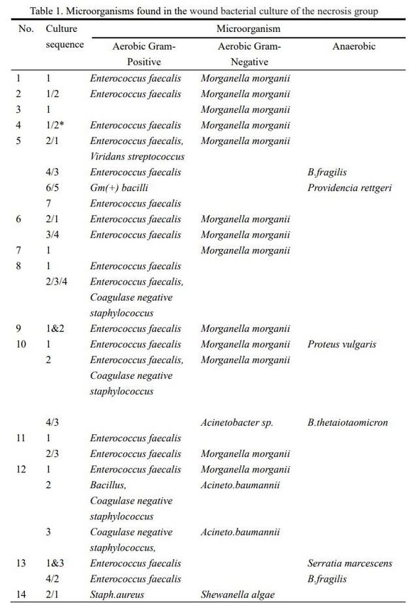
Preprints (www.preprints.org) | NOT PEER-REVIEWED | Posted: 27 January 2021

                                 1. Introduction
                                       Taiwan, a subtropical island, has many types of snakes. Clinically, six species of ven-
                                 omous snake are considered important, namely Naja atra (Taiwan cobra or Chinese cobra),
                                 Bungarus multicinctus, Protobothrops mucrosquamatus, Trimeresurus stejnegeri, Deinagkistro-
                                 don acutus, and Daboia siamensis. Among these, N. atra are responsible for approximately
                                 20% of venomous snakebites in Taiwan [1]. Although Taiwan cobra bites do not cause
                                 neurological complications (bites from other cobra species do), they cause wound tissue
                                 damage/necrosis and confer a high risk of wound infection [2–4]. To our knowledge, alt-
                                 hough some studies have investigated the bacteriology of cobra bite wounds [5,6], guide-
                                 lines for differentiating patients based on whether they have wound infections are lacking.
                                 Wound necrosis is a common complication associated with cobra bites [2], and affected
                                 patients may be treated with antibiotics. Therefore, knowledge on whether nonnecrotic
                                 cobra wounds are infected is important. Furthermore, to provide appropriate antibiotics
                                 to patients with wound infections, wound bacteriology must be investigated. Therefore,
                                 this study investigated the wound bacteriology of necrosis wounds and infected wounds
                                 without necrosis to develop a clinically useful wound infection prediction rule [Cobra
                                 Bites/Bacteriology of Infections in Taiwanese snake Envenomation (Cobra BITE) study] to
                                 guide antibiotic use (Figure 1).

                                       Figure 1. Study flow diagram for Cobra BITE score.

                                 2. Results

                                 2.1. Patient Characteristics
                                      In total, data on 195 patients with cobra bites were retrieved from data on 8,295,497
                                 emergency department (ED) visits from January 2001 to May 2017. The XXXXX XXXX
                                 Research Database (CGRD) contains data on 1,128 patients with snakebites (0.01% of total
                                 ED visits). Men accounted for the majority of this study population (n = 144, 73.8%; mean
                                 age: 49.97 ± 17.42 years). Generally, two vials (median) of freeze-dried neurotoxic anti-
                                 venin (interquartile range: 1–4) were administered to each patient, suggesting that these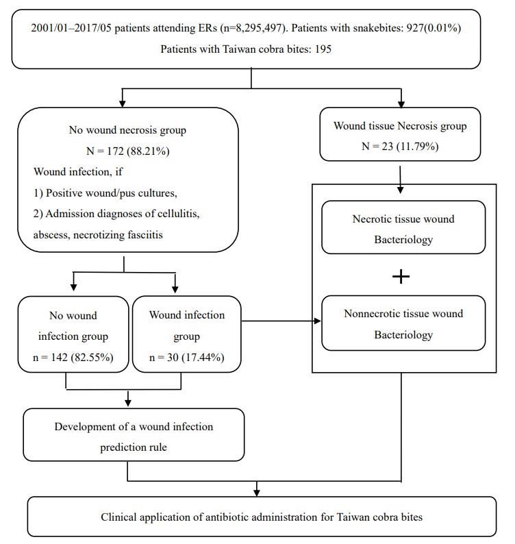
Preprints (www.preprints.org) | NOT PEER-REVIEWED | Posted: 27 January 2021

                                 patients were mostly mildly to moderately envenomed. A total of 74 patients (37.9%) re-
                                 quired hospital admission.
                                      Of these 195 patients, 53 (27.2%) developed wound infections. Among them, 23 and
                                 30 patients had wound tissue necrosis and wound infections, respectively. These 53 pa-
                                 tients with wound infections received larger antivenin doses [median (interquartile range)
                                 = 2 (1–3) vs 2 (1–4), p = 0.002], had more admissions (n = 47 vs 27, p < 0.0001), and had
                                 longer hospital stays (3.07 ± 2.67 vs 16.09 ± 13.99, p < 0.0001) than patients without in-
                                 fected wounds, implying that their clinical conditions were severe. Patients with Taiwan
                                 cobra bites lacked neurological complications but had the most severely infected wounds,
                                 and they underwent the most serious envenomation and surgical procedures, such as deb-
                                 ridement, fasciotomy, and graft. Of the 195 patients with cobra bites, 23, 16, and 15 pa-
                                 tients received debridement, fasciotomy, and graft, respectively.

                                 2.2. Wound Conditions of the Tissue Necrosis Group
                                       Of the 23 patients, 14 had positive wound culture findings (positive culture rate,
                                 60.87%), and among them, 8 patients received all the following surgical procedures: deb-
                                 ridement, fasciotomy, and graft. Of these 14 patients, 3, 2, and 1 accepted debride-
                                 ment/graft, debridement, and debridement/fasciotomy, respectively. Swab cultures for
                                 both aerobic and anaerobic bacteria were taken during debridement/fasciotomy proce-
                                 dures. If prominent pus or wound discharge was noted, swab cultures were collected and
                                 analyzed for both aerobic and anaerobic bacteria. Of these 14 patients with wound necro-
                                 sis, 6 and 8 had monomicrobial and polymicrobial infections, respectively. The most com-
                                 monly cultured aerobic Gram-negative and Gram-positive bacteria were Morganella mor-
                                 ganii and Enterococcus faecalis, respectively, and both were found in 11 of the 14 patients.
                                 Coagulase-negative staphylococcus was the third leading pathogen in this study. How-
                                 ever, anaerobic pathogens were identified in the cultures of only six patients (four and
                                 two in the necrosis and nonnecrosis groups, respectively).
                                       Notably, wound cultures of some patients were obtained several times of even up to
                                 seven times during their hospitalization course. E. faecalis and M. morganii were the two
                                 most commonly obtained bacteria in first-time wound cultures. Moreover, anaerobic bac-
                                 teria such as Bacteroides fragilis, Providencia rettgeri, Proteus vulgaris, and Serratia marcescens
                                 were observed in patients whose wound cultures were obtained multiple times when they
                                 underwent several surgical procedures (Table 1). The results of the antibiotic sensitivity
                                 tests are included in Table S1.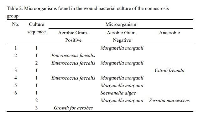
Preprints (www.preprints.org) | NOT PEER-REVIEWED | Posted: 27 January 2021
2.3. Wound Conditions of the Nonnecrosis Group
In contrast to the wound necrosis group, only six patients had positive wound cul-
tures in nonnecrosis group (n = 172), with three each having monomicrobial and polymi-
crobial infections. Five cultures showed M. morganii growth and two showed E. faecalis
growth (Table 2). Among these six patients, only one (Case 6) received fasciotomy and
graft because of clinically diagnosed compartment syndrome.
2.4. Development of the Clinical Prediction Rule for Wound Infections (Cobra BITE Score)
Among the 195 patients with cobra bites, 23 with tissue necrosis who required anti-
biotics for wound management were not included in the analysis of Cobra BITE score. In
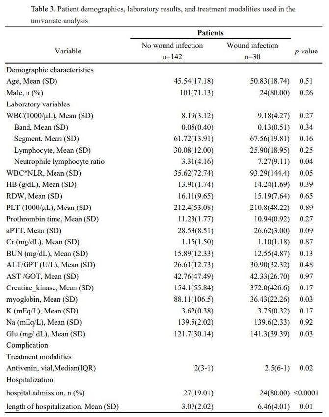
total, 172 patients without tissue necrosis were enrolled and divided into wound infection
and no wound infection groups. Men accounted for the majority of the patients in both
groups, but no statistical differences were observed in both age and sex. The wound in-
fection group had higher white blood cell (WBC) counts, neutrophil counts (band and
segment), and neutrophil–lymphocyte ratios (NLRs), but no statistical differences were
observed among them, except in NLR (p = 0.04). Moreover, the wound infection group
had high levels of myoglobin and blood glucose (p = 0.03 for both). However, no statistical
differences were noted in the following clinical characteristics: platelet count, prothrom-
bin time (PT), activated partial thromboplastin time (APTT), blood urea nitrogen (BUN),
creatinine (Cr), sodium, and potassium. In terms of treatment and outcome, patients in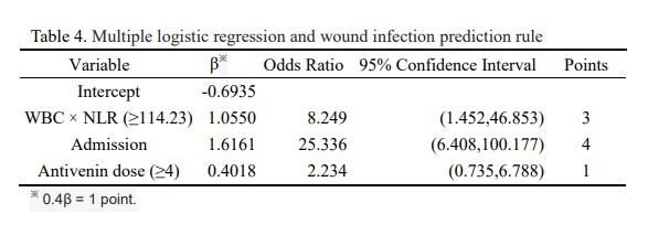
Preprints (www.preprints.org) | NOT PEER-REVIEWED | Posted: 27 January 2021

                                 the wound infection group received more antivenin (p = 0.02), had a higher admission rate
                                 (p < 0.0001), and had a longer hospitalization time (p = 0.01) than those in the noninfection
                                 group (Table 3).

                                      Clinical characteristics with a p value of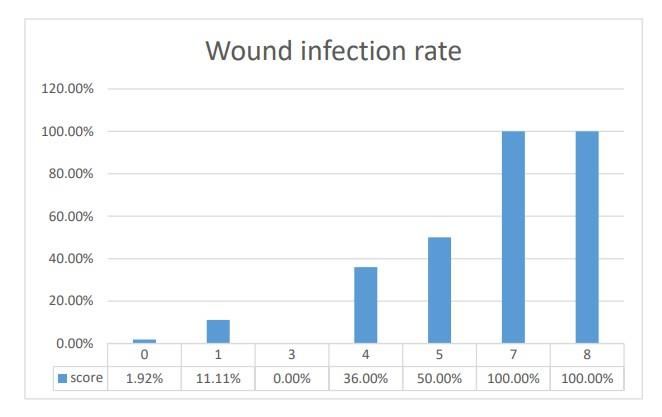
Preprints (www.preprints.org) | NOT PEER-REVIEWED | Posted: 27 January 2021
Preprints (www.preprints.org) | NOT PEER-REVIEWED | Posted: 27 January 2021

                                      Figure 2. Cobra BITE scores and wound infection rates.

                                      Figure 3. Receiver operator characteristic curve of Cobra BITE score

                                 3. Discussion
                                       Taiwan cobra bites account for 20% of venomous snakebites in Taiwan [1]. The inci-
                                 dence of cobra bites in our study was 21.03%. This was consistent with previous studies
                                 [1, 7, 8]. To our knowledge, our study is the largest multicenter study to examine the bac-
                                 teriology and prediction factors of cobra bite wounds. The infection rate of Taiwan cobra
                                 bites varied has varied from 28% [3] to 68% [4] and as high as 77% [5] in different studies.
                                 We found that 27.17% of our patients with cobra bites had wound infections; this rate was
                                 low compared with those in previous studies. One of the reasons for the discrepancy in
                                 infection rates may be the different geographic distribution of the Taiwan cobra. The high-
                                 est cobra bite wound infection rate was in central Taiwan, which had the highest cobra
                                 bite incidence [1]. The second reason may be that the study hospital is the main center in
                                 central Taiwan where patients with snakebites are referred, and therefore, patients with
                                 severe cobra envenomation were transferred there, giving a falsely high infection rate. In
                                 our study, we found that patients with severe envenomation had wound infection.

                                 3.1. Bacteriology, Culture Times, and Choice of Antibiotics for Patients with Taiwan Cobra Bites
Preprints (www.preprints.org) | NOT PEER-REVIEWED | Posted: 27 January 2021
Considering the bacterial culture of wound infections, irrespective of whether the
patients had wound tissue necrosis, snakebite wound infection is usually polymicrobial,
with M. morganii and E. faecalis being the most often identified pathogens [3,5,6,9]. There-
fore, using gentamicin, ceftriaxone, ciprofloxacin, or levofloxacin as the first-line mono-
therapy drug is reasonable for managing Taiwan cobra bite wound infections. However,
the cytotoxins of the Taiwan cobra cause considerable local tissue necrosis with swelling
[10, 11], and patients may receive multiple surgical procedures with the use of metroni-
dazole, augmentin, or piperacillin/tazobactam to cover both aerobic and anaerobic micro-
organisms [12]. In this study, Pseudomonas aeruginosa was not observed similar to previous
studies [3,5,6]. This finding suggested that the choice of antibiotics used should be based
on local bacterial patterns according to drug susceptibility tests. Therefore, ureidopenicil-
lins such as piperacillin should be reserved for patients prone to P. aeruginosa infection. In
conclusion, we recommended the stepwise use of antibiotics and the employment of ap-
propriate surgical interventions, such as debridement, to treat wound infections from Tai-
wan cobra bites successfully.
3.2. Factors Related to Cobra BITE Score and Its Association with a Secondary Bacterial
Infection from Taiwan Cobra Bites
Although moderate to high superimposed bacterial infections have been observed in
patients with Taiwan cobra bites, no clinical rules have been compiled to acknowledge
this. Some studies have suggested that snakebite wound infections should be considered
a special infection type that requires objective measurements [5, 13]. Our Cobra BITE score
was conceived for this purpose.
The Cobra BITE score has three components: WBC × NLR, hospital admission, and
antivenin dose. Generally, these three items are associated with severity degree, as sug-
gested by other studies, and moderate or patients with severe snakebite envenomation
often experience wound infections [14–16]. Using this score is convenient because
WBC/NLR, hospital admission data, and antivenin dose can be determined in the early
stage of snakebite treatment. Thus, appropriate antibiotics can be administrated to suita-
ble patients.
In this study, hospital admission was the most predictor of wound infections. Doctors
admit patients with severe bites (such as those who received a high antivenin dose) to
hospital for further care because patients with moderate or severe snakebites often de-
velop wound infections. Therefore, secondary wound infections in patients with snake-
bites are sufficiently serious to warrant hospital admission.
Second, both WBC and NLR are biomarkers routinely used to determine infection.
Several diseases and clinical conditions, such as trauma, infection, sepsis, operation, ma-
lignancy, and emotional instability, can cause leukocytosis. Generally, any factor that re-
sults in stress can induce leukocytosis [17]. Moreover, a study showed WBC elevation to
be a risk factor for post-snakebite compartment syndrome [18]. Conversely, compared
with leukocytosis, NLR increase can be detected earlier (when patients have a proinflam-
matory state), and this can make clinicians aware of potential infections and systemic in-
flammation [19]. In experimental studies using dogs, neutrophil concentration increased
and a left shift occurred between 4 and 12 h after bacterial inoculation [20]. As patients
with snakebites often seek medical help within 4 hours in Taiwan, using NLR to predict
wound infection is reasonable. Some studies have indicated that an NLR increase is re-
lated to vascular endothelium injury, which can result in poor wound healing [21]. NLR
has been used to evaluate diabetes mellitus–related foot infections [22], vascular ischemia
of the limbs [23], and sepsis [24,25]. Therefore, we integrated WBC and NLR to evaluate
wound healing and inflammation precisely. If additional studies can confirm that some
other local/circulatory inflammatory mediators or venom concentration are sensitive and
precise for predicting wound infections, such inflammatory mediators can be incorpo-
rated into the Cobra BITE score to further improve its utility at predicting wound infec-
tions in patients with snakebites.Preprints (www.preprints.org) | NOT PEER-REVIEWED | Posted: 27 January 2021
3.3. Utility of the Cobra BITE Score
Our score system has some advantages for clinical decision making. First, its primary
use is to enable the judicious use of antibiotics in patients with Taiwan cobra bites without
wound tissue necrosis. According to local bacteriological data, antibiotic use can be suit-
able for patients with cobra bite wound infections with and without tissue necrosis. Sec-
ond, these decision-making processes can be extended to patients bitten by other Naja
snakes. Naja bites are major contributors to the snakebite burden globally [26]. Bites from
different Naja species share common features of local tissue swelling, inflammation/infec-
tion, and substantial tissue necrosis. These Naja species are N. atra (Taiwan cobra) [27],
Naja siamensis (Thai spitting cobra) [28], Naja kaouthia (monocellate cobra) [29,30], Naja naja
(Indian cobra) [31,32], and Mozambique spitting cobra (Naja mossambica) [33,34]. Our Co-
bra BITE score offers the medical community a framework for stratifying the risk of
wound infection after cobra bites, and it can be improved upon and refined further. Future
studies on the aforementioned Naja species can be conducted to develop individual Cobra
BITE scores, enabling the judicious use of antibiotics in patients with cobra bites. Further-
more, studies can be conducted to investigate the use of antibiotic prophylaxis in patients
with cobra bites at a high risk of wound infections.
3.4. Limitations
Our study has some limitations. This was a retrospective study, and the study design
has inherent limitations (such as recall bias). However, we attempted to overcome these
innate limitations through systematically retrieving data from the CGRD, which is based
on original electronic medical records. However, the results should be interpreted with
caution. The second limitation of this study is that the Cobra BITE score has not yet been
validated. Nevertheless, the Hosmer–Lemeshow analysis of our Cobra BITE score sup-
ports the model’s stability.
4. Conclusions
We offer a treatment and study framework for cobra bite wound infections. Our rec-
ommendations are as follows: First, use antibiotics in a stepwise manner for all patients
with wound tissue necrosis. Gentamicin, ceftriaxone, ciprofloxacin, and levofloxacin are
reasonable first-line monotherapies. Second, if patients receive multiple surgical proce-
dures, then the use of metronidazole, augmentin, and piperacillin/tazobactam is encour-
aged to cope with the possible anaerobic wound infection. Third, ureidopenicillin should
be reserved for patients at risk of Pseudomonas spp. infection. Fourth, the Cobra BITE score
should be used to identify patients at a high risk of wound infection, and antibiotics
should be used in a stepwise manner as aforementioned. Last, our Cobra BITE score can
also be employed in further studies regarding the Naja spp.-associated wound infections.Preprints (www.preprints.org) | NOT PEER-REVIEWED | Posted: 27 January 2021
5. Materials and Methods
5.1. Ethics Statement
To meet research ethics standards, this study was conducted after permission was
received from the XXXXX XXX Memorial Hospital (XXX) Research Ethical Committee,
Taoyuan, Taiwan (Approval No: 201800736B0, date of approval: May 21, 2018). The re-
quirement of consent from study participants was waived in accordance with relevant
guidelines and regulations.
5.2. Data Resource and Setting
The XXXX network, a private hospital network of seven hospitals in Taiwan, estab-
lished in 1976, is located in the northeastern and southern regions of Taiwan. The CGMH
network is the largest medical network in Taiwan, and this medical group has approxi-
mately 10,070 beds and handles >280,000 admissions every year. Thus, 10.2% of patients
admitted to a hospital in Taiwan each year attend a XXXX branch. Outpatient visits and
ED visits to CGMH branches are approximately 8,500,000 and 500,000 per year, respec-
tively [35].
The variables analyzed in this study were retrieved from the CGRD, a computerized
deidentified database, which is systematically updated to include new data generated an-
nually. The CGRD was derived from original XXXX medical records. Its overall coverage
of 21.2% and 12.4% of outpatient and inpatient records, respectively, means its data can
provide a good foundation for high-quality and scientifically sound studies [36].
5.3. Patients Enrolled
All patient with Taiwan cobra bites who presented to the ED of a XXXX and received
at least one vial of freeze-dried neurotoxic antivenin (the designated antivenin for Taiwan
cobra bites) between January 2001 and May 2017 were identified using the International
Statistical Classification of Diseases, 9th and 10th revision codes for diagnosing snakebites
or determining antivenin administration. Patients receiving any other antivenin medica-
tion were excluded.
The following variables were collected: demographic characteristics (patient age and
sex); laboratory variables such as complete blood count with differential count, NLR, he-
moglobin, red blood cell distribution width, platelet count, PT, APTT, BUN, Cr, alanine
aminotransferase, aspartate aminotransferase, myoglobin, potassium, sodium, and blood
glucose. Data on the bacteriology of wounds and pus culture, such as antimicrobial drug
susceptibility of microorganisms obtained from swabs of snakebite wounds, were also
retrieved. Information on treatment modalities such as antivenin doses, type of surgical
procedure (debridement, fasciotomy, or graft), hospital admission, and hospitalization
length was retrieved. Based on a previous study, the term polymicrobial infection was
used to describe the growth of two or more microorganisms on the same infected wound
[37].
5.4. Management Protocol for Patients with Venomous Snakebite
The management protocol for patients with venomous snakebites was adequately
described in our previous work [9]. A short-term study revealed that all patients who
presented with a snakebite to our EDs were managed in accordance with World Health
Organization guidelines [38]. After the culprit snake was identified (through a pictorial
chart available in our EDs and compatible symptoms and signs), antivenin was appropri-
ately administrated to patients. Patients were then monitored in the ED for 24 h post-bite
until clinical improvement was observed in limb pain and swelling, after which those who
were hemodynamically stable were allowed to go home. If limb swelling reappears after
adequate antivenin administration, or if clinical signs of cellulitis were observed, these
patients were diagnosed as having a wound infection and were admitted to our wards.Preprints (www.preprints.org) | NOT PEER-REVIEWED | Posted: 27 January 2021
Patients with wound necrosis may receive surgical procedures such as debridement, fasci-
otomy, or tissue graft after thorough clinical consideration. This protocol remained con-
sistent throughout our study period.
5.5. Definitions of Wound Infection and Wound Necrosis
Patients were considered to have infected snakebite wounds if they satisfied one of
the following criteria: (1) had positive wound cultures, (2) had admission diagnoses of
cellulitis, abscess, or necrotizing fasciitis, or (3) underwent surgical wound debridement.
We defined patients who received debridement as those who had wound necrosis because
such patients require this procedure.
5.6. Statistical Analysis and Development of the Cobra BITE Score
Categorical variables are reported as frequencies and percentages, whereas continu-
ous variables are expressed as mean ± standard deviation, unless otherwise indicated. For
univariate analyses, we conducted Student’s t-test and a chi-squared test to evaluate nu-
merical and categorical variables separately. To assess the strength of the association be-
tween two groups and express statistical differences, odds ratios and 95% confidence in-
tervals were respectively used. Variables with p < 0.1 were identified as possible predictors
for further multivariate analysis using multiple logistic regression. We used Youden’s in-
dex or the most optimal value(s) to determine the cut-off points for Cobra BITE score var-
iables. Through weighting these variables according to their β coefficients, the Cobra BITE
score was then created. A Hosmer–Lemeshow analysis was used to test the model’s sta-
bility. Finally, the ROC curves and area under the curve were used to determine the accu-
racy of this prediction model. A p value ofPreprints (www.preprints.org) | NOT PEER-REVIEWED | Posted: 27 January 2021
Conflicts of Interest: The authors have no conflicts of interest to declare. The funders had no role
in the design of the study; the collection, analysis, or interpretation of the data; the writing of the
manuscript; or the decision to publish the results.
References
1. Chen, C.K.; Lin, C.C.; Shih, F.Y.; Chaou, C.H.; Lin, J.C.C.; Lai, T.I.; Tseng, C.Y.; Fang, C.C. Population-based study of venomous
snakebite in Taiwan. J. Acute Med. 2015, 5, 38–42.
2. Mao, Y.C., et al., Naja atra snakebite in Taiwan. Clin Toxicol (Phila), 2018. 56(4): p. 273-280.
3. Huang, L.W.; Wang, J.D.; Huang, J.A.; Hu, S.Y.; Wang, L.M.; Tsan, Y.T. Wound infections secondary to snakebite in central
Taiwan. J. Venom. Anim. Toxinsincl Trop. 2012, 18, 272–276.
4. Hsieh, Y.H.; Hsueh, J.H.; Liu, W.C.; Yang, K.C.; Hsu, K.C.; Lin, C.T.; Ho, Y.Y.; Chen, L.W. Contributing factors for complications
and outcomes in patients with snakebite: Experience in a medical center in southern Taiwan. Ann. Plast. Surg. 2017, 78, S32–S36
5. Mao, Y.C., et al., Bacteriology of Naja atra Snakebite Wound and Its Implications for Antibiotic Therapy. Am J Trop Med Hyg, 2016.
94(5): p. 1129-35.
6. Chen, C.M.; Wu, K.G.; Chen, C.J.; Wang, C.M. Bacterial infection in association with snakebite: A 10-year experience in a north-
ern Taiwan medical center. J. Microbiol. Immunol. Infect. 2011, 44, 456–460.
7. Tu, T.M. Statistical studies on victims of poisonous snakebites in Formosa. J. Formos. Med. Assoc. 1941, 40, 1477–1824.
8. Sawai, Y.; Tseng, C.S. Snakebites on Taiwan. Snake 1969, 1, 9–18.
9. Lin, C.-C.; Chen, Y.-C.; Goh, Z.N.L.; Seak, C.-K.; Seak, J. .-Y.; Shi-Ying, G.; Seak, C.-J.; SPOT Investigators. Wound Infections of
Snakebites from the Venomous Protobothrops mucrosquamatus and Viridovipera stejnegeri in Taiwan: Bacteriology, Antibiotic Sus-
ceptibility, and Predicting the Need for Antibiotics—A BITE Study. Toxins 2020, 12, 575.
10. Liu, C.C., et al., Pathogenesis of local necrosis induced by Naja atra venom: Assessment of the neutralization ability of Taiwanese freeze-
dried neurotoxic antivenom in animal models. PLoS Negl Trop Dis, 2020. 14(2): p. e0008054
11. Hung DZ, Liau MY, Lin-Shiau SY. The clinical significance of venom detection in patients of cobra snakebite. Toxicon.
2003;41(4):409–15. Epub 2003/03/27. pmid:12657310
12. Brook I. Treatment of anaerobic infection. Expert Rev Anti Infect Ther. 2007 Dec;5(6):991-1006. doi: 10.1586/14787210.5.6.991.
PMID: 18039083.
13. Blokhuis--Arkes MH, Haalboom M, van der Palen J, Heinzle A, Sigl E, Guebitz G, Beuk R, 2015. Rapid enzyme analysis as a
diagnostic tool for wound infection: comparison between clinical judgment, microbiological analysis and enzyme analysis.
Wound Repair Regen 23: 345–352.
14. Sachett, J.A.G.; Silva, I.M.d.; Alves, E.C.; Oliveira, S.S.; Sampaio, V.S.; Vale, F.F.d.; Romero, G.A.S.; Santos, M.C.D.; Marques,
H.O.; Colombini, M.; et al. Poor efficacy of preemptive amoxicillin clavulanate for preventing secondary infection from Both-
rops snakebites in the Brazilian Amazon: A randomized controlled clinical trial. PLoS Negl. Trop. Dis. 2017, 11, e0005745.
15. Bucaretchi, F.; Herrera, S.R.; Hyslop, S.; Baracat, E.C.; Vieira, R.J. Snakebites by Bothrops spp. in children in Campinas, São Paulo,
Brazil. Rev. Inst. Med. Trop. Sao Paulo 2001, 43, 329–333.
16. Resiere, D.; Mehdaoui, H.; Névière, R.; Olive, C.; Severyns, M.; Beaudoin, A.; Florentin, J.; Brouste, Y.; Banydeen, R.; Cabié, A.;
et al. Infectious complications following snakebite by bothrops lanceolatus in martinique: A case series. Am. J. Trop. Med. Hyg.
2020, 102, 232–240.
17. Riley, L.K. and J. Rupert, Evaluation of Patients with Leukocytosis. Am Fam Physician, 2015. 92(11): p. 1004-11.
18. Hsu, C.P., et al., Predictors of the development of post-snakebite compartment syndrome. Scand J Trauma Resusc Emerg Med, 2015.
23: p. 97.
19. Zahorec, R. Ratio of neutrophil to lymphocyte counts–rapid and simple parameter of systemic inflammation and stress in crit-
ically ill. Bratisl. Lek. Listy 2001, 102, 5–14.
20. Marsh JC, Boggs DR, Cartwright GE, Wintrobe MM. Neutrophil kinetics in acute infection. J Clin Invest 1967; 46:1943-1953.
21. Maruyama, Y., et al., Neutrophil-lymphocyte ratio and platelet-lymphocyte ratio as predictors of wound healing failure in head and neck
reconstruction. Acta Otolaryngol, 2017. 137(1): p. 106-110.
22. Altay, F.A., et al., Predicting diabetic foot ulcer infection using the neutrophil-to-lymphocyte ratio: a prospective study. J Wound Care,
2019. 28(9): p. 601-607.
23. Gary, T., et al., Neutrophil-to-lymphocyte ratio and its association with critical limb ischemia in PAOD patients. PLoS One, 2013. 8(2):
p. e56745.
24. Liu, X., et al., Prognostic Significance of Neutrophil-to-Lymphocyte Ratio in Patients with Sepsis: A Prospective Observational Study.
Mediators Inflamm, 2016. 2016: p. 8191254.
25. Honda, T.; Uehara, T.; Matsumoto, G.; Arai, S.; Sugano, M. Neutrophil left shift and white blood cell count as markers of bacte-
rial infection. Clin. Chim. Acta 2016, 457, 46–53.
26. Kasturiratne, A.; Wickremasinghe, A.R.; Silva, N.D.; Gunawardena, N.K.; Pathmeswaran, A.; Premaratna, R.; Savioli, L.; Lalloo,
D.G.; Silva, H.J.D. The global burden of snakebite: A literature analysis and modelling based on regional estimates of enven-
oming and deaths. PLoS Med. 2008, 5, e218.
27. Lin CC, Chaou CH, Tseng CY. An investigation of snakebite antivenom usage in Taiwan. J Formos Med Assoc. 2016;115(8):672-
7.Preprints (www.preprints.org) | NOT PEER-REVIEWED | Posted: 27 January 2021
28. Pochanugool C, Limthongkul S, Wilde H. Management of thai cobra bites with a single bolus of antivenin. Wilderness Environ
Med. 1997;8(1):20-3.
29. Wongtongkam N, Wilde H, Sitthi-Amorn C, Ratanabanangkoon K. A study of Thai cobra (Naja kaouthia) bites in Thailand. Mil
Med. 2005;170(4):336-41.
30. Faiz MA, Ahsan MF, Ghose A, Rahman MR, Amin R, Hossain M, et al. Bites by the Monocled Cobra, Naja kaouthia, in Chitta-
gong Division, Bangladesh: Epidemiology, Clinical Features of Envenoming and Management of 70 Identified Cases. Am J Trop
Med Hyg. 2017;96(4):876-84.
31. Kularatne SA, Budagoda BD, Gawarammana IB, Kularatne WK. Epidemiology, clinical profile and management issues of cobra
(Naja naja) bites in Sri Lanka: first authenticated case series. Trans R Soc Trop Med Hyg. 2009;103(9):924-30.
32. Kumar V, Maheshwari R, Verma HK. Toxicity and symptomatic identification of species involved in snakebites in the Indian
subcontinent. Journal of Venomous Animals and Toxins including Tropical Diseases. 2006;12:3-18.
33. Warrell DA, Greenwood BM, Davidson NM, Ormerod LD, Prentice CRM. Necrosis, Haemorrhage and Complement Depletion
Following Bites by the Spitting Cobra (Naja nigricollis). QJM: An International Journal of Medicine. 1976;45(1):1-22.
34. Tilbury CR. Observations on the bite of the Mozambique spitting cobra (Naja mossambica mossambica). South African medical
journal = Suid-Afrikaanse tydskrif vir geneeskunde. 1982;61(9):308-13.
35. Chang Gung Memorial Hospital. Available online: http://www.chang-gung.com/en/m/about.aspx?id=11& bid=1 (accessed on
14 November 2020).
36. Tsai, M.; Lin, M.H.; Lee, C.P.; Yang, Y.; Chen, W.C.; Chang, G.H.; Tsai, Y.T.; Chen, P.C.; Tsai, Y.H. Chang Gung research data-
base: A multi-institutional database consisting of original medical records. Biomed. J. 2017, 40, 263–269.
37. Lipsky, B.A.; Berendt, A.R.; Cornia, P.B.; Pile, J.C.; Peters, E.J.; Armstrong, D.G.; Deery, H.G.; Embil, J.M.; Joseph, W.S.;
Karchmer, A.W.; et al. Infectious diseases society of america. 2012 infectious diseases society of america clinical practice guide-
line for the diagnosis and treatment of diabetic foot infections. Clin. Infect. Dis. 2012, 54, e132–e173.
38. WHO/Regional Office for South-East Asia. Guidelines for the Management of Snakebites, 2nd ed.; Warrell, D., Ed.; SEARO Publi-
cations: New Delhi, India, 2016; ISBN 9789290225300. Available online: https://www.who. int/snakebites/re-
sources/9789290225300/en/ (accessed on 29 August 2020).You can also read