The Great Gut Mimicker: A case report of MIS-C and appendicitis clinical presentation overlap in a teenage patient - BMC Pediatrics
←
→
Page content transcription
If your browser does not render page correctly, please read the page content below
Hwang et al. BMC Pediatrics     (2021) 21:258
https://doi.org/10.1186/s12887-021-02724-x

 CASE REPORT                                                                                                                                        Open Access

The Great Gut Mimicker: A case report of
MIS-C and appendicitis clinical presentation
overlap in a teenage patient
Michelle Hwang, Kelsey Wilson, Lisa Wendt, Joshua Pohlman, Emily Densmore, Caitlin Kaeppler,
Kyle Van Arendonk and Sarah Yale*

  Abstract
  Background: Abdominal pain and other gastrointestinal symptoms are common presenting features of
  multisystem inflammatory syndrome in children (MIS-C) and can overlap with infectious or inflammatory abdominal
  conditions, making accurate diagnosis challenging.
  Case Presentation: We describe the case of a 16-year-old female who presented with clinical symptoms
  suggestive of appendicitis and an abdominal computed tomography (CT) that revealed features concerning for
  appendicitis. After laparoscopic appendectomy, histopathology of the appendix demonstrated only mild serosal
  inflammation and was not consistent with acute appendicitis. Her overall clinical presentation was felt to be
  consistent with MIS-C and she subsequently improved with immunomodulatory and steroid treatment.
  Conclusions: We note that MIS-C can mimic acute appendicitis. This case highlights MIS-C as a cause of abdominal
  imaging with features concerning for appendicitis, and MIS-C should be considered in the differential for a patient
  with appendicitis-like symptoms and a positive COVID-19 IgG. Lab criteria, specifically low-normal white blood cell
  count and thrombocytopenia, appears to be of high relevance in differing MIS-C from acute appendicitis, even
  when appendix radiologically is dilated.
  Keywords: Appendicitis, appendectomy, MIS-C, COVID-19, radiologic appendicitis mimic

Background                                                                             shock. Laboratory features include elevated inflamma-
Studies of coronavirus disease 2019 (COVID-19) in chil-                                tory     markers,    lymphopenia,     neutrophilia,   and
dren have demonstrated an overall milder clinical course                               thrombocytopenia. [4–6] The presence of high fever,
and more favorable outcomes compared to adults. [1–3]                                  rash, conjunctivitis, severe abdominal pain, and neck
However, there is a subset of pediatric patients who de-                               pain, with a history of SARS-CoV-2 exposure are “red
velop multisystem inflammatory syndrome in children                                    flags” for MIS-C. [5].
(MIS-C), a hyperinflammatory state that is temporally                                     Up to 84 % of patients with MIS-C have gastrointes-
related to a recent infection with severe acute respiratory                            tinal symptoms (abdominal pain, nausea, vomiting, diar-
syndrome coronavirus 2 (SARS-CoV-2). [3] These pa-                                     rhea) as a prominent presenting characteristic. [6, 7]
tients may exhibit features of Kawasaki disease, signs of                              Several reports describe patients with MIS-C whose
systemic inflammation, end organ dysfunction and                                       presentation is concerning for a surgical diagnosis,
                                                                                       prompting abdominal imaging and/or operative inter-
                                                                                       vention. [6–9] In this case report, we describe an adoles-
* Correspondence: syale@mcw.edu
Department of Pediatrics, Medical College of Wisconsin, Children’s Corporate           cent whose presentation with MIS-C included clinical
Center Suite 560, 999 North 92nd Street, Wisconsin 53226 Milwaukee, USA

                                        © The Author(s). 2021 Open Access This article is licensed under a Creative Commons Attribution 4.0 International License,
                                        which permits use, sharing, adaptation, distribution and reproduction in any medium or format, as long as you give
                                        appropriate credit to the original author(s) and the source, provide a link to the Creative Commons licence, and indicate if
                                        changes were made. The images or other third party material in this article are included in the article's Creative Commons
                                        licence, unless indicated otherwise in a credit line to the material. If material is not included in the article's Creative Commons
                                        licence and your intended use is not permitted by statutory regulation or exceeds the permitted use, you will need to obtain
                                        permission directly from the copyright holder. To view a copy of this licence, visit http://creativecommons.org/licenses/by/4.0/.
                                        The Creative Commons Public Domain Dedication waiver (http://creativecommons.org/publicdomain/zero/1.0/) applies to the
                                        data made available in this article, unless otherwise stated in a credit line to the data.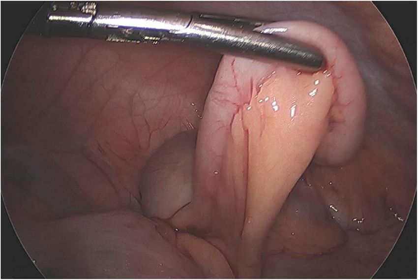
Hwang et al. BMC Pediatrics (2021) 21:258 Page 2 of 5
and radiologic signs of appendicitis but had a negative one month prior (she was asymptomatic but underwent
appendectomy. testing due to several family members testing positive).
Additional labs were obtained with concern for MIS-C
Case Presentation and were significant for a positive SARS-CoV-2 IgG
A previously healthy 16-year-old female presented with antibody test and normal troponin and N-terminal Pro-
a four-day history of abdominal pain, vomiting, fever, Brain Natriuretic Peptide. Given fever, positive SARS-
headache, myalgias and cough. Her initial vital signs in CoV-2 IgG, laboratory evidence of inflammation, and
the referring emergency department were temperature multisystem involvement she was hospitalized for fur-
39.4° Celsius, pulse 154, respiratory rate 16, blood pres- ther monitoring and treatment of MIS-C.
sure 115/61, and oxygen saturation 96 %. Physical exam- Overnight, the patient was persistently febrile and
ination was notable for pallor and right lower quadrant tachycardic. She also reported increasing RLQ pain and
(RLQ) abdominal tenderness without guarding or re- exhibited new abdominal rebound tenderness. The at-
bound. Initial laboratory testing significant for white tending pediatric radiologist’s review of the prior CT
blood cell (WBC) count 5.8 10^3/uL (reference range 4- concluded that the imaging was consistent with acute
10.5) with 93 % neutrophils, hemoglobin 11.9 g/dL (12– appendicitis as there was dilation of the appendix, meas-
15), platelets 102 10^3/uL (150–450), C-reactive protein uring 8 mm, mild appendiceal mucosal hyperenhance-
11 mg/dL (0–1.0), erythrocyte sedimentation rate 26 ment and adjacent mesenteric fat stranding (Fig. 1). The
mm/hr (0–20), and procalcitonin 0.50 ng/mL (< 0.09). patient’s care was re-discussed with pediatric surgery
Urine hCG negative. Urinalysis showed trace leukocyte and together the multidisciplinary team was unable to
esterase (negative), negative nitrites, and 1–5 WBCs (0). definitively rule out appendicitis as a concurrent
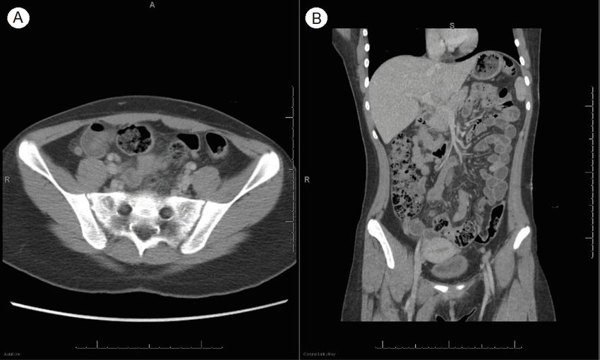
Contrast-enhanced computed tomography (CT) of the pathology. While a diagnosis of MIS-C generally requires
abdomen/pelvis showed mesenteric edema, dilation of exclusion of other etiologies, it was felt that she could
the appendix (8mm), and fat stranding throughout the have appendicitis and MIS-C simultaneously. Empiric
lower abdomen and pelvis. She received two intravenous treatment for appendicitis was started with piperacillin-
(IV) fluid boluses, anti-pyrectics and was transferred to tazobactam. The surgical team reviewed the risks and
our pediatric hospital. benefits of non-operative management with continued
On arrival she was evaluated by the pediatric surgery antibiotics versus diagnostic laparoscopy and appendec-
team who felt her presentation was not consistent with tomy. Surgical management was selected. Echocardio-
acute appendicitis. Further history and exam revealed gram was obtained to assess for cardiac involvement and
that she had mild bilateral conjunctival injection and showed normal cardiac function and no dilation of the
neck tenderness in addition to RLQ pain and had a posi- coronary arteries. She remained stable on the acute floor
tive SARS-CoV-2 polymerase chain reaction (PCR) test on hospital day two and therapy with intravenous
Fig. 1 CT abdomen/pelvis. (A) Appendix (red solid arrow) wall thickness > 3mm. Lack of luminal air, appendix fluid depth > 2.6mm, and
hyperdensity. Red dashed arrow shows peri-appendiceal fat stranding. (B) A diameter of 8mm (blue solid arrow) with inflammatory fat stranding
(blue dashed arrow), most pronounced around the cecumHwang et al. BMC Pediatrics (2021) 21:258 Page 3 of 5
immune globulin (IVIG) and aspirin were ordered for on daily aspirin with close cardiology follow up to moni-
treatment of MIS-C. However, given the timing of when tor progression of her coronary involvement.
surgery could take her to the operating room, she under-
went diagnostic laparoscopy and appendectomy first. Discussion and Conclusions
She was found to have a grossly normal appendix with There is increasing recognition of gastrointestinal in-
no inflammation to suggest appendicitis (Fig. 2). As a volvement in patients with COVID-19 and MIS-C. Sev-
surgeon’s intra-operative “eyeball” assessment of appen- eral case reports describe patients who present with
dicitis is not perfect and the risk of appendectomy while typical symptoms of appendicitis who are also found to
undergoing laparoscopy is low the decision was made to be infected with SARS-CoV-2 virus or meet the diagno-
complete appendectomy. Histologic findings revealed sis for MIS-C. [10–12] Studies have demonstrated that
only mild chronic serosal inflammation and edematous most children with MIS-C will present with gastrointes-
mesothelium; it did not show the transmural acute in- tinal (GI) symptoms. [4, 7] MIS-C should be high on the
flammation diagnostic of appendicitis. Piperacillin- differential for patients who present with GI symptoms
tazobactam was discontinued and the patient was and a history of recent SARS-CoV-2 exposure or infec-
returned to the acute care floor for post-surgical moni- tion, even if findings seem consistent with other GI
toring and MIS-C treatment with IVIG infusion (2 g/kg) pathologies such as appendicitis, infection, or inflamma-
and aspirin (81 mg). tory bowel disease (IBD).
On hospital day three she completed IVIG therapy, The current case demonstrates MIS-C as a radiologic
and her blood pressure and fever curve improved. Re- mimic of acute appendicitis on CT in a patient who
peat labs showed worsening lymphocytopenia, anemia, underwent negative appendectomy. A prior report de-
and thrombocytopenia and hypoalbuminemia. She then scribed a case of MIS-C that mimicked appendicitis clin-
developed tachycardia and hypotension refractory to ically and on ultrasound imaging, prompting IV
fluid resuscitation and given concern for refractory MIS- antibiotics and surgery (open appendectomy and resec-
C, she was started on corticosteroid therapy (2 mg/kg tion of an inflamed segment of ileum) before treatment
twice daily). She was transferred to the intensive care with IVIG and steroids was initiated. Pathology did not
unit and started on norepinephrine (0.02 mcg/kg/min) show appendicitis but did show necrotic mesenteric
for hemodynamic support, which she required for lymphangitis and vasculitis. [13] A South African series
24 hours. The patient stabilized and was then transferred described four children with appendicitis, confirmed by
back to the acute care floor with improvement of her surgical findings, in the setting of SARS-CoV-2-positive
pancytopenia over the next two days. She was discharged PCR. MIS-C was diagnosed in three of these children
home in stable condition on day seven of hospitalization after appendectomy. [14] Other case series have de-
to complete a course of low dose aspirin and a steroid scribed appendicitis in the setting of acute SARS-CoV-2
taper. infection. [11, 12, 14] In addition, an increase in the rate
At her follow-up cardiology appointment four weeks of appendiceal perforation in children without SARS-
after discharge, she was asymptomatic, but echocardio- CoV-2 infections has been found during the COVID-19
gram showed moderately dilated right and left coronary pandemic, presumably due to reluctance to present to
arteries with normal ventricular function. She remained the hospital for evaluation [15]. The hyperinflammatory
state seen in COVID-19 and MIS-C may play a role in
the pathogenesis of intestinal involvement. There is a
similar known association with Kawasaki disease and
gastrointestinal manifestations, including appendicitis.
[16, 17] Others hypothesize the contributory role of the
angiotensin-converting enzyme 2 (ACE2) receptor that
is expressed in the intestine, especially the terminal
ileum, [18] allowing SARS-CoV-2 to invade gastrointes-
tinal cells. [11, 12].
Our patient had some findings of appendicitis with
RLQ tenderness, fever, nausea, emesis, and decreased
appetite. Many of the classic signs of appendicitis (fever,
anorexia, nausea, guarding, and migration of pain from
the umbilical region to the RLQ) may be absent in chil-
dren with appendicitis; these findings also may be
Fig. 2 Intraoperative photograph revealing a grossly normal
present with diseases other than appendicitis. [19] Up to
appendix without signs of inflammation
20 % of patients with appendicitis present withoutHwang et al. BMC Pediatrics (2021) 21:258 Page 4 of 5
leukocytosis, [19] and our patient presented with an ini- syndrome coronavirus 2; RLQ: Right lower quadrant; WBC: White blood cell;
tially normal WBC count. IV: Intravenous; PCR: Polymerase chain reaction; IVIG: Intravenous immune
globulin; GI: Gastrointestinal; IBD: Inflammatory bowel disease;
The patient’s CT had findings concerning for acute ap- ACE2: Angiotensin-converting enzyme 2
pendicitis. CT has a sensitivity of 94 % and specificity of
95 % for appendicitis. [19] Causes of false positive CT in- Acknowledgements
not applicable.
clude appendiceal neoplasms, IBD, cystic fibrosis, viral
infections, and tubo-ovarian infection. [20] In a study of Author contributions
radiologic findings in MIS-C, abdominal imaging showed MH, KW, LW, ED, CK and SY provided direct patient care, drafted the initial
manuscript, worked on literature review, and critically reviewed the
small-volume ascites (38 %), hepatomegaly (38 %), manuscript. KVA and JP provided direct patient care and revised the
echogenic kidneys (31 %), bowel wall thickening (19 %), manuscript. All authors read and approved the final manuscript as submitted
gallbladder wall thickening (19 %), mesenteric lymph- and agree to be accountable for all aspects of the case report.
adenopathy (13 %), splenomegaly (6 %), and bladder wall
Funding
thickening (16 %).[21] Appendiceal dilation and fat non-applicable (no funding source).
stranding on CT, diagnostic of appendicitis and present
in our patient, have not previously been described. Availability of data and materials
not applicable.
Non-operative management with antibiotics alone has
been quite successful in managing acute uncomplicated Declarations
appendicitis meeting strict inclusion criteria[22] and was a
Ethics approval and consent to participate
strong consideration in this patient. However, the success not applicable.
of antibiotics to treat appendicitis in the setting of concur-
rent treatment with IVIG and steroids for MIS-C is un- onsent for publication
Written consent was obtained from the patient’s parent for publication of
known with no outcomes data currently available in the this case report and accompanying images. A copy of the written consent is
literature. Additionally, given the high likelihood that the available for review by the Editor of this journal.
patient’s abdominal symptoms would continue given her
Competing interests
MIS-C (independent of the diagnosis of appendicitis), the
the authors have no financial or non-financial competing interests to declare.
team was concerned they would be unable to reliably fol-
low her abdominal exam to determine if her appendicitis Received: 1 April 2021 Accepted: 19 May 2021
was being adequately treated with antibiotics.
While our patient’s clinical presentation and radio- References
graphic findings necessitated surgical evaluation, her 1. Mehta NS, Mytton OT, Mullins EWS, et al. SARS-CoV-2 (COVID-19): What Do
surgical and pathologic findings ultimately were not We Know About Children? A Systematic Review. Clin Infect Dis. 2020;71(9):
2469–79.
consistent with appendicitis. Laboratory criteria may be 2. Henderson LA, Canna SW, Friedman KG, et al. American College of
important in helping to differentiate MIS-C from acute Rheumatology Clinical Guidance for Multisystem Inflammatory Syndrome in
appendicitis - specifically, lymphopenia, thrombocytope- Children Associated With SARS–CoV‐2 and Hyperinflammation in Pediatric
COVID‐19: Version 2. Arthritis Rheumatol. 2021;73:e13–29.
nia, and inappropriately normal WBC count. Our patient 3. Dufort EM, Koumans EH, Chow EJ, et al. Multisystem Inflammatory
had a progressive lymphopenia and thrombocytopenia Syndrome in Children in New York State. New Engl J Med. 2020;383(4):347–
that did not fit with the classic presentation of 58.
4. Lee PY, Day-Lewis M, Henderson LA, et al. Distinct clinical and
appendicitis. immunological features of SARS-CoV-2-induced multisystem inflammatory
Patients with MIS-C can present with findings character- syndrome in children. J Clin Invest. 2020;130(11):5942–50.
istic of acute appendicitis, including RLQ pain, fever, nau- 5. Carlin RF, Fischer AM, Pitkowsky Z, et al. Discriminating Multisystem
Inflammatory Syndrome in Children Requiring Treatment from Common
sea, emesis, anorexia, and radiographic evidence of Febrile Conditions in Outpatient Settings. J Pediatr. 2021;229:26–32 e22.
appendicitis. This presents a diagnostic challenge for clini- 6. Nakra N, Blumberg D, Herrera-Guerra A, Lakshminrusimha S. Multi-System
cians. Although difficult to exclude appendicitis in the case Inflammatory Syndrome in Children (MIS-C) Following SARS-CoV-2 Infection:
Review of Clinical Presentation, Hypothetical Pathogenesis, and Proposed
of CT evidence, certain laboratory criteria, namely relative Management. Children. 2020;7(7):69.
leukopenia and thrombocytopenia may be helpful in differ- 7. Miller J, Cantor A, Zachariah P, Ahn D, Martinez M, Margolis KG.
entiating these patients. However, the use of immunomod- Gastrointestinal Symptoms as a Major Presentation Component of a Novel
Multisystem Inflammatory Syndrome in Children That Is Related to
ulatory agents in the treatment of MIS-C may complicate Coronavirus Disease 2019: A Single Center Experience of 44 Cases.
the care of a patient with potential appendicitis, and ap- Gastroenterology. 2020;159(4):1571–4 e1572.
pendectomy may still be preferable as the lowest risk option 8. Tullie L, Ford K, Bisharat M, et al. Gastrointestinal features in children with
COVID-19: an observation of varied presentation in eight children. Lancet
despite a low clinical suspicion for appendicitis. Child Adolesc Health. 2020;4(7):e19–20.
9. Jackson RJ, Chavarria HD, Hacking SM. A Case of Multisystem Inflammatory
Syndrome in Children Mimicking Acute Appendicitis in a COVID-19
Abbreviations Pandemic Area. Cureus. 2020;12:e10722.
MIS-C: Multi-inflammatory syndrome in children; CT: Computer tomography; 10. Meyer JS, Robinson G, Moonah S, et al. Acute appendicitis in four children
COVID-19: Coronavirus disease 2019; SARS-CoV-2: Severe acute respiratory with SARS-CoV-2 infection. J Pediatr Surg Case Rep. 2021;64:101734.Hwang et al. BMC Pediatrics (2021) 21:258 Page 5 of 5
11. Suwanwongse K, Shabarek N. Pseudo-Appendicitis in an Adolescent With
COVID-19. Cureus. 2020;12:e9394.
12. Abdalhadi A, Alkhatib M, Mismar AY, Awouda W, Albarqouni L. Can COVID
19 present like appendicitis? IDCases. 2020:e00860.
13. Jackson RJ, Chavarria HD, Hacking SM. A Case of Multisystem Inflammatory
Syndrome in Children Mimicking Acute Appendicitis in a COVID-19
Pandemic Area. Cureus. 2020;12(9):e10722.
14. Lishman J, Kohler C, de Vos C, et al. Acute Appendicitis in Multisystem
Inflammatory Syndrome in Children With COVID-19. Pediatr Infect Dis J.
2020;39(12):e472–3.
15. Place R, Lee J, Howell J. Rate of Pediatric Appendiceal Perforation at a
Children's Hospital During the COVID-19 Pandemic Compared With the
Previous Year. JAMA Netw Open. 2020;3(12):e2027948.
16. Garnett GM, Kimball S, Melish ME, et al. Appendicitis as the presenting
manifestation of Kawasaki disease. Pediatr Surg Int. 2014;30(5):549–52.
17. Gwendolyn M, Sarah K, Melish ME, et al. Appendicitis as the presenting
manifestation of Kawasaki disease. Pediatric Surg Int. 2014;30(5):549–52.
18. Ni W, Yang X, Yang D, et al. Role of angiotensin-converting enzyme 2
(ACE2) in COVID-19. Crit Care. 2020;24(1):422.
19. Glass CC, Rangel SJ. Overview and diagnosis of acute appendicitis in
children. Semin Pediatr Surg. 2016;25(4):198–203.
20. Dietz KR, Merrow AC, Podberesky DJ, Towbin AJ. Beyond acute appendicitis:
imaging of additional pathologies of the pediatric appendix. Pediatr Radiol.
2013;43(2):232–42.
21. Blumfield E, Levin TL, Kurian J, Lee EY, Liszewski MC. Imaging Findings in
Multisystem Inflammatory Syndrome in Children (MIS-C) Associated With
Coronavirus Disease (COVID-19). Am J Roentgenol. 2021;216(2):507–17.
22. Minneci PC, Hade EM, Lawrence AE, et al. Association of Nonoperative
Management Using Antibiotic Therapy vs Laparoscopic Appendectomy
With Treatment Success and Disability Days in Children With
Uncomplicated Appendicitis. JAMA. 2020;324(6):581–93.
Publisher’s Note
Springer Nature remains neutral with regard to jurisdictional claims in
published maps and institutional affiliations.You can also read