Intra-diploic epidermoid cyst: An iceberg lesion - Nepal ...
←
→
Page content transcription
If your browser does not render page correctly, please read the page content below
Neuro View Box                                                                Nepal Journal of Neuroscience 2021;18(3):69-72

Intra-diploic epidermoid cyst: An iceberg lesion
Prashant Punia MCh1 , Ashish Chugh MCh2 , Sarang Gotecha MCh3
1,2,3
      Department of Neurosurgery, Dr. D.Y. Patil Medical College and Hospital, Pimpri, Pune, Maharashtra,
India
Date of submission: 22nd January 2021             Date of acceptance: 16th August 2021       Date of publication: 1st September 2021

Abstract
Intraosseous epithelial inclusion cysts of the skull, presenting as lytic defects, constitute a very small percentage of
the primary intracranial tumours. We report a 45-year-old female patient presented with a small scalp swelling in the
occipital region. During initial exploration, the surgeon suspected an intracranial extension due to a large bony defect
at base and inability to reach the inferior margin of the swelling, consequently the procedure was abandoned. Imaging
revealed a well-defined, mixed density lesion with sharp marginated bone defect involving both the outer and inner
tables of the occipital bone. Subsequently the lesion was approached through a right occipital craniotomy. Pearly
white, flaky contents of the lesion along with the capsule was excised completely. The case is presented by virtue of
not only the rarity of the variant but also to highlight the importance of timely intervention by a neurosurgeon after
adequate investigation and in a tertiary care setting.
Key words: Epidermoid, Intra-diploic, Scalp lesions.

                        Introduction                                                        Case Report

I  ntraosseous epithelial inclusion cysts of the skull,
   presenting as lytic defects, constitute a very small
percentage of the primary intracranial tumors.1 These
                                                                            A 45-year-old female patient presented to a local
                                                                       Primary Healthcare Centre (PHC) with a small scalp
                                                                       swelling in the occipital region. She was not advised
cysts are derived from ectodermal cells of cranium and                 any imaging and after routine blood examination,
are lined by stratified squamous epithelium. Although                  excision of swelling was planned under local anaesthesia.
rare, their common locations include frontal, parietal and             Intraoperative identification of intracranial extension
occipital bones.2 We intend to report a case of intra-diploic          was made by the surgeon as the inferior margin of the
epidermoid cyst of occipital bone presenting as a small                swelling could not be reached and also by palpation of
scalp swelling in a middle aged female patient. The case               the huge bone defect following which the procedure was
is presented by virtue of not only the rarity of the variant           abandoned midway and the patient was referred to our
and site of occurrence but also to highlight the importance            centre for further management.
of timely intervention by a neurosurgeon after adequate                     On examination, a residual swelling which was
investigation and in a tertiary care setting.                          partially excised in its superficial portion was noticed
                                                                       along with an open cavity extending intracranially via the
                                                                       bone defect. There was active bleeding from the residual
Access this article online                                             lesion which was sutured and the patient was shifted for
Website: https://www.nepjol.info/index.php/NJN
                                                                       imaging.
DOI: https://doi.org/10.3126/njn.v18i3.34433
HOW TO CITE                                                            Imaging
Punia P, Chugh A, Gotecha S. Intra-diploic epidermoid cyst: An             Contrast Enhanced Computerised Tomography
iceberg lesion. NJNS. 2021;18(3):69-72.
                                                                       (CECT) revealed a well-defined, mixed density lesion
Address for correspondence:                                            with hypodense and an isodense component in the right
Dr. Prashant Punia                                                     occipital region. Lesion measured 4.2 (Cranio caudal)
Department of Neurosurgery,                                            x 3.3 (Antero posterior) x 3.6 (transverse) cm. A sharp
Dr. D.Y. Patil Medical College and Hospital,                           marginated bone defect was noted involving both the
Pimpri, Pune, Maharashtra, India.                                      outer and inner tables of the occipital bone.
E-mail: getdrprashant@gmail.com
                                                                           Lesion displayed a thin, mildly enhancing membrane
Phone: +91-9049755538
                                                                       on the inner aspect causing mild mass effect on right
Copyright © 2021 Nepalese Society of Neurosurgeons (NESON)             posterior parietal and occipital lobes. Enhancing dural
ISSN: 1813-1948 (Print), 1813-1956 (Online)                            vessels were seen along this membrane (Figure 1).
                                                                           Magnetic Resonance Imaging (MRI) of the brain
              This work is licensed under a Creative Commons
              Attribution-Non Commercial 4.0 International License.
                                                                       confirmed the CECT findings in terms of size and site of

                                    Nepal Journal of Neuroscience, Volume 18, Number 3, 2021                             69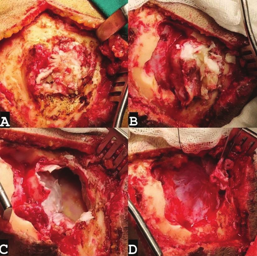
Punia et al
location and revealed a T1 hyperintense and T2 mixed craniotomy wherein the margins of bone defect were
intensity lesion with peripherally enhancing membrane nibbled away to gain a wide access to the lesion. Pearly
on the inner aspect. Restricted diffusion was evident white, flaky contents of the lesion along with the capsule
on Diffusion Weighted Imaging (DWI). Thinning and were identified and excised completely. Dura mater was
destruction of inner and outer tables was noted and there intact and the capsule was easily separable from the dura.
was no perilesional oedema. Keeping in mind the clinical Patient underwent cranioplasty using artificial cranial
picture and imaging findings, a diagnosis of an intra bone graft (Figure 2).
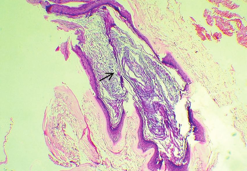
diploic epidermoid cyst in the right occipital region was Histopathological examination (HPE) revealed
made and the patient was taken up for definitive surgery. fibrocollagenous tissue and stratified squamous epithelium
with a granular layer composed of keratinous flaky
Intraoperative findings material. Mild haemorrhage and lymphocytic infiltration
Lesion was approached through a right occipital was also noted (Figure 3).
Figure 1a: Contrast Enhanced Computed tomography (CECT) brain, axial view showing non contrast enhancing lesion in
the right parieto occipital region with minimal peripheral contrast enhancement.
1b: 3D reconstructed bony window of CT brain showing bone defect at the site of lesion.
1c: T1 weighted Magnetic Resonance Imaging (MRI), axial view showing a hyperintense lesion in the right parieto occipital
region.
1d: Diffusion Weighted Imaging (DWI) showing restricted diffusion in the lesion.
70 Nepal Journal of Neuroscience, Volume 18, Number 3, 2021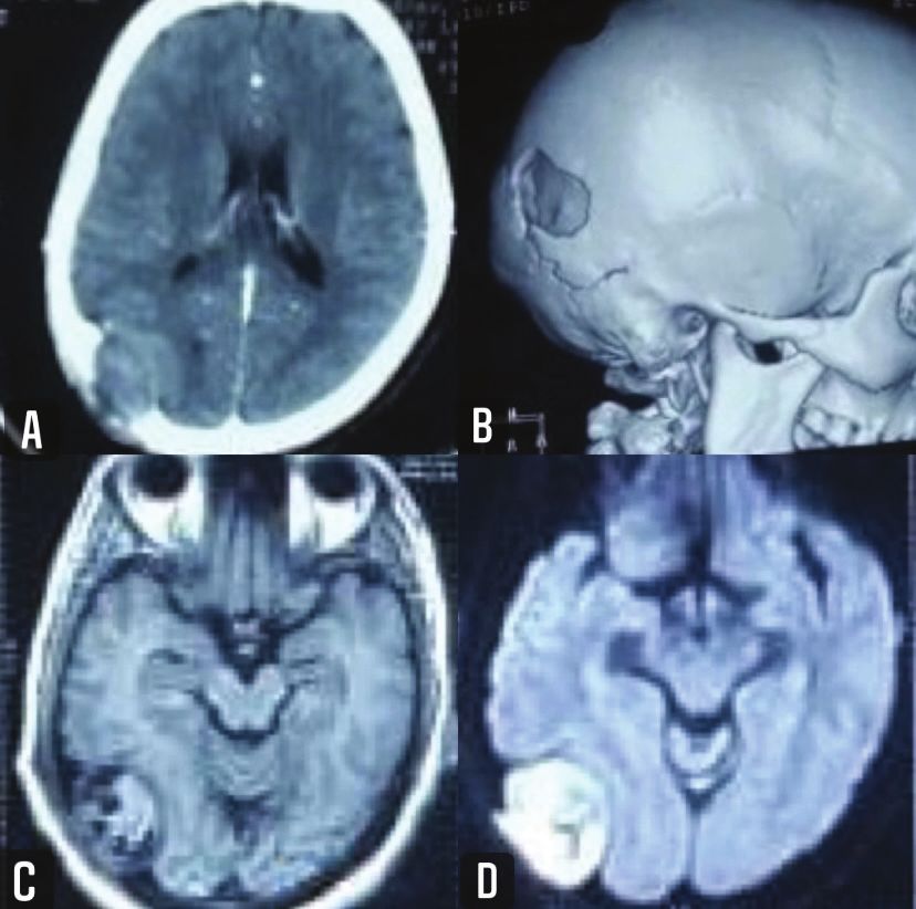
Intra-diploic epidermoid cyst: An iceberg lesion

Figure 2a: Intraoperative photograph showing exposure of the scalp lesion, its size and whitish contents.
2b: Intraoperative photograph after widening of bone defect via craniotomy, thus revealing the intracranial part of the
lesion.
2c: Intraoperative photograph showing pearly, white capsule of the lesion after decompression and evacuation of its
contents.
2d: Intraoperative photograph showing capsule of lesion being dissected off dura and thinned out dura.

                                                                   Intra-diploic epidermoid cysts are very rare,
                                                              accounting for
Punia et al

entrapment of the surface ectoderm.7                               Conflict of Interest: None
     CECT and MRI are the best investigations to make              Source(s) of support: None
an accurate diagnosis and these lesions typically appear
as hypodense, non-enhancing lesions and on MRI                                     References
demonstrate high signal intensity in T1 weighted images
and a variable T2 weighted signal and sometimes, the cyst     1. Kalgutkar A, Kini S, Jambhekar N, Das S. Intradiploic
contents can be hyperdense, mimicking a haemorrhage.9            primary epithelial inclusion cyst of the skull. Annals
No or minimal peripheral contrast enhancement may be             of Diagnostic Pathology. 2006;10:20-23. https://doi.
seen. Diffusion restriction is typically seen on DWI and         org/10.1016/j.anndiagpath.2005.07.007
it is considered the best imaging sequence in diagnosing      2. Ciapetta P, Artico M, Salvati M, Raco A, Gagliardi
epidermoid cysts.6                                               FM. Intradiploic epidermoid cysts of the skull:
     Gross total excision remains a cornerstone for the
                                                                 report of 10 cases and review of the literature.
treatment of epidermoid cysts. As proposed by Cushing,
                                                                 Acta Neurochirurgica. 1990;102:33-7. https://doi.
the aim of surgery is complete removal of tumour, together
                                                                 org/10.1007/BF01402183
with its capsule, which must be carefully dissected from
                                                              3. Cushing H. A large epidermal cholesteatoma of the
the bone and dura mater.3 Complete resection is to be
                                                                 parietotemporal region deforming the hemisphere
aimed, as the only living and growing part of the intra-
diploic epidermoid cyst is its capsule which must be             without cerebral symptoms. Surg Gynecol Obstet.
excised in its entirety to avoid recurrence.6 Skardowa           1922;34:557-66.
in his article on benign paediatric cranial vault tumours     4. Prior A, Anania P, Pacetti M, Secci F, Ravegnani M,
observed constant enlargement of tumours in 1/3rd of their       Pavanello M, et al. Dermoid and epidermoid cysts
patients and advocated complete tumour excision as it not        of the scalp: Case series of 234 consecutive patients.
only provides a complete recovery but also the material          World Neurosurgery. 2018;120:119-24. https://doi.
for an ultimate diagnosis.10                                     org/10.1016/j.wneu.2018.08.197
     This case also brings forth the importance of proper     5. Ichimura S, Hayashi T, Yazaki T, Yoshida K, Kawase
clinical and radiological evaluation not only to assess          T. Dumbbell shaped intradiploic epidermoid cyst
the tumour in its entirety but also aid in proper planning.      involving the dura mater and cerebellum. Neurol Med
Had the patient been subjected to imaging at the first           Chir (Tokyo). 2008;48;83-5. https://doi.org/10.2176/
presentation, the full extent of lesion would have been          nmc.48.83
demonstrated and an unnecessary surgery would have            6. Oommen A, Govindan J, Peroor DS, Azeez CR,
been avoided. The surgeon was probably misled by the             Rashmi R. Giant occipital intradiploic epidermoid
smaller extracranial sized scalp lesion whereas the much         cyst. Asian J Neurosurg. 2018;13(2);514-7. https://
larger and aggressive intracranial part was hidden in plain      doi.org/10.4103/1793-5482.181146
sight like an iceberg and the identification of the same      7. Swisher RC Jr, Tesluk H. Epidermoid cyst of the
intraoperatively led to abandonment of the procedure.            skull displacing the brain. JAMA. 1969;210:1280-1.
     The authors stress on the use of radio imaging even         https://doi.org/10.1001/jama.1969.03160330080020
for visibly small lesions on scalp to gain all around         8. Khalid S, Ruge J. Considerations in the
knowledge about the tumour and extrapolate the same to
                                                                 management of congenital cranial dermoid
the surgical modality to aid in complete resection.
                                                                 cysts. J Neurosurg. 2017;20:30-4. https://doi.
                                                                 org/10.3171/2017.2.PEDS16701
                      Conclusion                              9. Gaivas S, Rotariu D, Dumitrescu G, Iliescu b, Apetrei
                                                                 C, Poeata I. Intradiploic epidermoid cyst of the skull.
     Cranial epidermoid is a fairly common entity and
                                                                 Case report. Romanian Neurosurgery. 2011;18(2):
intra-diploic variant of the same is not commonly seen
                                                                 1-5.
in neurosurgical practise. These lesions may present as a
                                                              10. Skadorwa T, Bogdan C. Clinical characteristics
small scalp lesion which should not be judged based on its
                                                                 of benign pediatric cranial vault tumors: Surgical
apparent size as these lesions are not infrequently known
to have a bigger intracranial extension like an iceberg          considerations based on 100 cases. Pediatr Neurosurg.
and they should incite a doubt in the surgeon’s mind to          2017;52(1):13-9. https://doi.org/10.1159/000448045
hold the knife and further investigate the patient as being
forewarned is being forearmed.

      72                       Nepal Journal of Neuroscience, Volume 18, Number 3, 2021
You can also read