Case Report Furuncular myiasis in Italian traveler returning from Kenya - JIDC
←
→
Page content transcription
If your browser does not render page correctly, please read the page content below
Case Report

Furuncular myiasis in Italian traveler returning from Kenya

Ester Oliva1, Graziano Bargiggia1, Gianpaolo Quinzan2, Paola Lanza2, Claudio Farina1
1
    UOC Microbiologia e Virologia, ASST “Papa Giovanni XXIII”, Bergamo, Italia
2
    UOC Malattie Infettive, ASST “Papa Giovanni XXIII”, Bergamo, Italia

Abstract
Myiasis has been defined as the infestation of organs and/or tissues with dipterous larvae. They are especially widespread in tropical and
subtropical areas. Cutaneous myiasis is its most frequent clinical presentation. This report presents a case of furuncular myiasis caused by the
larva of Cordylobia anthropophaga in a 22-year-old girl living in Bergamo, Northern Italy, who returned from Kenya (Watamu) with a big,
painful furuncle in her right gluteus. The patient accidentally removed the larva from a large pimple and took it to the infectious disease
ambulatory clinic at the ASST “Papa Giovanni XXIII” Hospital, Bergamo. In the Microbiology and Virology Department of the same hospital,
a larva of C. anthropophaga was identified and the diagnosis of myiasis was confirmed.

Key words: Furuncular myiasis; Tumbu fly; Cordylobia anthropophaga; Kenya; traveler.

J Infect Dev Ctries 2020; 14(1):114-116. doi:10.3855/jidc.11560

(Received 11 April 2019 – Accepted 13 december 2019)

Copyright © 2020 Oliva et al. This is an open-access article distributed under the Creative Commons Attribution License, which permits unrestricted use,
distribution, and reproduction in any medium, provided the original work is properly cited.

Introduction                                                                      contaminated by animal urine or feces, or on dirty
     Myiasis is a parasitic infestation of a live mammal's                        and/or non-ironed clothes. The flies may also be
organs and/or tissues by larvae of dipterous flies, which                         stimulated for oviposition by the soiled napkins of
can be living or partially/completely necrotic. They                              babies. Flies never deposit their eggs directly on the
feed on their host’s tissue to grow and mature [1].                               human skin. The eggs hatch and larvae can survive
Especially widespread in tropical and subtropical areas                           without food for approximately 9 days. Upon exposure
(including Central America, South America, and                                    to warmth or vibration, they penetrate into the skin
tropical Africa), but often in temperate climates too, this                       areas covered by the contaminated clothes [4].
parasitosis is classified based on two main systems:                                   The penetration of C. anthropophaga into the skin
anatomical and ecological. The anatomical                                         is usually asymptomatic, although the area of
classification is more useful for practical diagnosis and                         penetration can be slightly itchy up to two days after the
for identifying the organ affected by the parasitosis.                            infestation. In a few days, a reddish papule develops and
This classification system divides myiasis into four                              takes on a boil-like appearance when fully developed
groups: sanguinivorous or bloodsucking, cutaneous                                 [2,4]. An intense inflammatory reaction in the tissue
(furuncular and migratory), wound, and cavitary                                   surrounding the lesions develops over a period of six
myiasis (ocular, ear-nose-mouth, urogenital, intestinal                           days [2]. Once the larvae have penetrated into the skin,
and cerebral) [2].                                                                their development may take 8 to 12 days to proceed
     Its most frequent form, furuncular myiasis, is                               through three stages of larval development prior to
defined as the penetration of the larvae in the healthy                           entering the prepupal stage. The pre-pupa then leaves
skin and subsequent development of a boil-like nodule.                            the host and drops to the ground to develop later into a
It is common in tropical countries and can be caused by                           fly [5,6]. The recommended preventive measures
the human botfly (Dermatobia hominis) in Latin                                    against myiasis are to avoid hanging laundry outside in
America [3], and by the Tumbu fly (Cordylobia                                     the shade and to iron it when it is left outside to dry on
anthropophaga) and Lund’s fly (Cordylobia rodhaini)                               the soil (as practiced in many endemic countries).
in Africa [2,4].                                                                       We present here a case of furuncular myiasis in an
     In particular, C. anthropophaga incidentally affects                         Italian patient returning from Kenya, East Africa. The
humans mainly during the rainy season. Adult flies lay                            larva was accidentally removed from a boil-like lesion
their eggs on dry sand or shady soil, which can be often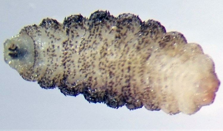
Oliva et al. – Myiasis from Kenya J Infect Dev Ctries 2020; 14(1):114-116.
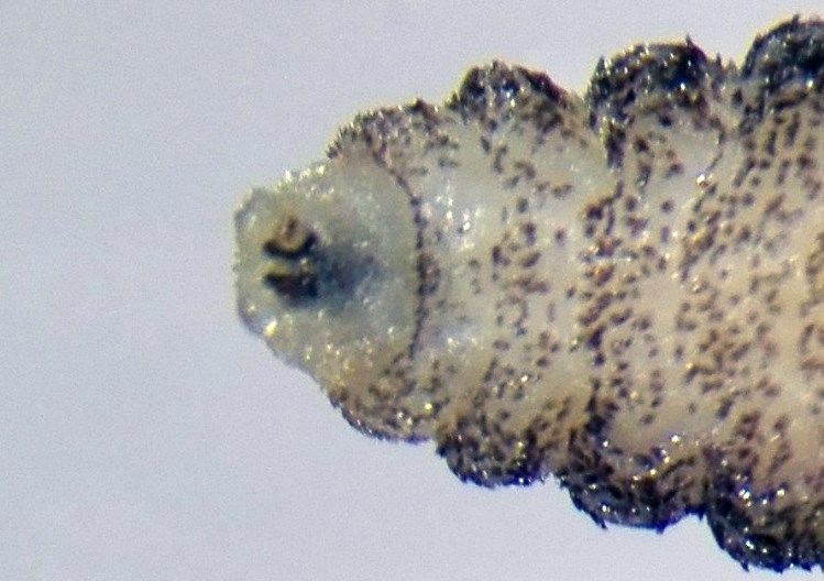
of the patient’s buttocks and was identified as Figure 1. The larva of C. anthropophaga removed from the
Cordylobia anthropophaga. lesion.
Case report
A 22-year-old Caucasian girl came to the infectious
disease ambulatory of the Azienda Socio-sanitaria
Territoriale (ASST) “Papa Giovanni XXIII” Hospital in
Bergamo (Northern Italy) with a still-living larva. The
patient lives in Bergamo with her family, where she
works as a maid. She does not smoke and does not drink
alcohol, and had no comorbidities.
At the interview with the doctor, the patient
reported that she had been in a tourist village in
Watamu, Kenya (Eastern Africa) from 23 November
2017 to 01 December 2017, with a-two-day-long safari
(25 and 26 November 2017) into Tsavo National Park.
Since November 29, she had noticed a small, itchy
red papule similar to an insect bite on the right gluteus. Figure 2. The pair of curved black mouth-hooks of the larva of
In the following days, it became more voluminous and C. anthropophaga according to the laboratory diagnosis.
the surrounding skin became erythematous and sore.
On December 4, the girl squeezed what looked like
a large pimple and a whitish larva leaked out. The larva
was immediately sent to the laboratory of Microbiology
and Virology of ASST “Papa Giovanni XXIII” Hospital
where it has been identified as Cordylobia
anthropophaga.
The larva was identified as follows: it was 6 × 3 mm
(Figure 1) in size, yellowish color with a cylindrical
body, and had 11 segments; segments III-VIII had
numerous small black, scattered spines, while IX-XI
were almost bare. The anterior end had two black
mouth-hooks with curved and sharp tips, without a
cluster of conic denticles (Figure 2).
The posterior spiracles of the larva (Figure 3)
carried three pairs of curved spiracular openings.
These morphological features were consistent with
those described for C. anthropophaga. Figure 3. The posterior spiracles of the larva of C.
anthropophaga.
At clinical examination, patient presented in good
general condition, without systemic symptoms or
objective alterations. On the right gluteus, an
erythematous area was still evident, infilled about 2
centimeters with a small papule in the center. After
manual removal of the larva, the patient took
amoxicillin/clavulanic acid for five days (1 g, 3
times/day), to treat secondary bacterial skin infection.
The lesion was completely resolved.
Conclusion
We report one case of Cordylobia anthropophaga
furuncular myiasis with a typical clinical pattern
appearing in Northern Italy but acquired during a trip to
Kenya, East Africa.
115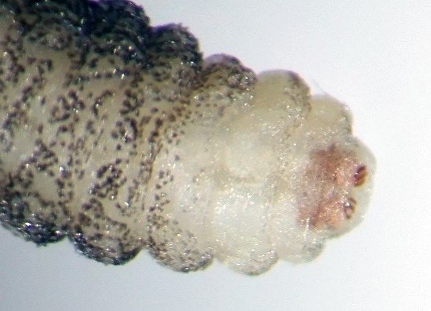
Oliva et al. – Myiasis from Kenya                                                            J Infect Dev Ctries 2020; 14(1):114-116.

    This case shows that the increase in international                   Valdovinos M, Maravilla P, Martinez-Hernandez F (2018)
travel, both for tourism and for business, underlines the                Imported and autochthonous cases of myiasis caused by
                                                                         Dermatobia hominis: Taxonomic identification using the
need to improve the awareness of diseases that are not                   internal transcribed spacer region. Am J Trop Med Hyg 99:
endemic in our country, especially those caused by                       940-944.
infectious agents. Skin diseases, together with systemic            4.   Song SM, Kim SW, Goo YK, Hong Y, Ock M, Cha HJ, Chung
febrile illness and acute diarrhea, are the leading causes               DI (2017) A case of furuncular myiasis due to Cordylobia
                                                                         anthropophaga in a Korean traveler returning from Uganda.
of health problems in travelers and account for 8% to                    Korean J Parasitol 55: 327-331.
12% of all tourist-related medical problems. Myiasis is             5.   Zumpt F (1965) Myiasis in man and animals in the Old World:
usually among the five most common dermatologic                          a textbook for physicians, veterinarians and zoologists,
conditions, representing 7.3% to 11% of cases [4,7].                     London: Butterworths, 267 pp.
    Diagnosis of furuncular myiasis is easily done                  6.   Mandell GL, Bennett JE, Dolin R (1995) Myiasis and tungiasis,
                                                                         chapter 274, In Mandell GL, Douglas and Bennett editors.
based solely on clinical grounds, especially in regions                  Principles and practice of infectious diseases. Philadelphia,
where the disease is endemic [2]. The patient's travel                   PA: Churchill, Livingstone, Elsevier, pp 2562-2564.
history may help to identify possible predisposing                  7.   Villalobos G, Vega-Memije ME, Maravilla P, Martinez-
factors. In this case, thanks to travel anamnesis (this                  Hernandez F (2016) Myiasis caused by Dermatobia hominis:
                                                                         countries with increased risk for travelers going to neotropic
type of myiasis is endemic in Africa) and the patient's                  areas. Int J Dermatol 55: 1060-1068.
ability to remove the larva without damaging it. Thanks
to these features we were able to correctly identify the
larva of C. anthropophaga and confirm the diagnosis.                Corresponding author
                                                                    Claudio Farina, MD.
                                                                    UOC Microbiologia e Virologia
                                                                    ASST “Papa Giovanni XXIII”
References
                                                                    Piazza OMS, 1, 24127
1.   How EH, Yap D, Mbakada N (2017) An exotic abscess within
                                                                    Bergamo, Italia
     the United Kingdom the Gambia: a case report. J Med Case
                                                                    Tel: +39.0352673666
     Rep 11: 310.
                                                                    Fax: +39.0352674921
2.   Francesconi F, Lupi O (2012) Myiasis. Clin Microbiol Rev 25:
                                                                    E-mail: cfarina@asst-pg23.it
     79-105.
3.   Toussaint-Caire S, Woroszylski-Yoselevitz A, Vega-Memije
     ME, Villalobos G, Rivas N, Alejandre-Aguilar R, Romero-        Conflict of interests: No conflict of interests is declared.

                                                                                                                                        116
You can also read