Pulmonary laceration and contusion in a young male patient due to a motorcycle accident
←
→
Page content transcription
If your browser does not render page correctly, please read the page content below
Case Report                                                                                                                                                              PNEUMON

Pulmonary laceration and contusion in a young male
patient due to a motorcycle accident
Vasiliki Apollonatou1,2, Galateia Verykokou1,2, Aggeliki Lazaratou1, Andriana I. Papaioannou1, Mirto Kardara3, Ioannis
Papadiochos3, Veroniki Papakosta3, Stavros Vassiliou3, Eugenia Koursoumi4, Panteleimon Messaropoulos5, Christina
Kontopoulou5, Stelios Loukides1, Effrosyni D. Manali1*, Spyros A. Papiris1*

ABSTRACT                                                                                                                          AFFILIATION
                                                                                                                                  1 2nd Pulmonary Medicine Department,
Chest trauma injuries are one of the main causes of death in young people
                                                                                                                                  Attikon University General Hospital, School
and include lung contusions, lacerations, pneumothorax, hemothorax, rib                                                           of Medicine, National and Kapodistrian
fractures and tracheobronchial injuries. Pulmonary contusions are the most                                                        University of Athens, Athens, Greece
common identified entities after trauma, and they result in alveolar hemorrhage                                                   2 Western Attica General Hospital Agia
without loss of the physiological structure of lung parenchyma. On the other                                                      Barbara, Athens, Greece
                                                                                                                                  3 Clinic of Oral & Maxillofacial Surgery,
hand, pulmonary lacerations, which are often associated with contusions,
                                                                                                                                  Attikon University General Hospital, School
result in rupture of the alveoli causing formation of cavities. Patients present                                                  of Medicine, National and Kapodistrian
symptoms ranging from minimal to severe, including cough, chest pain,                                                             University of Athens, Athens, Greece
hemoptysis, dyspnea, tachypnea, and hypoxemia. Findings may not be apparent                                                       4 2nd Department of Anesthesiology,
immediately after injury and chest CT is the most sensitive imaging technique                                                     Attikon University General Hospital, School
for diagnosis. Contusions usually resolve with supportive care in 5–7 days. In                                                    of Medicine, National and Kapodistrian
                                                                                                                                  University of Athens, Athens, Greece
this report, we present a case of lung contusion and laceration in a 19-year-old                                                  5 2nd Department of Radiology, Attikon
patient after a motorcycle accident.                                                                                              University General Hospital, School of
                                                                                                                                  Medicine, National and Kapodistrian
                                                                                                                                  University of Athens, Athens, Greece
                                                                                                                                  *Contributed equally

                                                                                                                                  CORRESPONDENCE TO
                                                                                                                                  Effrosyni D. Manali. 2nd Pulmonary Medicine
                                                                                                                                  Department, National and Kapodistrian
                                                                                                                                  University of Athens, School of Medicine,
                                                                                                                                  Attikon University General Hospital, 1 Rimini
                                                                                                                                  Street, Athens, 12462, Greece. E-mail:
                                                                                                                                  fmanali@otenet.gr

                                                                                                                                  KEYWORDS
                                                                                                                                  trauma, lung contusion, lung laceration

                                                                                                                                  Received: 7 February 2021
                                                                                                                                  Accepted: 9 February 2021

INTRODUCTION                                                                                      CASE PRESENTATION
Chest trauma injuries are one of the main causes of                                               A 19-year-old patient, non-smoker, without previous
death in young people and include lung contusions,                                                medical history, presented to the emergency room due to
lacerations, pneumothorax, hemothorax, rib fractures                                              fever and pain at the right periorbital area of the face after
and tracheobronchial injuries 1,2. Pulmonary contusion                                            a motorcycle collision twenty-four hours ago. The patient
is the most common identified entity after trauma and                                             was examined initially by general surgeons. He was febrile
usually results from blunt chest trauma (traffic accidents,                                       (38℃) and hemodynamically stable. His oxygen saturation
falls from great heights), shock waves associated with                                            was normal (SatO2: 98% breathing room air) and he had
penetrating chest injury, or explosion injuries 3,4. Unlike                                       normal breath sounds in auscultation. From physical
contusion, pulmonary laceration results in disruption of                                          examination, he presented with bruise injuries in the right
the architecture of the lung and could potentially cause                                          side of the face and a right periorbital hematoma. His
more serious damage. Pulmonary lacerations are commonly                                           laboratory examinations revealed normal hemoglobin (15.9
caused by penetrating trauma and result in formation of                                           g/dL), elevated white blood cell count (14.90 K/μL with
one or multiple cavities filled with air, blood, or both5.                                        78.7% neutrophils), elevated creatine kinase (956 U/L) and
In this report, we present a case of lung contusion and                                           elevated C reactive protein (96.9 mg/L). After exclusion of
laceration in a 19-year-old patient after a motorcycle                                            SARS-Cov-2 infection, he underwent computed tomography
accident.                                                                                         (CT) of the head which showed fracture displacement

Published by European Publishing. © 2021 Apollonatou V. et al. This is an Open Access article distributed under the terms of the Creative Commons Attribution NonCommercial 4.0 International
License. (http://creativecommons.org/licenses/by-nc/4.0)

Pneumon 2021;34(1):1
https://doi.org/10.18332/pne/136153

                                                                                              1
Case Report PNEUMON
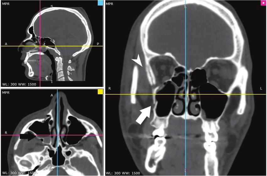
Figure 1. CT of the head which revealed fracture displacement of the right maxillary sinus and orbitally
wall (white arrow), air-fluid level within injured sinus (black arrow) and right periorbital hematoma
(arrow head)
of the right maxillary sinus and orbitally wall, air-fluid Figure 2. Chest X-ray on admission showing
level within injured sinus and right periorbital hematoma a cavitary lesion in the right middle lung zone
(Figure 1). The patient was admitted to the Oral and (white arrow) and a fracture displacement of the
Maxillofacial Surgery Department for further management right clavicle (black arrow)
with reconstructive surgery. Upon admission, a chest X-ray
revealed a cavitary lesion in the right middle lung zone and a
fracture displacement of the right clavicle (Figure 2). Chest
CT confirmed a fracture displacement of the right clavicle
without pneumothorax as well as three cavitary lesions with
air-fluid level within the minor and major fissure of the right
lung with maximum diameter of about 3.7 cm and adjacent
ground glass opacities (Figure 3). Pulmonary preoperative
evaluation was performed and the working diagnosis of
pulmonary laceration with contusion post-acute chest
trauma was made. Thorough evaluation for common and
specific pathogens for lower respiratory tract infection proved
insignificant. The patient received empirical antimicrobial
treatment with ampicillin/sulbactam and azithromycin
for superinfection, oxygen therapy with high fractions of
inspired oxygen to increase the resolution rate of the cystic
lesions and was systematically monitored. Multidisciplinary
discussion between pulmonologists, the treating surgeons,
Pneumon 2021;34(1):1
https://doi.org/10.18332/pne/136153
2Case Report PNEUMON
Figure 3. CT of the chest which revealed three cavitary lesions with air-fluid level within the minor and
major fissure of the right lung with maximum diameter of about 3.7 cm (a, b, d) and adjacent ground
glass opacities (c)
Figure 4. Chest X-ray, 2 weeks later, showing the anesthesiologists and the thoracic surgeons led to
complete remission of the cavitary lesion the decision to postpone imminent surgical management
in order to minimize the risk of pneumothorax under
positive pressure due to generalized anesthesia. Surgical
reconstructive management was undertaken two weeks later
without any complications after a new chest X-ray and CT
revealed remission of the laceration and contusion lesions
(Figures 4 and 5). The patient was discharged from hospital
in a very good condition.
DISCUSSION
A pulmonary contusion is an injury to the lung parenchyma
which usually occurs from blunt chest trauma and results in
alveolar hemorrhage. As a pathology, it was first described
in 1761 by an Italian anatomist, Morgagni 6, while the
term pulmonary contusion was coined in the 19th century
by Dupuytren, a French military surgeon7. Widespread use
Pneumon 2021;34(1):1
https://doi.org/10.18332/pne/136153
3Case Report PNEUMON
Figure 5. Chest CT, 2 weeks later, which revealed remission of the laceration and contusion lesions
of explosives during the time of World War I and II led to diagnosis of contusion12,13. Ground glass opacities may be
increased recognition of contusion due to blast injuries4. present in mild cases whereas widespread consolidations
The most common causes are traffic accidents, pedestrian may indicate severe injury14. Specific treatment is often not
injuries, falls from great heights, explosions and sports required and supportive care with prevention of respiratory
injuries3. The mechanism is the disruption of alveoli and failure, pain control, management of airway drainage and
capillaries due to rapid compression and decompression adequate intravenous fluid replacement are the primary
of the chest wall. Alveolar hemorrhage and few hours later aims of therapy 4. In severe cases with development of
interstitial oedema develop in the afflicted parenchyma6,8. acute respiratory distress syndrome (ARDS) non-invasive
Patients can present with symptoms ranging from positive pressure ventilation or invasive ventilation may be
minimal to severe, including cough, chest pain, hemoptysis, used3. Surgical stabilization may be required in the case of
dyspnea, tachypnea, and hypoxemia. Breath sounds may be multiple rib fracture/flail chest3. Contusions usually resolve
decreased, while hematoma and subcutaneous emphysema with supportive care in 5–7 days3, while findings in X-rays
may be present9. In chest X-rays, findings may not be may disappear after 10 days8. Pulmonary lacerations are
apparent until 48 hours post-injury10, while chest CT is a often associated with pulmonary contusions15. They are
more sensitive imaging technique 11. Studies have also usually caused by penetrating chest trauma but can be seen
shown that chest ultrasonography could be useful for the after blunt trauma as well16,17. They were first described in
Pneumon 2021;34(1):1
https://doi.org/10.18332/pne/136153
4Case Report PNEUMON
1940 by Fallon18. The mechanism is rupture to alveoli due Μεσσαρόπουλος5, Χριστίνα Κοντοπούλου5, Στέλιος Λουκίδης1,
to compressive forces applied to the lung parenchyma and Ευφροσύνη Δ. Μάναλη1, Σπύρος Α. Παπίρης1
the concomitant retraction of the surrounding elastic tissue 1
Β΄ Πανεπιστημιακή Πνευμονολογική Κλινική, Πανεπιστημιακό
resulting in the formation of small cavities that are filled Γενικό Νοσοκομείο «Αττικόν», Ιατρική Σχολή, Εθνικό και
with air or blood19. These entities are mostly observed in Καποδιστριακό Πανεπιστήμιο Αθηνών
patients agedCase Report PNEUMON
7. K a r m y - J o n e s R , J u r k o v i c h G J . B l u n t c h e s t
trauma. Curr Probl Surg. 2004;41(3):211-380.
doi:10.1016/j.cpsurg.2003.12.004
8. Allen GS, Cox CS Jr. Pulmonary contusion in children: diagnosis
and management. South Med J. 1998;91(12):1099-1106.
doi:10.1097/00007611-199812000-00002
9. Miller DL, Mansour KA. Blunt traumatic lung injuries. Thorac Surg
Clin. 2007;17(1):57-61. doi:10.1016/j.thorsurg.2007.03.017
10. Ganie FA, Lone H, Lone GN, et al. Lung Contusion: A Clinico-
Pathological Entity with Unpredictable Clinical Course. Bull
Emerg Trauma. 2013;1(1):7-16. Accessed February 7, 2021.
https://www.ncbi.nlm.nih.gov/pmc/articles/PMC4771236/
pdf/bet-1-007.pdf
11. Schild HH, Strunk H, Weber W, et al. Pulmonary contusion:
CT vs plain radiograms. J Comput Assist Tomogr.
1989;13(3):417-420.
12. Soldati G, Testa A, Silva FR, Carbone L, Portale G, Silveri
NG. Chest ultrasonography in lung contusion. Chest.
2006;130(2):533-538. doi:10.1378/chest.130.2.533
13. Xirouchaki N, Georgopoulos D. The use of lung ultrasound: A
brief review for critical care physicians and pneumonologists.
Pneumon. 2007;20(2):134-141.
14. Miller LA. Chest wall, lung, and pleural space
trauma. Radiol Clin North Am. 2006;44(2):213-224.
doi:10.1016/j.rcl.2005.10.006
15. Gavelli G, Canini R, Bertaccini P, Battista G, Bnà C, Fattori R.
Traumatic injuries: imaging of thoracic injuries. Eur Radiol.
2002;12(6):1273-1294. doi:10.1007/s00330-002-1439-6
16. Magret M. Lung trauma. Clin Pulm Med. 2010;17(2):75-81.
doi:10.1097/CPM.0b013e3181d269aa
17. Papagiannis A, Gaziotis G, Anastasiadis K. Post-traumatic
pulmonary pseudocyst: an unusual complication of blunt
chest injury. Pneumon. 2005;18(2):228-232.
18. Fallon M. Lung injury in intact thorax with report of case. Br J
Surg. 1940;28(109):39-49. doi:10.1002/bjs.18002810905
19. Tsitouridis I, Tsinoglou K, Tsandiridis C, Papastergiou
C, Bintoudi A. Traumatic pulmonary pseudocysts: CT
findings. J Thorac Imaging. 2007;22(3):247-251.
doi:10.1097/RTI.0b013e3180413e2a
20. Sorsdahl OA, Powell JW. CAVITARY PULMONARY LESIONS
FOLLOWING NONPENETRATING CHEST TRAUMA IN
CHILDREN. Am J Roentgenol Radium Ther Nucl Med.
1965;95(1):118-124. doi:10.2214/ajr.95.1.118
21. Carroll K, Cheeseman SH, Fink MP, Umali CB, Cohen
IT. Secondary Infection of Post-traumatic Pulmonary
Cavitary Lesions in Adolescents and Young Adults: Role
of Computed Tomography and Operative Debridement
and Drainage. J Trauma. 1989;29(1):109-112.
doi:10.1097/00005373-198901000-00024
22. Mavarez-Martinez A, Soghomonyan S, Sandhu G, Rankin
D. Intraoperative Tension Pneumothorax in a Patient
With Remote Trauma and Previous Tracheostomy.
J Investig Med High Impact Case Rep. 2016;4(1).
doi:10.1177/2324709616636397
Pneumon 2021;34(1):1
https://doi.org/10.18332/pne/136153
6You can also read