Crohn's Disease with Atypical Extra-Intestinal Manifestations Developing Under Treatment with Vedolizumab
←
→
Page content transcription
If your browser does not render page correctly, please read the page content below
European Journal
                                                                           of Case Reports in
                                                                       Internal Medicine

  Crohn’s Disease with Atypical Extra-Intestinal Manifestations Developing
                                                Under Treatment with Vedolizumab
                              Aasem Abu Shtaya1,2, Shai Cohen1,2, Yana Kogan2,3, Michal Shteinberg2,3, Ori Sagool2,4
                                     1
                                         Department of Internal Medicine “B”, Lady Davis Carmel Medical Center, Haifa, Israel
                               2
                                   Technion – Israel Institute of Technology, The B. Rappaport Faculty of Medicine, Haifa, Israel
                                     3
                                         Pulmonology Institute and CF center, Lady Davis Carmel Medical Center, Haifa, Israel
                                         4
                                             Department of Gastroenterology, Lady Davis Carmel Medical Center, Haifa, Israel

                                    Doi: 10.12890/2021_002265- European Journal of Case Reports in Internal Medicine - © EFIM 2021

Received: 03/01/2021
Accepted: 20/01/2021
Published: 19/02/2021

How to cite this article: Abu Shtaya A, Cohen C, Kogan Y, Shteinberg M, Sagool O. Crohn's disease with atypical extra-intestinal manifestations developing
under treatment with vedolizumab. EJCRIM 2021;8: doi:10.12890/2021_002265.

Conflicts of Interests: The Authors declare that there are no competing interests.
This article is licensed under a Commons Attribution Non-Commercial 4.0 License

ABSTRACT
Crohn’s disease is a chronic inflammatory bowel disease that can affect any part of the GI tract, which is frequently associated with extra-
intestinal manifestations. Pulmonary parenchymal disease is very uncommon and usually considered to be debilitating and harder to
diagnose. Pulmonary granulomas are rarely described in the literature as a complication of Crohn’s disease. Here, we present a patient with
Crohn’s disease exacerbation who developed granulomatous lung disease under treatment with vedolizumab. Our case may add evidence to
the emerging theory that gut-selective biologic agents could lead to upregulation of some pro-inflammatory factors leading to the evolution
of pulmonary disease.

LEARNING POINTS
• Pulmonary parenchymal diseases are rare in Crohn’s disease but they can be debilitating and life-threatening as they are usually tardily
   diagnosed; awareness of this association is of high value and could potentially shorten the time to a definite diagnosis.
• Pulmonary manifestations of Crohn’s disease could be subclinical without any respiratory complaints and not diagnosed with
   conventional imaging modalities such as chest x-ray.
• Gut-selective biologic agents could lead to the emergence of extra-intestinal manifestations due to upregulation of multiple pro-
   inflammatory cytokines.

KEYWORDS
Crohn’s disease, granuloma, vedolizumab, infliximab

CASE DESCRIPTION
This case involves a 31-year-old male patient, diagnosed with small intestinal Crohn’s disease at the age of 18, who had previously been
treated with adalimumab, 6-mercaptopurine and 5-aminosalicylic acid with a partial response to the 3 treatments and frequent relapses
requiring treatment with systemic steroids. He suffered from severe anaemia and hypoalbuminaemia secondary to his illness and had
severe osteoporosis with a T-score of -4.5 due to frequent steroid use.
He was admitted to our department due to a febrile illness, abdominal pain and bloody diarrhoea that started 3 weeks prior to admission.
Physical examination was significant for cachexia and sarcopenia; the patient weighed 60 kg at admission (BMI=23). Abdominal examination
revealed diffuse tenderness. Testing for inflammatory biomarkers and stool calprotectin, along with stool samples for culture, parasites

DOI: 10.12890/2021_002265                                                                   European Journal of Case Reports in Internal Medicine © EFIM 2021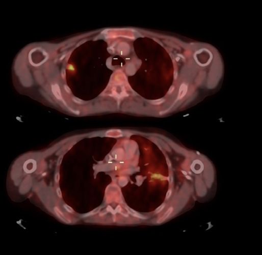
European Journal
of Case Reports in
Internal Medicine
and Clostridium toxin were negative, while stool multiplex PCR assessment for an infectious process was also negative. A chest x-ray
on admission was normal (Fig. 1) and abdominal CT enterography demonstrated minimal fat stranding. Upper endoscopy revealed mild
gastritis. Colonoscopy, as well as biopsies along the colon and the terminal ileum were normal. With a working hypothesis of Crohn’s disease
exacerbation enteral feeding was withheld and the patient was treated with ceftriaxone, metronidazole and azithromycin, without response.
We did not initiate steroidal therapy at that stage due to his severe osteoporosis.
.
Figure 1. Evolution of pleural effusion, at day 10 (A, with iatrogenic pneumothorax)
and 1 month later (B)
In order to better evaluate his intestinal disease, the patient underwent magnetic resonance enterography, which did not yield any significant
findings.
After 10 days of antibiotic therapy the febrile illness continued, along with severe diarrhoea, weight loss and a deteriorating general medical
condition. Antibiotic therapy was stopped, total parenteral nutrition was initiated and the patient received a single loading dose of gut-
selective vedolizumab. A few days later the patient reported improvement of abdominal pain and the frequency of diarrhoea, but his febrile
illness persisted. We intensified the investigations, including repeat blood and urine cultures, viral and immunological serology testing,
cryoglobulin measurement, echocardiography, ophthalmological evaluation, chest x-ray, abdominal x-ray and ultrasonography. All tests
returned normal. Due to further deterioration the patient received a course of steroidal therapy without any improvement.
After being treated with antibiotics, vedolizumab and steroids, fever continued on a daily basis. Hence, the patient underwent PET-CT that
demonstrated increased metabolic activity in both lungs without any enhancement along the GI tract (Fig. 2), while a repeat chest x-ray
(that was normal a few days previously) revealed new bilateral infiltrates on both lungs and a new left pleural effusion (Fig. 3). Thoracentesis
was performed revealing an exudative effusion; the effusion tested negative for an infectious process through multiplex PCR panels and
cytology assessment was also negative.
A bronchoscopy with bronchoalveolar lavage revealed no endobronchial abnormality; the lavage fluid was also negative for infectious
causes (including COVID-19 PCR testing). A transbronchial biopsy yielded findings consistent with acute non-specific pneumonitis. The
patient received an additional antibiotic course of levofloxacin for 1 week without any improvement, and continued with daily febrile peaks.
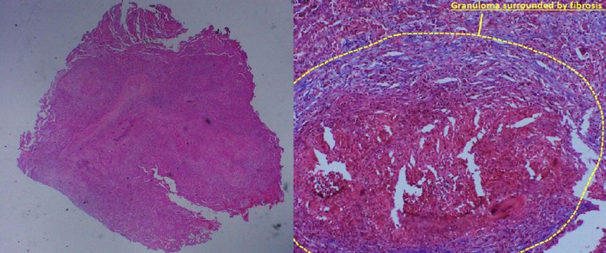
A thoracoscopy with open lung biopsy and wedge resection of the two largest infiltrates was performed, revealing confluent ill-defined
granulomas mostly with purulent centres surrounded by an organizing pneumonia pattern, without evidence of vasculitis and with negative
stains for infectious agents (Fig. 4).
In light of the finding of multiple granulomas on lung tissue, we considered an atypical pulmonary manifestation of Crohn’s disease, and
hence, treatment with non-selective infliximab was initiated. With this treatment the patient reported improved well-being with complete
resolution of the fever, abdominal pain and diarrhoea. Four weeks later the pulmonary infiltrates on chest x-ray resolved completely.
DOI: 10.12890/2021_002265 European Journal of Case Reports in Internal Medicine © EFIM 2021European Journal
of Case Reports in
Internal Medicine
.
Figure 2. Top: PET-CT showing high intensity uptake in the periphery of the upper lobe of Figure 3. A chest x-ray revealing new bilateral infiltrates on both lungs and a new left
the right lung. Bottom: PET-CT showing similar uptake in the lower lobe of the left lung pleural effusion
Figure 4.Left: lung parenchyma with many granulomas (H&E 20x). Right: granuloma surrounded by fibrosis (Masson’s trichrome 100x)
DISCUSSION
Crohn’s disease is associated with extra-intestinal manifestations mainly involving the skin, joints, eyes and the hepatobiliary system. Less
commonly, it can involve blood vessels, kidneys and the respiratory system.
Pulmonary involvement is relatively rare; however, there is a wide array of manifestations ranging from airway disease and pleural disease
to lung parenchymal disease and drug-related diseases. The most frequently occurring of these is bronchial inflammation and suppuration
of the upper airways with or without bronchiectasis [1].There are multiple probable mechanisms by which the lung is injured; there is large
similarity in the immune systems of both the pulmonary and intestinal mucosa [2]. In addition, there is evidence of circulating immune
complexes and autoantibodies in patients with Crohn’s disease that could lead to pulmonary injury [3].
DOI: 10.12890/2021_002265 European Journal of Case Reports in Internal Medicine © EFIM 2021European Journal
of Case Reports in
Internal Medicine
Several forms of lung parenchymal involvement in Crohn’s disease are recognized, including interstitial lung diseases such as cryptogenic
organizing pneumonia, unspecified interstitial lung disease, non-caseating granulomatous inflammation and fibrosis [1].
Chew et al. described a case of a pulmonary nodule that was biopsied and was found to contain both necrotizing and non-necrotizing
granulomas; further work-up led to the diagnosis of colonic Crohn’s disease, and treatment with infliximab led to resolution of both
pulmonary and GI symptoms [4].
Another case was described by Lissner et al. [5] of a 28-year-old patient assumed to have ulcerative colitis at presentation, who had received
3 infusions of vedolizumab, presenting with respiratory complaints and bilateral infiltrates on a chest CT scan. An open lung biopsy revealed
non-caseating granulomas, while a repeat colonoscopy revealed similar granulomas along the GI tract, leading to reclassification of the
diagnosis to Crohn’s disease with atypical pulmonary disease under vedolizumab treatment.
Vedolizumab is a monoclonal antibody that reduces chronically inflamed GI parenchymal tissue associated with ulcerative colitis and Crohn’s
disease by binding specifically to the α4β7 integrin receptor and blocking its interaction with mucosal addressin cell adhesion molecule 1.
This inhibits the movement of memory T lymphocytes across the endothelium into inflamed GI tissue.
Lissner et al. analyzed integrin expression in whole blood samples from patients, showing neutralization of α4β7 on naive and memory T
lymphocytes as expected with vedolizumab treatment; interestingly, they observed a simultaneous increase in other integrins such as β1
integrin in the lungs that led to altered migrational behaviour of leucocytes into organs other than the gut [5].
Our case highlights some important points. First, pulmonary parenchymal manifestations are rare in Crohn’s disease. They may be
subclinical and difficult to diagnose. Our patient’s chest x-ray was completely normal multiple times during his hospitalization, and he had
no pulmonary-specific complaints until a few weeks into his illness. Pulmonary granulomas are an even rarer manifestation among Crohn’s
disease patients but they can be life-threatening if not diagnosed promptly.
We cannot be certain that there is a direct connection between treatment with vedolizumab and the emergence of the lung consolidations,
although the chronological order of events strongly suggests this possibility. The resolution of GI symptoms and the evolution of respiratory
disease occurred directly after treatment with vedolizumab, similar to the case presented by Lissner et al. [5] described earlier.
We hypothesize that gut-selective treatments such as vedolizumab could unmask subclinical pulmonary involvement. The fact that stopping
vedolizumab and replacing it with infliximab (anti-TNF) led to complete clinical and radiological resolution also supports this theory.
REFERENCES
1. Lu DG, Ji XQ, Liu X, Li HJ, Zhang CQ. Pulmonary manifestations of Crohn’s disease. World J Gastroenterol 2014;20(1):133–141.
2. MacDermott RP, Nash GS, Nahm MH. Antibody secretion by human intestinal mononuclear cells from normal controls and inflammatory bowel disease patients. Immunol Invest
1989;18(1–4):449–457.
3. Bienenstock J, McDermott M, Befus D, O’Neill M. A common mucosal immunologic system involving the bronchus, breast and bowel. Adv Exp Med Biol 1978;107:53–59.
4. Chew MT, Chak E, Matsukuma K. A rare cause of pulmonary nodules. Case Rep Gastroenterol 2016;10(3):633–639.
5. Lissner D, Glauben R, Allers K, Sonnenberg E, Loddenkemper C, Schneider T, et al. Pulmonary manifestation of Crohn’s disease developed under treatment with vedolizumab.
Am J Gastroenterol 2018;113(1):146–148.
DOI: 10.12890/2021_002265 European Journal of Case Reports in Internal Medicine © EFIM 2021You can also read