Spindle cell hemangioma of nasal passage and ethmoidal sinus in a 4-month old infant
←
→
Page content transcription
If your browser does not render page correctly, please read the page content below
Case report Arch Argent Pediatr 2021;119(1):e36-e40 / e36
Spindle cell hemangioma of nasal passage and ethmoidal
sinus in a 4-month old infant
Tugba Tastemel Ozturk, M.D.a, Ahmet E. Suslu, Professorb, Altan Kavuncuoglu, M.D.c, Ekim Gumeler, M.D.d,
Kemal Kosemehmetoglu, Associated Professorc and Bilgehan Yalcin, Professore
ABSTRACT INTRODUCTION
Spindle cell hemangioma (SCH) is a benign unusual vascular
Spindle cell hemangioma (SCH) is an unusual
neoplasm. It does not have gender predilection and can occur
at all ages. The disease affects dermis and subcutis of distal vascular lesion described in 1986 by Weiss and
extremities predominantly; head and neck involvement is Enzinger as spindle cell hemangioendothelioma
very rare, paranasal sinus involvement has not been reported resembling cavernous hemangioma and Kaposi
before. Herein we present a 4-month-old infant with nasal
sarcoma. 1 This lesion was defined as a low-
obstruction since two weeks of age due to a mass in ethmoid
sinus obliterating the nasal passage. After the histopathological grade angiosarcoma initially, but owing to lack
diagnosis of SCH, the tumor was partially resected. In the sixth of metastasis and excellent prognosis, it was
month follow-up, there was minimal regression of residual considered as a benign vascular neoplasm despite
lesions. In the imaging studies performed 30 months after the
the high rate of local recurrences, and termed as
surgery, the residual mass was found to be disappeared. SCH
is not frequent in the head and neck, and presentation of some SCH in 1996. 1,2 Spindle cell hemangioma does
patients may not suggest the diagnosis. Histopathology is not have gender predilection and can occur
important for differential diagnosis and to orientate treatment. at all ages. It affects dermis and subcutis of
Awareness of SCH may increase the reported cases.
distal extremities predominantly. 2 Head and
Key words: pediatrics, head and neck neoplasms, paranasal sinuses,
spindle cell hemangioma. neck region involvement is very rare. To our
knowledge, 11 cases of SCH have been reported
http://dx.doi.org/10.5546/aap.2021.eng.e36 in the head and neck region so far, and paranasal
sinus involvement has not been reported before.3
Herein we present a 4-month-old baby with nasal
To cite: Tastemel Ozturk T, Suslu AE, Kavuncuoglu A, Gumeler E, et al.
Spindle cell hemangioma of nasal passage and ethmoidal sinus in a obstruction due to a SCH mass in ethmoid sinus
4-month old infant. Arch Argent Pediatr 2021;119(1):e36-e40. obliterating the nasal passage.
CASE REPORT
A 4-month-old baby boy was admitted to the
otorhinolaryngology clinic with the complaint of
nasal obstruction. His parents said he had slight
swelling on his left eyelid and a nasal obstruction
from the second week after birth. They had
noticed a mass in the nose in the second month
after birth. The patient was taken to another
hospital where he underwent a surgical excision
and most of the mass was removed, which was
found to obstruct the left nasal passage.
a. Department of Pediatrics.
b. Department of Otolaryngology. We didn’t have any information concerning
c. Department of Pathology. the initial histopathology of the resected tissues.
d. Department of Radiology. Considering the prominent vascular nature of
e. Division of Pediatric Oncology, Department of Pediatrics. the lesion, oral propranolol had been started at
Hacettepe University Faculty of Medicine, Ankara, Turkey.
the previous hospital. The patient was admitted
E-mail address: to our hospital when he experienced nasal
Tugba Tastemel Ozturk, M.D.: t_tastemel@hotmail.com obstruction again two months after surgery.
He was assessed in the Otorhinolaryngology
Funding: None. Department, and an incisional biopsy of the
mass in the left nasal passage was performed.
Conflict of interest: None.
Histopathological examination revealed a spindle
Received: 2-26-2020 cell tumor with solid and cleft-like vascular spaces
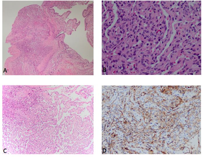
Accepted: 8-28-2020 beneath the respiratory epithelium (Figure 1A). OnCase report / Arch Argent Pediatr 2021;119(1):e36-e40 / e37 high magnification, the tumor consisted of bland Magnetic resonance imaging (MRI) revealed epithelioid endothelial cells that did not display a lobulated mass in the left ethmoid sinus, cytological atypia and contained intracytoplasmic extending inferiorly to the middle and superior vacuoles. Erythrocyte extravasation was present nasal meatus, superiorly to left frontal sinus (Figure 1B). The tumor was also accompanied via frontoethmoidal recesses. The medial and by hyalinized papillary areas lined by superior orbital extraconal area was infiltrated single layer endothelial cells consistent with through lamina papyracea and inferior wall papillary endothelial hyperplasia (Figure 1C). of the frontal sinus. Scalloping of the superior Immunohistochemical studies showed that the wall of the frontal sinus was present. The mass tumor cells were positive with CD31 supporting was hyperintense on T2 weighted images and its vascular origin (Figure 1D). The tumor was hypointense on T1 weighted images, enhanced negative with HHV-8, D2-40, GLUT1, and desmin, vividly but heterogeneously after gadolinium- respectively helping differentiation from Kaposi based contrast media injection on T1 weighted sarcoma, Kaposiform hemangioendothelioma, images (Figure 2). Vascular flow voids were infantile hemangioma, and rhabdomyosarcoma. present in the T2 weighted images and Based on these findings, the patient was postcontrast T1 weighted images. diagnosed with SCH and papillary endothelial The patient was under follow-up hyperplasia. Figure 1. Representative images of pathologic slices from the biopsy of the mass in the left nasal passage (A) The tumor is seen beneath the respiratory epithelium and contains vascular clefting (hematoxylin and eosin staining, x4). (B) Endothelial cell proliferation with intracytoplasmic vacuoles and accompanying erythrocyte extravasation (hematoxylin and eosin staining, x40). (C) Areas of papillary endothelial hyperplasia (hematoxylin and eosin staining, x10). (D) The tumor cells were positive with CD31 immunohistochemically.
e38 / Arch Argent Pediatr 2021;119(1):e36-e40 / Case report

conservatively since surgery was deemed risky                     follow-up, and in the imaging studies performed
and oral propranolol treatment was continued.                     30 months after surgery, it was found that the
But, his nasal obstruction complaint didn’t                       residual mass had disappeared.
improve, and he started to have frequent                              Informed consent was received from the
nosebleeds. Propranolol was discontinued when                     family.
he was 8-month-old, and surgical resection was
decided. Under general anesthesia, the mass was                   DISCUSSION
removed endoscopically. The mass was fragile                          Spindle cell hemangioma is an uncommon
and vascular and occupied the ethmoid region.                     benign lesion that has a predilection to extremities
The anterior skull base and orbit were explored,                  and may present as solitary or multifocal, painless
the lamina papyracea was defective. The mass                      or painful mass. 2,4 Spindle cell hemangioma
was dissected from periorbitis and skull base                     commonly appears as a nodule on extremities.2
under endoscopic vision. It was removed in four                   Although 11 cases of SCH localized in the head
pieces, the largest one was 3 x 2.5 x 1.3 cm, and                 and neck region have been reported previously,3
the specimen was macroscopically brown and                        our case is interesting and unusual because, to our
irregular. Histopathological examination revealed                 knowledge, it is the first case with paranasal sinus
spindle cell hemangioma similar to the initial                    involvement. While most of the reported cases
biopsy result.                                                    were asymptomatic, our patient had swelling
    An MRI performed three months after surgery                   in the left eyelid, nasal obstruction, and nasal
revealed a 1.5 x 1.7 x 1 cm residual lesion in the                bleeding. Spindle cell hemangioma can be seen
left frontal sinus and frontoethmoidal recess,                    at any age, 2 and our case can be considered
with the same aforementioned imaging features                     congenital because the swelling in the left eyelid
(Figure 2). In the sixth month follow-up, there                   had been noticed since birth and nasal obstruction
was minimal regression of residual lesions.                       since the second week of age.
There were no significant complaints during the                       While most SCH lesions are smaller than

Figure 2. MRI images of the patient before (A, B) and after surgery (C, D, E, F, G, H).

Axial fat-suppressed (FS) T2 weighted image shows a hyperintense mass with scalloping-thinning of lamina paprycea
and extending to extraconal orbital fat (A). Postcontrast FS coronal T1 weighted image reveals intense but heterogeneous
enhancement of the mass (B). Axial T2 weighted image (C) and postcontrast FS coronal T1 weighted image (D), three months
after surgery, show residual mass at frontoethmoidal recess (arrows). The residual mass shows gradual decrease in size six
months after the surgery (arrows), shown on axial T2 weighted image (E) and postcontrast FS coronal T1 weighted image
(F). On the latest follow-up MRI, 30 months after the surgery, axial T2 weighted image (G) and postcontrast FS coronal T1
weighted image (H) show no sign of active disease (arrows).
Case report / Arch Argent Pediatr 2021;119(1):e36-e40 / e39
2 cm, 5 in our case, it was remarkable that the on the ankle and foot had been successfully
surgically removed material was much bigger. treated with sclerotherapy.13 Radiotherapy is not
Histopathological examination of spindle recommended because a case with malignant
cell hemangioma consists of cavernous vascular transformation and lymph node metastasis has
spaces that may have thrombus and lined by been reported after radiotherapy.1 So, we planned
endothelial cells and solid spindle cell areas. to follow the patient closely in terms of recurrence
Spindle cell areas also have epithelioid endothelial after spontaneous regression.
cells with cytoplasmic vacuoles. Nuclear atypia Spindle cell hemangioma is not a frequent
is generally low in SCH, and it rarely shows tumor in the head and neck region, and presenting
mitotic activity.6 But, this case of SCH also had complaints may not suggest the diagnosis, as in
papillary endothelial hyperplasia areas, which our patient. Since this entity is less well known
was rarely reported in the literature.5,7,8 In the and has clinical potential to be misdiagnosed
immunohistochemical examination of SCH, as hemangiomas or other vascular lesions, the
capillary and cavernous vessels are positive histopathological diagnosis of such lesions
for CD31, and spindle cell area is positive for has great importance for treatment planning.
smooth muscle actin.9 Although the lesion was Awareness of this diagnosis may increase the
thought to originate from the blood vessels, a reported cases. n
recent study showed endothelial cells of SCH
are positive for PROX-1 (expressed in lymphatic REFERENCES
proliferation) and suggested SCH as a lymphatic 1. WeissSW,EnzingerFM.Spindlecellhemangioendothelioma.
A low-grade angiosarcoma resembling a cavernous
malformation. 10 Recently, somatic mosaic
hemangioma and Kaposi’s sarcoma. Am J Surg Pathol. 1986;
mutations in IDH1 (isocitrate dehydrogenase) 10(8):521-30.
and IDH2 were described in SCH and not in 2. Perkins P, Weiss SW. Spindle cell hemangioendothelioma.
other vascular lesions. Kurek et al, showed 71 % An analysis of 78 cases with reassessment of its pathogenesis
of SCH had IDH1/IDH2 mutation. 11 Another and biologic behavior. Am J Surg Pathol. 1996; 20(10):1196-
204.
study revealed that 16/17 of SCH cases had
3. French KE, Felstead AM, Haacke N, Theaker J, et al.
IDH1/IDH2 mutation, and these mutations were Spindle cell haemangioma of the tongue. J Cutan Pathol.
not found in other vascular anomalies involving 2016; 43(11):1025-7.
lymphatic malformation. So, it was concluded 4. Hakozaki M, Tajino T, Watanabe K, Yamada H, et al.
that this mutation is highly specific for SCH and Intraosseous spindle cell hemangioma of the calcaneus: a
case report and review of the literature. Ann Diagn Pathol.
can be used for diagnosis.12
2012; 16(5):369-73.
Multiple lesions of SCH may be associated 5. Tosios KI, Gouveris I, Sklavounou A, Koutlas IG. Spindle
with Maffucci’s syndrome, Klippel-Trenaunay cell hemangioma (hemangioendothelioma) of the head
syndrome, congenital lymphedema, and varicose and neck: case report of an unusual (or underdiagnosed)
veins. 2,7 Our patient had no findings of these tumor. Oral Surg Oral Med Oral Pathol Oral Radiol Endod.
syndromes. 2008; 105(2):216-21.
6. Marušić Z, Billings SD. Histopathology of Spindle Cell
Although SCH is a benign lesion, recurrences Vascular Tumors. Surg Pathol Clin. 2017; 10(2):345-66.
up to 58 %, have been reported. 2 More than 7. Fletcher CD, Beham A, Schmid C. Spindle cell
half of the lesions are localized partially or haemangioendothelioma: a clinicopathological and
totally intravascular, and local recurrences occur immunohistochemical study indicative of a non-neoplastic
lesion. Histopathology. 1991; 18(4):291-301.
through these vessels in the region where the
8. Tosios K, Koutlas IG, Kapranos N, Papanicolaou SI. Spindle-
primary lesion is located. The overall prognosis cell hemangioendothelioma of the oral cavity. A case report.
is excellent with total surgical excision. No J Oral Pathol Med. 1995; 24(8):379-82.
metastases and no deaths due to SCH have been 9. Tsukamoto S, Honoki K, Shimada K, Fujii H, et al. Periosteal
reported. 2 Cases with spontaneous regression spindle cell hemangioma of the fibula: a case report. Skeletal
Radiol. 2013; 42(8):1165-8.
have also been reported.7 Endoscopic transnasal
10. Wang L, Gao T, Wang G. Expression of Prox1, D2-40, and
surgery was performed to our patient considering WT1 in spindle cell hemangioma. J Cutan Pathol. 2014;
the advantages of low morbidity, no need for 41(5):447-50.
an incision, and providing detailed vision. 11. Kurek KC, Pansuriya TC, van Ruler MA, van den Akker
However, total excision could not be performed B, et al. R132C IDH1 mutations are found in spindle
cell hemangiomas and not in other vascular tumors or
due to the localization of the mass. Fortunately,
malformations. Am J Pathol. 2013; 182(5):1494-500.
in our patient, the residual mass regressed 12. Ten Broek RW, Bekers EM, de Leng WWJ, Strengman E, et
spontaneously and disappeared. Also, it was al. Mutational analysis using Sanger and next generation
reported that a 10-year-old girl with SCH nodules sequencing in sporadic spindle cell hemangiomas: A studye40 / Arch Argent Pediatr 2021;119(1):e36-e40 / Case report
of 19 cases. Genes Chromosomes Cancer. 2017; 56(12):855-60. spindle cell hemangioma: Report of two cases. Indian J
13. Kramer D, Downey C, Vargas P, Castro A. Multifocal Dermatol Venereol Leprol. 2016; 82(1):93-5.You can also read