Minimally Invasive Repair of a Delayed Recurrent Traumatic Cerebrospinal Fluid Rhinorrhea: A Case Report of Dislocation of the Bony and Dural Defects
←
→
Page content transcription
If your browser does not render page correctly, please read the page content below
Minimally Invasive Repair of a Delayed Recurrent
Traumatic Cerebrospinal Fluid Rhinorrhea: A Case
Report of Dislocation of the Bony and Dural Defects
Xiaofei Liu
University of South China
Ping Chen
The Second Hospital,University of South China
Bing Wang ( wangb46@mail2.sysu.edu.cn )
Second hospital,University of South China
Case Report
Keywords: Cerebrospinal uidrhinorrhea, Craniocerebral trauma, Recurrent, Minimally invasive repair
DOI: https://doi.org/10.21203/rs.3.rs-32281/v1
License: This work is licensed under a Creative Commons Attribution 4.0 International License.
Read Full License
Page 1/8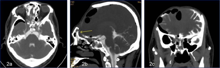
Abstract
Background Dural and bony defects mostly occur in the same position in the cerebrospinal uid CSF
rhinorrhea of anterior cranial base fractures,and a few cases of delayed CSF leakage after repair are also
reported.

Case presentation We report a case in which a pedicled temporoparietal fascial ap was used to repair
the comminuted fracture of the anterior skull base with CSF leakage. Delayed CSF leakage occurred 45
days after the operation.A minimally invasive approach through an eyebrow incision was performed for
reoperation,it was found that the bony defect was located in the right frontal sinus and the dural defect
was located in the right ethmoid plate.

Conclusions:This case suggests that delayed traumatic CSF rhinorrhea after reconstructive surgery is
more complex than usual,and appropriate approach should be adopted to repair the dural and bony
defects , the transeyebrow approach is a good choice.

1 Background
Cerebrospinal uid(CSF)rhinorrhea, commonly observed in anterior cranial base fractures,requires
surgical treatment when conservative measures fail. Dural and bony defects mostly occur in the same
position. Few cases of delayed secondary CSF leakage requiring surgical treatment are
reported,dislocation of the bony and dural defects is rare. Due to the scarcity of such cases, limited data
exist to guide the management of this complication. We have reported the case of a patient with a
bilateral comminuted anterior skull base who underwent a rst-stage craniotomy with pedicled fascial
 aps. Despite this, the patient suffered delayed CSF rhinorrhea. During the second operation, it was found
that bony and dural defects were not contiguous. The defects were repaired with a minimally invasive
approach using multiple layers of free fat and fascia.

2 Case Presentation
2.1 Patient Imformation
A 33-year-old man was admitted for posture-related CSF rhinorrhea, which was prominent in lateral and
head-down positions. 45 days ago, this patient was sent to emergency department with a consciousness
disorder and a nasal leakage due to a fall injury after an electric shock. He also suffered from multiple
skin burns on his limbs and perineum,with no signi cant past history. Continuous dynamic CT scan
revealed an increase in bilateral frontal hematoma(Fig. 1).The GCS score dropped from 8 to 6.Then he
underwent bilateral craniotomy,bilateral temporoparietal fascia was used to repair the anterior skull base
and the open frontal sinus was closed with bone wax. After the surgery, the patient’s GCS improved, CSF
rhinorrhea stopped without any intracranial infection. He was then transferred to the burn department for

                                                  Page 2/8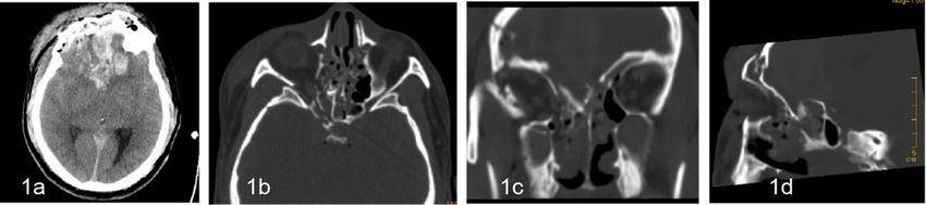
skin graft surgery. He then started to have posture-related CSF rhinorrhea and was transferred to
neurosurgery again.

2.2 Investigations
CT scan showed intracranial pneumocephalus and the quantitative determination of glucose in nasal
leakage was 4.5 mmol/L. CT cisternography showed that the contrast agent owed from the frontal
sinus into the nasal cavity. (Fig. 2). The CSF collected from lumbar puncture showed normal results
without any indication of intracranial infection.

2.3 Diagnosis

  1. Delayed CSF rhinorrhea ( stula located in right frontal sinus).
  2. Post-operation of bilateral anterior skull base reconstruction.

2.4 Treatment
The patient underwent craniotomy through the right trans-eyebrow approach. During the operation, the
frontal sinus was found to be open, and no obvious epidural leakage was found near the frontal sinus.
Further exploration of the anterior cranial base along the epidural was conducted, and the dural defect
was found in the right cribriform plate, with a size of about 10*10 mm (Fig. 3). We used a multi-layer
reconstruction method with autologous fat and fascia to repair the leak. The patient was discharged on
the 7th day.

2.5 Outcome and Follow-Up
At 6 months’ follow-up, the patient had no CSF leakage, no intracranial infection, and could live a normal
life.

3 Discussion
Traumatic CSF leakage occurs in 2% of head trauma patients, and accounts for 12–30% of all skull base
fractures[1]. Persistent CSF leaks often require surgical repair. Following repair, early recurrence can occur
whereas the occurrence of delayed stula is less common.

The qualitative diagnosis of CSF rhinorrhea is relatively simple,compared with the determination of sugar
content[2, 3], the determination of beta-transferrin [4], is more sensitive and speci c. However, the di culty
lies in the determination of the location of CSF leakage, which is of great signi cance to clinical decision-
making. The detction of bony defect is not di cult, and dural defect may be indicated by indirect signs. In
most cases, the defects are contiguous. The most signi cant displacement of fracture slices are mostly
the location of the stula [5]. Patients with chronic traumatic CSF leakage can be inferred from the
presence of meningeal encephalocele on MRI T2 images and abnormal accumulation of CSF[6]. CT

                                                    Page 3/8
cisternography is a good method to show the stula. However, it has been noted that no imaging
techniques are particularly useful[7].In this case, CT cisternography[8] suggests that the bony defect is in
the frontal sinus. However, this method is static and does not dynamically show CSF ow,that is, the
dural defect cannot be clearly shown. In this case, the dural defect was found to be non-contiguous with
the bone fracture.
Delayed post-traumatic CSF rhinorrhea is more inclined to surgical treatment to reduce the risk of
intracranial infection[9]. There are two kinds of surgical repair methods: craniotomy and transnasal
approach[10, 11]. Common craniotomy incisions include unilateral frontal incision and bilateral frontal
coronal incision[12]. It is suitable for patients with large skull base defects and extensive repairs, and for
patients with frontal sinus opening. The transnasal approach is more suitable for those leakages in the
sphenoid sinus, planum sphenoidale, tuberculum sellae, and the cribriform plate. These defects are
generally small [13]. Therefore, identifying the stula, including dural and bony defects is the most
important task.
In this case, the stula was considered to be in the frontal sinus. Considering that the blood supply of the
pedicle could have been compromised if the original incision had been taken. Hence,we chose the
transeyebrow incision to expose the defects. This minimally invasive approach is widely used in the
resection of lesions in the anterior skull base[14–17], including tumors and aneurysms. It can effectively
expose the bone and the dura of the anterior skull base, and is feasible for the exploration and repair of
the bony and dural defects.Unexpectedly, the dural and bony defects are discontinous, and the choice of
transnasal repair may lead to the failure of the operation. To analyze the causes of inconsistent stula,we
infer that the anterior skull base was covered with pedicled temporoparietal fascial aps in the rst
operation, thus the bony defect was repaired, while the dural defect perhaps remained patent. Delayed
CSF leakage may occur when the frontal sinus was not closed properly or when the bone wax contracted
and displaced[18]. This case also suggests that the sealing of the frontal sinus may not be su cient with
bone wax alone, but pedicled periosteal aps may be required.Since there is no pedicled vascular tissue
available for the second operation, we chose free fat and fascia for multilayer repair, which proved to be
reliable.
4 Conclusions
This case suggests that delayed traumatic CSF rhinorrhea after reconstructive surgery is more complex
than usual,CT cisternography is not reliable,especially for identifying the dural defect,and appropriate
approach should be adopted to repair the dural and bony defects, the transeyebrow approach is a good
choice.
Abbreviations
CSF
Page 4/8Cerebrospinal uid; CT:Computed tomography; GCS:Glasgow Coma Scale;MRI:Magnetic resonance
imaging
Declarations
Acknowledgements
Not applicable.
Authors’ contributions
Xiaofei Liu and Ping Chen participated in the treatment of this patient with assistance from Bing Wang
and was involved in the development of the conclusions. Bing Wang wroteand edited the draft. All
authors had read and approved the nal manuscript.
Funding
This work was supported by grants from the Natural Science Foundation of Hunan province
(2018JJ3461).
Availability of data and materials
Not applicable.
Ethics approval and consent to participate
Not applicable.
Consent for publication
Written informed consent for publication of the clinical details and clinical images was obtained from the
patient.
Competing interests
The authors declare that they have no competing interests.
Author details
Page 5/81Department of Neurosurgery, The Second Hospital, University of South China,Hengyang 421001, China.
References
1. Friedman JA, Ebersold MJ, Quast LM. Persistent posttraumatic cerebrospinal uid leakage.
Neurosurg Focus. 2000;9(1):e1.
2. Ziu M, Savage JG, Jimenez DF. Diagnosis and treatment of cerebrospinal uid rhinorrhea following
accidental traumatic anterior skull base fractures. Neurosurg Focus. 2012;32(6):E3.
3. Oakley GM, Alt JA, Schlosser RJ, Harvey RJ, Orlandi RR. Diagnosis of cerebrospinal uid rhinorrhea:
an evidence-based review with recommendations. Int Forum Allergy Rhinol. 2016;6(1):8–16.
4. Mantur M, Łukaszewicz-Zając M, Mroczko B, et al. Cerebrospinal uid leakage–reliable diagnostic
methods. Clin Chim Acta. 2011. 412(11–12): 837 – 40.
5. Reddy M, Baugnon K. Imaging of Cerebrospinal Fluid Rhinorrhea and Otorrhea. Radiol Clin North Am.
2017;55(1):167–87.
6. Bonetto N, Manara R, Citton V, Cagnin A. Spinal subtraction MRI for diagnosis of epidural leakage in
SIH. Neurology. 2011;77(21):1873–6.
7. Kranz PG, Tanpitukpongse TP, Choudhury KR, Amrhein TJ, Gray L. Imaging Signs in Spontaneous
Intracranial Hypotension: Prevalence and Relationship to CSF Pressure. AJNR Am J Neuroradiol.
2016;37(7):1374–8.
8. Nursal GN, Yapar AF. Demonstration of cerebrospinal uid leakage on radionuclide cisternography by
SPECT/CT. Clin Nucl Med. 2015;40(1):e55-7.
9. Liao KH, Wang JY, Lin HW, et al. Risk of death in patients with post-traumatic cerebrospinal uid
leakage–analysis of 1773 cases. J Chin Med Assoc. 2016;79(2):58–64.
10. Liu P, Wu S, Li Z, Wang B. Surgical strategy for cerebrospinal uid rhinorrhea repair. Neurosurgery.
2010;66(6 Suppl Operative):281–5. discussion 285-6.
11. Kreatsoulas DC, Shah VS, Otto BA, Carrau RL, Prevedello DM, Hardesty DA. Surgical outcomes of the
endonasal endoscopic approach within a standardized management protocol for repair of
spontaneous cerebrospinal uid rhinorrhea. J Neurosurg. 2020: 1–7.
12. Safavi-Abbasi S, Komune N, Archer JB, et al. Surgical anatomy and utility of pedicled vascularized
tissue aps for multilayered repair of skull base defects. J Neurosurg. 2016;125(2):419–30.
13. Clavenna MJ, Turner JH, Chandra RK. Pedicled aps in endoscopic skull base reconstruction: review
of current techniques. Curr Opin Otolaryngol Head Neck Surg. 2015;23(1):71–7.
14. Reisch R, Marcus HJ, Hugelshofer M, Koechlin NO, Stadie A, Kockro RA. Patients' cosmetic
satisfaction, pain, and functional outcomes after supraorbital craniotomy through an eyebrow
incision. J Neurosurg. 2014;121(3):730–4.
15. Chiappini A, Marchi F, Reinert M, Robert T. Supraorbital approach through eyebrow skin incision for
aneurysm clipping: how I do it. Acta Neurochir (Wien). 2018;160(6):1155–8.
Page 6/816. He H, Li W, Liang C, et al. Eyebrow Incision for Combination Supraorbital Minicraniotomy with Orbital
Osteotomy: Application to Cranio-Orbital Lesions. World Neurosurg. 2018;114:e631–40.
17. Lee YM, Park HJ, Kim SD, Cho KS. Mini Osteoplastic Flap Through Supra-Eyebrow Incision for
Primary Frontal Sinus Squamous Cell Carcinoma. J Craniofac Surg. 2020;31(2):517–9.
18. Das JM. Bone Wax in Neurosurgery: A Review. World Neurosurg. 2018;116:72–6.
Figures
Figure 1
CT scan before the rst operation.1a shows contusion and laceration of bilateral frontal lobe with
hematoma formation, 1b-1d show comminuted fracture of frontal bone and anterior skull base, especially
on the right side.
Figure 2
Image of CT Cisternography.CT cisternography shows contrast agent leaks from right frontal sinus to
nasal cavity.The arrow in gure 2b shows a leak of contrast material from the frontal sinus.
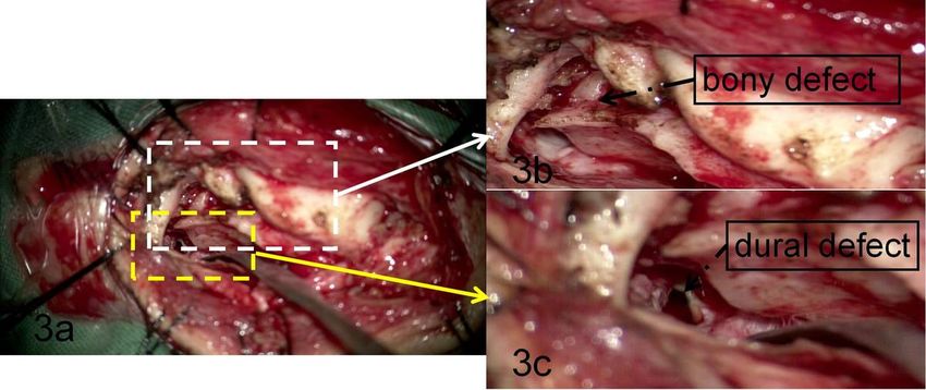
Page 7/8Figure 3
Screenshot of the second operation video.The right transeyebrow approach was used.3a shows the
intraoperative exploration of CSF leakage. 3b and 3c show bony and dural defect, respectively.
Page 8/8You can also read