Anatomic and Embryologic Analysis of the Dural Branches of the Ophthalmic Artery
←
→
Page content transcription
If your browser does not render page correctly, please read the page content below
Published January 7, 2021 as 10.3174/ajnr.A6939

                                                                                                                                     REVIEW ARTICLE
                                                                                                                                         ADULT BRAIN

 Anatomic and Embryologic Analysis of the Dural Branches of
                                    the Ophthalmic Artery
                                                                                               S. Bonasia,   S. Smajda,       G. Ciccio, and    T. Robert

      ABSTRACT
      SUMMARY: The ophthalmic artery has one of the most fascinating embryologic developments among the craniofacial arteries.
      Most of the ophthalmic artery orbital branches develop from the formation and regression of the stapedial artery and share their
      origin with dural branches of the ophthalmic artery. The concomitant embryologic development of the ophthalmic artery and mid-
      dle meningeal artery explains adequately the important varieties of anastomosis between these 2 arteries. It also explains the pres-
      ence of many dural branches from the ophthalmic artery. In this review, we focused on dural branches of the ophthalmic artery
      with the description of rare variations possible, in particular the ophthalmic artery origin of the middle meningeal artery and the
      ophthalmic artery origin of the marginal tentorial artery.

      ABBREVIATIONS: dAVF ¼ dural arteriovenous fistula; ECA ¼ external carotid artery; MMA ¼ middle meningeal artery; MTA ¼ marginal tentorial artery; OA ¼
      ophthalmic artery; PDOA ¼ primitive dorsal ophthalmic artery; PVOA ¼ primitive ventral ophthalmic artery; SA ¼ stapedial artery

T   he ophthalmic artery (OA) is a very fascinating artery for its
    complex embryologic development and also for numerous vas-
cular anastomoses developed with branches of the external
                                                                                     Informed consent was obtained from all individual partici-
                                                                                     pants included in the study.

carotid artery (ECA). The role of the OA in supplying the
                                                                                     History
dura is not well-known, but the understanding of the dural
                                                                                     Meyer,1 in 1887, considered a pioneer in the orbital vascular anat-
function of the OA and also of its possible variations is a cor-
                                                                                     omy, was the first to precisely describe all branches of the oph-
nerstone for surgical and endovascular treatment of dural
                                                                                     thalmic artery, including its dural territory. A few years before,
pathologies (dural arteriovenous fistulas (dAVFs), skull base
                                                                                     Curnow,2 in 1873, had already described 3 cadaveric cases of var-
meningiomas, chronic subdural hematoma embolization). In
                                                                                     iations in the origin of the OA. One of these 3 cases was the first
this review, we will focus on the dural branches of the OA
                                                                                     description of an OA origin of the MMA. With the advent of the
with rare variations, in particular the OA origin of the middle
                                                                                     DSA, Kuru,3 in 1967, gave a detailed description of the OA me-
meningeal artery (MMA) and the OA origin of the marginal
                                                                                     ningeal branches, and after him, a few authors4-10 focused on the
tentorial artery (MTA). All procedures performed in the
                                                                                     variants of the OA and the MMA. Lasjaunias et al8,9,11 gave their
studies involving human participants were in accordance
                                                                                     crucial contribution to the comprehension of the orbital and me-
with the ethical standards of the institutional and/or national
research committee and with the 1964 Helsinki Declaration                            ningeal vascular supply, combining their knowledge of the em-
and its later amendments or comparable ethical standards.                            bryology with an accurate angiographic analysis. The last author
                                                                                     to give a comprehensive description of dural vascularization was
                                                                                     Rhoton,12 in 2002, based on a large cadaveric dissection
Received July 18, 2020; accepted after revision September 17.                        experience.
From the Department of Neurosurgery (S.B., T.R.), Neurocentral of Southern
Switzerland, Regional Hospital of Lugano, Lugano, Switzerland; University of
Southern Switzerland (S.B., T.R.), Lugano, Switzerland; and Department of            Embryology. The OA starts its development when the embryo is
Interventional Neuroradiology (S.S., G.C.), Rothschild Foundation Hospital, Paris,   about 4 mm and reaches its adult configuration at about 40 mm.
France.
                                                                                     Its development is strictly connected with the arterial embryology
Please address correspondence to Sara Bonasia, MD, Department of Neurosurgery,
Neurocentral of Southern Switzerland, Regional Hospital of Lugano, Via Tesserete     of the primitive ICA, the stapedial artery (SA), and the pharyn-
46, CH-6903 Lugano, Switzerland; e-mail: sara.bonasia@gmail.com                      geal artery system. In the past, 2 authors were interested in the
     Indicates open access to non-subscribers at www.ajnr.org                        comprehension of the complex events of the OA formation:
     Indicates article with online supplemental data.                                Padget,13 who formulated her theory after the dissection of 22
http://dx.doi.org/10.3174/ajnr.A6939                                                 embryos, and Lasjaunias et al,11 who added to Padget’s
                                                                                                  AJNR Am J Neuroradiol :      2021   www.ajnr.org     1

                                       Copyright 2021 by American Society of Neuroradiology.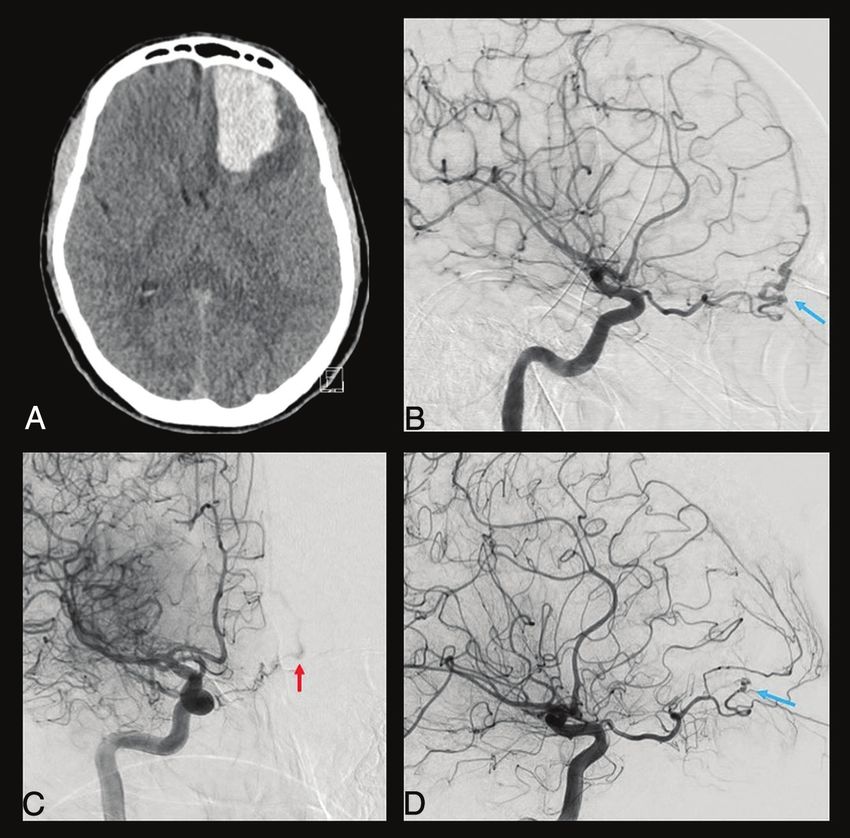
supraorbital artery. The supraorbital ar-
                                                                                                       tery enters the orbit through the supe-
                                                                                                       rior orbital fissure and gives off 2
                                                                                                       branches: the ethmoid nasal and the lac-
                                                                                                       rimal artery.
                                                                                                           The relationship among the PDOA,
                                                                                                       the PVOA, and the SA is most impor-
                                                                                                       tant in the last 2 stages (stage 5: embryo
                                                                                                       of 20 mm; stage 6: embryo of 40 mm).
                                                                                                       During stage 5, an anastomotic ring
                                                                                                       appears around the optic nerve, formed
                                                                                                       by the anastomosis among the PVOA,
                                                                                                       the PDOA, and the supraorbital artery
FIG 1. Theories of Padget and Lasjauinias et al about OA origin migration. When the embryo is
about 18 mm, the OA reaches its definitive origin on the supraclinoid ICA. This phenomenon is           (through the ethmoid nasal artery).
explained by Padget13 by the cranial elongation of the ICA during this stage with the consequent       However, in stage 6, this ring is ventrally
movement of the PDOA origin (black arrows in A). On the other hand, Lasjaunias et al11 hypothe-        interrupted to give the definitive config-
sized the presence of an intradural anastomosis between the PVOA and the primitive ICA (black          uration of the OA. The part of the anas-
arrow in B) in correspondence with the future origin with successive regression of the original        tomotic ring that regresses is crucial to
stem. HA indicates hyaloid artery; MM, maxillo-mandibulary artery; SOrbA, supraorbital artery.
                                                                                                       determine which of the 2 primitive OAs
                                                                                                       persists to form the adult OA.
                                                                                                       According to Padget,13 it is the PDOA
knowledge an accurate angiographic evaluation. These authors               that persists, with consequent PVOA proximal regression. On the
agreed that the definitive features of the OA depend mostly on 2           other hand, Lasjaunias et al9,11,14 wrote that the distal portion of
embryonic arteries, the primitive dorsal ophthalmic artery                 the PDOA regresses and its proximal part is destined to form the
(PDOA) and the primitive ventral ophthalmic artery (PVOA).                 future inferolateral trunk. Thus, their opinion is that the PVOA
                                    13
    According to Padget’s theory, the embryologic development              mostly contributes to the formation of the definitive OA.
of the OA could be divided into 6 stages, which are summarized                 At the same time, the extraorbital part of the supraorbital ar-
in the Online Supplemental Data. The PDOA appears when the                 tery regresses to let the lacrimal artery to be annexed by the OA.
embryo is about 4–5 mm (stage 1), originating from the bifurca-
tion of the primitive internal carotid artery. In the following stage      Dural Branches of the OA
2 (9-mm embryos), while the PDOA enlarges through the optic                Different dural branches of the OA and their possible anastomo-
cup as plexiform channels, the PVOA arises from the cranial divi-          ses with other dural arteries are listed in Table 1; their respective
sion of the primitive ICA. The PDOA and PVOA are then des-                 dural territories are illustrated in Fig 2.
tined to elongate following the ventral shifting of the optic cup
                                                                           Deep Recurrent Ophthalmic Artery. The deep recurrent oph-
and the dorsal shifting of the cerebral hemispheres. In embryos of
                                                                           thalmic artery arises from the first segment of the OA and has a
14–17 mm, one can note the appearance of 2 branches from the
                                                                           recurrent course through the medial part of the superior orbital
PDOA artery: the primitive hyaloid artery and the common tem-
                                                                           fissure. This artery supplies the dura of the lateral wall of the cav-
poral ciliary artery (future lateral posterior ciliary artery). The
                                                                           ernous sinus. It consistently anastomoses with the anteromedial
PVOA gives off, at the same time, the common nasal ciliary ar-
                                                                           branch of the inferolateral trunk and often with the cavernous
tery (future medial posterior ciliary artery).
                                                                           branch of the middle meningeal artery and with the accessory
    In the embryo of about 18 mm (stage 3), the OA undergoes a
                                                                           meningeal artery. It is considered as a remnant of the primitive
process that will cause migration of its origin on the supraclinoid
                                                                           dorsal ophthalmic artery.9
ICA. This phenomenon is explained by Padget by the cranial
elongation of the ICA during this stage with the consequent                Superficial Recurrent Ophthalmic Artery. The superficial recur-
movement of the PDOA. On the other hand, Lasjaunias et                     rent ophthalmic artery is a meningeal branch that takes its origin
al9,11,14 explained this migration by the presence of an intradural        from the proximal part of the lacrimal artery or directly from the
anastomosis between the PVOA and the primitive carotid artery              second segment of the OA.9,11,15 This artery passes through the
in correspondence of the future origin, with consequent regres-            lateral part of the superior orbital fissure to reach the dura over
sion of the original stem. The 2 theories of OA origin migration           the anterior clinoid process and the cavernous sinus roof.3,16 The
are illustrated in Fig 1.                                                  superficial recurrent ophthalmic artery also supplies the intra-
    From stage 1 to 4, another artery grows at the same time and           dural part of the third and fourth cranial nerves. This artery is the
contributes to the adult configuration of the orbital arteries: the        orbital remnant of the supraorbital branch of the stapedial
SA. In the first stages, the optic cup is supplied on its ventral side     artery.11
by the primitive maxillary artery. However, it starts to regress at
the end of stage 2, to be substituted by the SA in its orbital terri-      Posterior Ethmoidal Artery. The posterior ethmoidal artery is a
tory. This latter gives off 2 branches that follow the 3 divisions of      small meningeal branch that originates from the third segment of
the trigeminal nerve: the maxillomandibular artery and the                 the OA, which exits the orbit through the posterior ethmoidal
2    Bonasia    2021   www.ajnr.org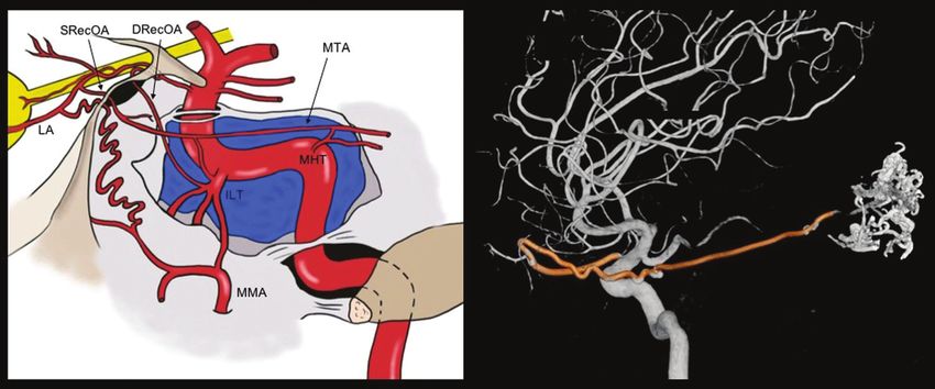
Table 1: Different dural branches of the OA with their respective supply and anastomoses
                       Origin from                                                     Possible                 Clinical Consequences in Case
    OA Branches           the OA          Foramen            Dural Territory         Anastomosis                          of Embolism
 Deep recurrent       First segment Superior orbital     Superior orbital       Inferolateral trunk            Cerebrovascular accident
   OA                                   fissure              fissure (lateral        (ICA)
                                                            part), sphenoid
                                                            wing
 Superficial           Second          Superior orbital   Anterior clinoid       Posterior ethmoidal            Cerebrovascular accident, loss
   recurrent OA          segment        fissure              process                artery                        of vision
                                                          Lesser sphenoid        MMA (anterior
                                                            wing                   division)
                                                          Middle fossa           Medial tentorial
                                                            (anteromedial          artery (ICA)
                                                            portion)
 Anterior ethmoidal Third segment Anterior               Anterior convexity     Contralateral
   artery                               ethmoidal canal     (anterior              anterior
                                                            meningeal artery)      ethmoidal artery
                                                          Anterior cranial       Bilateral MMAs
                                                            fossa (medial          Posterior
                                                            third)                 ethmoidal artery
                                                          Anterior falx cerebri Olfactory branch
                                                            (anterior falcine      (ACA)
                                                            artery)
 Posterior            Third segment Posterior            Anterior cranial       Contralateral
   ethmoidal artery                     ethmoidal canal     fossa (medial          posterior
                                                            third)                 ethmoidal artery
                                                          Anterior clinoid       Anterior ethmoidal
                                                            process                artery
                                                          Chiasmatic groove      MMA (anterior
                                                                                   division)
Note:—ACA indicates anterior cerebral artery.

                                                                              canal.15 Its average diameter is 0.4 mm, and it is usually in bal-
                                                                              ance with the diameter of the anterior ethmoidal artery.17 This ar-
                                                                              tery supplies the dura of the planum sphenoidale, the posterior
                                                                              cribriform plate, and the anterior clinoid process.11 Martins et
                                                                              al,16 in 2005, showed that the posterior ethmoidal artery often
                                                                              anastomoses with dural branches of the internal carotid artery,
                                                                              middle meningeal artery, and anterior ethmoidal artery. When
                                                                              absent (approximately 20%), its meningeal territory is taken over
                                                                              by these 3 other arteries.16

                                                                              Anterior Ethmoidal Artery. The anterior ethmoidal artery is a
                                                                              more constant artery, which has been found in .90% of the
                                                                              orbits if the OA crosses over the optic nerve, and in 80% of
                                                                              cases when the OA crosses under the nerve. It originates
                                                                              from the distal part of the OA and could give off from 1 to 5
                                                                              little branches that pass through the anterior ethmoidal
                                                                              canal. Other than the nasal septum and nasal fossa, its me-
                                                                              ningeal territory is limited to the anterior part of the cribri-
FIG 2. Dural territories of OA branches. A, Territory of the deep             form plate, the medial part of the orbital roof, and the
recurrent ophthalmic artery (green), which exits from the medial part         anterior third of the falx cerebri. The anterior ethmoidal ar-
of the superior orbital fissure and supplies the dura of the lateral wall      tery gives off a branch, well-described angiographically by
of the cavernous sinus. B, Dural territory of the superficial recurrent
                                                                              Kuru,3 in 1967, along the falx cerebri, called the anterior fal-
ophthalmic artery (pink), which passes through the lateral part of the
superior orbital fissure to reach the dura over the anterior clinoid           cine artery or the artery of the falx cerebri. This anterior fal-
process and the cavernous sinus roof. C, The posterior ethmoidal ar-          cine artery could be present bilaterally, but usually 1 side is
tery (orange) passes through the posterior ethmoidal canal to reach           predominant. If the anterior ethmoidal artery is well-devel-
the dura of the planum sphenoidale, the posterior cribriform plate, and       oped, it can give off some branches called “anterior menin-
the anterior clinoid process. D, The anterior ethmoidal artery (light
blue) passes through the anterior ethmoidal canal, and its meningeal
                                                                              geal arteries,” that differ from the anterior falcine artery
territory consists of the anterior part of the cribriform plate, the medial   because of their paramedial course and can supply the dura
part of the orbital roof, and the anterior third of the falx cerebri.         of the anterior convexity.
                                                                                           AJNR Am J Neuroradiol :    2021   www.ajnr.org    3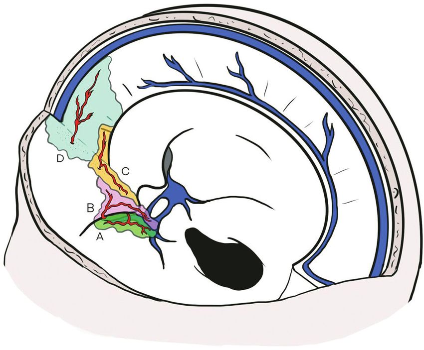
FIG 3. MMA origin from the OA. Anterior-posterior and lateral view angiograms (A and B) show a rare case of complete MMA origin from the
OA. The OA, through the superficial recurrent OA, gives rise to the MMA, which passes through the lateral part of the superior orbital fissure
and gives off its anterior (red arrow) and posterior divisions (blue arrow). In the angiograms C, D, and E, a rare case of partial origin of the MMA
from the OA is shown. The angiograms D and E show a left ICA injection in frontal and lateral views, where the posterior branch of the MMA
(blue arrow) originates from the OA and feeds a tentorial arteriovenous fistula. After the ECA injection (C), only the anterior branch of the
MMA is enhanced (red arrow). Reproduced from Bonasia et al.27

Dural Supply of the OA                                                      been described in the literature. The first case was presented by
The 4 meningeal branches previously described supply the dura               Curnow,2 in 1873, and in the same period, Meyer,1 in 1887, also
of the cribriform plate: the planum, the anterior clinoid process,          cited 4 cadaveric cases originally described by Zuckerkandl in
the superior orbital region, the roof and the lateral part of the cav-      1876 during a congress presentation.18 Two rare cases of this var-
ernous sinus, the medial part of the orbital roof, and the anterior         iation are shown in Fig 3. This vascular anomaly is considered
part of the falx cerebri. This vascular territory is variable, and the      the consequence of 2 different embryologic processes. The first
OA is in balance with other meningeal arteries of this region like          is the failure of the supraorbital branch (stapedial artery) regres-
the middle meningeal artery (cavernous ramus), the accessory                sion. The second is the absence of anastomosis between the max-
meningeal artery, and the inferolateral trunk.                              illomandibular branch of the stapedial artery and the internal
                                                                            maxillary artery. Consequently, the MMA originates from the
Variations of Dural Branches of the Ophthalmic Artery                       OA and passes through the lateral part of the superior orbital fis-
Ophthalmic Artery Origin of the Middle Meningeal Artery. In                 sure; thus, the foramen spinosum is usually absent. Maiuri et al,19
rare cases, the middle meningeal artery could originate from the            in 1998, proposed 3 different types of this vascular variation as
OA instead of the internal maxillary artery. The incidence of this          highlighted in Table 2. The first type is the complete MMA terri-
vascular variation was estimated to be 0.5% by Dilenge and                  tory taken over by the OA through the superficial recurrent OA.
Ascherl,10 in 1980, based on a large angiographic series. A few             In the second type, only the anterior branch of the MMA origin
cases of the middle meningeal artery arising from the OA have               from the OA and the posterior branch of the MMA retain their
4     Bonasia    2021   www.ajnr.org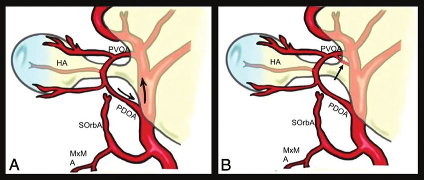
origin from the internal maxillary artery. The third type is not            the accessory meningeal artery or the inferolateral trunk, has
really an OA origin of the MMA but an anastomosis between the               also been described.
OA and the accessory meningeal artery (through the deep recur-
rent OA). The consequence is that the anterior meningeal terri-             Clinical Implications
tory is supplied by both the MMA and the OA without any                     The knowledge of the dural branches arising from the OA and
communication. It is still a matter of debate whether the MMA               their variations represents the cornerstone for interventional neu-
originates from the OA directly or from the proximal part of the            roradiologists and neurosurgeons who treat anterior and middle
lacrimal artery.                                                            cranial fossa pathologies. Two critical examples are cribriform
Ophthalmic Artery Origin of the Marginal Tentorial Artery.                  plate dAVFs and anterior and middle skull base meningiomas.
The marginal tentorial artery (or artery of the free margin of the
tentorium cerebelli) normally arises from the meningohypophy-               Cribriform Plate Dural Arteriovenous Fistulas. The cribriform
sary trunk, but its origin is variable, as illustrated in Fig 4. This ar-   plate dAVFs are usually mostly supplied by the anterior ethmoidal
tery supplies the medial third of the tentorium, partially the walls        artery and the MMA. A bilateral supply of the dAVF, found in
of the cavernous sinus, and also the transdural segment of the              approximately 10% of cases, is well-explained by the anastomoses
oculomotor and trochlear nerves.16 An OA origin of this artery              between the 2 anterior ethmoidal arteries within the dural or ethmoi-
has been described by Lasjaunias et al,11 in 2001, distinguish-             dal sinuses. Endovascular treatment of such pathologies consists of
ing 2 different types. The first is when the marginal tentorial             the embolization, usually through branches of the MMA. The neuro-
artery originates from the lacrimal artery. The second when                 radiologist has to consider the presence of dural MMA-OA anasto-
the marginal tentorial artery arises directly from the OA and               moses during the injection of the liquid agent to avoid retrograde
the lacrimal artery originates from the MMA (meningolacri-                  flow into ocular branches of the OA. In case of direct embolization of
mal type). A marginal tentorial artery arising from the MMA,                the dAVF through the ophthalmic artery, attention should be paid to
                                                                            the possibility of retrograde flow of the embolic agent into ocular
Table 2: Different types of OA origin of the MMA by Maiuri et               branches. Because the central retinal artery usually arises from the
al19                                                                        second segment of the OA, the injection should be performed as dis-
                                                 Foramen                    tal as possible to limit the eventual damage caused by the reflux.
 Type             Vascular Anatomy               Spinosum                        The surgical exclusion of a cribriform plate dAVF also
 I      Complete OA origin of the MMA         Absent                        necessitates a precise knowledge of dural branches of the
 II     Partial OA origin of the MMA          Reduced in size
          Anterior division from the OA
                                                                            OA. The aim of the treatment is to exclude the cortical ve-
          Posterior division from the IMA                                   nous drainage of the dAVF, clipping or coagulating the
 III    OA origin of the accessory meningeal Normal                         draining vein at its exit point from the dura. A case of a cri-
          artery                                                            briform plate dAVF treated surgically is shown in Fig 5. The
Note:—IMA indicates internal maxillary artery.                              knowledge of the arterioarterial anastomoses among the an-
                                                                            terior ethmoidal, posterior ethmoidal, and middle meningeal

FIG 4. Marginal tentorial artery origin and course. The marginal tentorial artery, also called artery of the free margin of the tentorium or artery
of Bernasconi and Cassinari, may have different origins, which are shown in the graphic representation. It can arise from the lacrimal artery (LA)
within the orbit, through the superficial recurrent ophthalmic artery (SRecOA), from the inferolateral trunk (ILT), and from the meningohypo-
physeal trunk (MHT). The artery courses posterolaterally along the free margin of the tentorium. Note a 3D-DSA reconstruction of a rare case
of MTA (highlighted in red) origin from the OA. The MTA exits the orbit through the superior orbital fissure (SOF) and is directed posteriorly to
feed an arteriovenous malformation. DRecOA indicatesdeep recurrent ophthalmic artery .
                                                                                         AJNR Am J Neuroradiol :      2021   www.ajnr.org     5
In case of an operation in a
middle cranial fossa meningioma,
the devascularization of the tumor
as a first step could be helpful to
better understand the arterial sup-
ply of the lesion and limit drasti-
cally blood loss.
Intra-arterial Injection of Chemo-
therapy for Retinoblastoma. The
classic technique used to inject
chemotherapeutic agents into the
OA for the treatment of retino-
blastoma requires the superselec-
tive catheterization of the OA.20
However, the knowledge of OA
dural branches acquires a more
important role when the direct
catheterization of the OA is not
possible, as can happen in chil-
dren due to its reduced size. In
these cases, alternative ways to
reach the OA indirectly have been
described,21 especially through
catheterization of the MMA. Thus,
the pharmacologic agents can be
injected through the anterior divi-
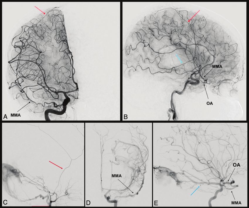
FIG 5. Clinical case of a ruptured cribriform plate dAVF. A 49-year-old man was admitted for sudden sion of the MMA, cannulating its
onset of unusual headache with nausea and vomiting. The CT scan performed in the emergency meningolacrimal branch. The
department (A) shows a left frontal basis intraparenchymal hematoma. The DSA highlighted a cribriform reflux into the OA could be from
plate dAVF with major feeders represented by the left anterior ethmoidal artery from the left OA (blue
anastomoses between MMA or-
arrow in B). The right ICA injection also showed a contribution from the contralateral OA through its eth-
moidal branches (red arrow in C). The venous drainage was represented by a single cortical vein directed bital branches and the recurrent
into the superior sagittal sinus (type III according to the Cognard-Lariboisière classification26). The patient branches of the OA, from the lac-
successfully underwent left supraorbital craniotomy and clipping of the dAVF (D), with no enhancement rimal artery, or sometimes from
of the dAVF on the postoperative DSA (blue arrow) and complete clinical recovery. the direct origin of the OA from
the MMA.
Surgical and Endovascular Treat-
arteries is necessary to understand the dAVF and the techni- ment of Refractory Epistaxis. Re-fractory epistaxis may be
cal difficulties of the treatment. Another case of a dAVF fed caused by many clinical conditions and occurs in about 60% of
by dural branches of the OA is shown in Fig 6, also with a the adult population, with most cases considered idiopathic.
contribution from the MTA. Among them, about 6% of the epistaxis is refractory to con-
The knowledge of the dural branches of the OA and MMA or- servative management and requires surgical or interventional
igin from the OA also adequately explains the possible participa- treatment.22
tion of OA branches in the supply of carotid-cavernous fistulas or The best way to understand the source of bleeding in case
tentorial pathologies (Figs 3 and 4). of refractory epistaxis is to perform a diagnostic DSA includ-
ing the ICA and ECA. The DSA allows identifying the so
Anterior and Middle Cranial Fossa Meningiomas. The surgical called “vascular blush,” an anastomotic plexus located in the
removal of cribriform plate or sphenoid wing meningiomas nasal septum, considered the source of 90% of epistaxis. The
requires a detailed knowledge of vascular normal anatomy and sphenopalatine artery represents its main blood supply, and
tumor vascular supply. Meningeal tumors of the anterior and it is most commonly responsible for refractory epistaxis, even
middle skull base are usually supplied by dural branches of the if, in rare cases, the ethmoidal arteries could also be
MMA, internal carotid artery, and the ophthalmic artery. It is of involved.23 If these latter are involved in the bleeding, they
paramount importance for interventional neuroradiologists who can be ligated through a surgical approach. On the other
plan an embolization, usually performed through the MMA, to hand, if the sphenopalatine artery is responsible for bleeding,
consider the possible variations in the supply of the skull base it can be occluded through an endonasal approach or it can
dura to avoid involuntary OA reflux of embolic liquid agent. be embolized.23
6 Bonasia 2021 www.ajnr.orgFIG 6. Clinical case of a dAVF fed by multiple OA dural branches. An 89-year-old woman, previously having undergone an operation for a pi-
tuitary adenoma, was admitted for unusual headache associated with vomiting. The CT scan shows an intraventricular hemorrhage with
mild hydrocephalus. The diagnostic DSA shows a complex dAVF (Cognard-Lariboisière grade IIa1b26) supplied by the OA through the
anterior and posterior ethmoidal arteries (AEtA, PEtA), with both direct and indirect shunts with the superior sagittal sinus (SSS).
Another point of shunt with the SSS is reached by the MTA and the posterior meningeal artery (PMA) and the MMA. Also, other
branches from the ECA contribute to the shunt, like the occipital artery (OccA) and the superficial temporal artery (STA). Because of
the patient’s age, the complexity of the dAVF, the high risk associated with every option of treatment, and the absence of alteration of
consciousness, we managed the dAVF conservatively.
The diagnostic DSA allows identifying possible dangerous vascular structures or as result of tumor necrosis after treat-
anastomoses between branches of the ECA and the OA, ment. In these cases, the symptoms can also cause hemoptysis
which can result in postembolization visual or central defi- due to the frequent nasopharyngeal localization of these
cits. The occurrence of cerebrovascular accidents and tumors. Endovascular treatment should be considered in
obstruction of the central retinal artery have been described these cases to treat uncontrollable epistaxis or hemoptysis.
to occur in about 0%–2% of cases.24 The most important Also benign tumors, like paragangliomas and nasopharyngeal
anastomoses to consider during such procedures are between angiofibromas, can benefit from endovascular embolization
the sphenopalatine and anterior ethmoidal arteries via the as a preoperative procedure to reduce the intraoperative
turbinate and infraorbital arteries23 and between the lacrimal blood loss. In these cases, the embolization of the sphenopa-
artery and the middle meningeal artery through the recurrent latine artery could be insufficient, and devascularization
meningeal artery.24 The relevance of these anastomoses and requires the embolization of the facial artery and ascending
the periprocedural risk can be estimated by analyzing the pharyngeal artery.23 In these cases, neuroradiologists should
“choroidal blush.” This blush is commonly visualized after pay attention to the known anastomoses between the facial
contrast injection into the ICA. However, if the anastomoses artery and the dorsal nasal artery (through the angular ar-
among the posterior ciliary arteries, the lacrimal artery, and tery).25 For these pathologies, the neuroradiologists should
the MMA are very consistent or if the lacrimal artery and the observe the same rules as previously described to avoid com-
OA branch directly from the MMA, the choroidal blush can plications due to ICA-ECA anastomoses.23
be seen after the ECA injection.24 In conclusion, the knowledge of the embryology and anatomy
of the dural branches of the OA is mandatory for treating pathol-
Embolization of Facial Tumors. Even if epistaxes are mostly idi- ogy of the dura mater located in the anterior and middle cranial
opathic, some cases can be due to neoplastic erosion of fossae. These arteries show high variability and supply territories
AJNR Am J Neuroradiol : 2021 www.ajnr.org 7in competition with the middle meningeal artery and internal ca- 15. Hayreh SS. The ophthalmic artery, III: branches. Br J Ophthalmol
rotid artery branches. 1962;46:212–47 CrossRef Medline
16. Martins C, Yasuda A, Campero A, et al. Microsurgical anatomy of
the dural arteries. Neurosurgery 2005;56:211–51; discussion 211–51
CrossRef Medline
REFERENCES 17. Lang J, Kageyama I. The ophthalmic artery and its branches, meas-
1. Meyer F. Zur anatomie der Orbitalarteien. Morph Jahr urements and clinical importance. Surg Radiol Anat 1990;12:83–90
1887;12:414–58 CrossRef Medline
2. Curnow J. Two instances of irregular ophthalmic and middle me- 18. Zuckerkandl E. Zur Anatomie der Orbita Arterien. Med Jahr
ningeal arteries. J Anat Physiol 1873;8:155–56 Medline 1876:343
3. Kuru Y. Meningeal branches of the ophthalmic artery. Acta Radiol 19. Maiuri F, Donzelli R, de Divitiis O, et al. Anomalous meningeal
Diagn (Stockh) 1967;6:241–51 CrossRef Medline
branches of the ophthalmic artery feeding meningiomas of the
4. Gabriele OF, Bell D. Ophthalmic origin of the middle meningeal ar-
brain convexity. Surg Radiol Anat 1998;20:279–84 CrossRef Medline
tery. Radiology 1967;89:841–44 CrossRef Medline
20. Yamane T, Kaneko A, Mohri M. The technique of ophthalmic arte-
5. Royle G, Motson R. An anomalous origin of the middle meningeal
artery. J Neurol Neurosurg Psychiatry 1973;36:874–76 CrossRef rial infusion therapy for patients with intraocular retinoblastoma.
Medline Int J Clin Oncol 2004;9:69–73 CrossRef Medline
6. McLennan JE, Rosenbaum AE, Haughton VM. Internal carotid ori- 21. Klufas MA, Gobin YP, Marr B, et al. Intra-arterial chemotherapy as
gins of the middle meningeal artery: the ophthalmic-middle me- a treatment for intraocular retinoblastoma: alternatives to direct
ningeal and stapedial-middle meningeal arteries. Neuroradiology ophthalmic artery catheterization. AJNR Am J Neuroradiol
1974;7:265–75 CrossRef Medline 2012;33:1608–14 CrossRef Medline
7. Vignaud J, Hasso AN, Lasjaunias P, et al. Orbital vascular anatomy 22. Christensen NP, Smith DS, Barnwell SL, et al. Arterial embolization
and embryology. Radiology 1974;111:617–26 CrossRef Medline in the management of posterior epistaxis. Otolaryngol Head Neck
8. Lasjaunias P, Moret J, Manelfe C, et al. Arterial anomalies at the Surg 2005;133:748–53 CrossRef Medline
base of the skull. Neuroradiology 1977;13:267–72 CrossRef Medline 23. Reyre A, Michel J, Santini L, et al. Epistaxis: the role of arterial
9. Lasjaunias P, Brismar J, Moret J, et al. Recurrent cavernous branches embolization. Diagn Interv Imaging 2015;96:757–73 CrossRef Medline
of the ophthalmic artery. Acta Radiol Diagn (Stockh) 1978;19:553– 24. Mames RN, Snady-McCoy L, Guy J. Central retinal and posterior
60 CrossRef Medline ciliary artery occlusion after particle embolization of the external
10. Dilenge D, Ascherl GF Jr. Variations of the ophthalmic and middle carotid artery system. Ophthalmology 1991;98:527–31 CrossRef
meningeal arteries: relation to the embryonic stapedial artery.
Medline
AJNR Am J Neuroradiol 1980;1:45–54 Medline
25. Bertelli E, Regoli M, Bracco S. An update on the variations of the or-
11. Lasjaunias P, Bereinstein A, Ter Brugge KG. Surgical Neuroangiography.
bital blood supply and hemodynamic. Surg Radiology Anat
Springer-Verlag; 2001
2017;39:485–96 CrossRef Medline
12. Rhoton AL Jr. The orbit. Neurosurgery 2002;51:S303–34 CrossRef
Medline 26. Cognard C, Gobin YP, Pierot L, et al. Cerebral dural arteriovenous
13. Padget DH. The development of cranial arteries in the human fistulas: clinical and angiographic correlation with a revised classi-
embryo. In: Corner G, ed. Contributions to Embryology. Carnegie fication of venous drainage. Radiology 1995;194:671–80 CrossRef
Institution; 1948:205–62 Medline
14. Lasjaunias P, Moret J, Mink J. The anatomy of the inferolateral 27. Bonasia S, Smajda S, Ciccio G, et al. Middle meningeal artery: anat-
trunk (ILT) of the internal carotid artery. Neuroradiology omy and variations. AJNR Am J Neuroradiol 2020;41:1777–85
1977;13:215–20 CrossRef Medline CrossRef Medline
8 Bonasia 2021 www.ajnr.orgYou can also read