VASCULARIZATION OF THE AORTIC ARCH IN - NEOTROPICAL OTTER (Lontra longicaudis, OLFERS 1818)
←
→
Page content transcription
If your browser does not render page correctly, please read the page content below
IUCN Otter Spec. Group Bull. 38(3) 2021

                                         ARTICLE

        VASCULARIZATION OF THE AORTIC ARCH IN
     NEOTROPICAL OTTER (Lontra longicaudis, OLFERS 1818)
       Procassia M. L. BARBOSA1,2, Pricila S. ESTEVES2, Alan L. SANTOS1,
                         Oldemar CARVALHO JUNIOR2
 1
    Universidade Federal de Santa Catarina; Departamento de Zootecnista e Desenvolvimento Rural,
                                 Rod. Admar Gonzaga, s/n, Itacorubi-SC
 2
   Instituto Ekko Brasil, Servidão Euclides João Alves, S/N, Lagoa do Peri, 88066-000, Florianópolis,
                           SC, Brazil. e-mail: atendimento@ekkobrasil.org.br

                      (Received 5th October 2020, accepted 6th December 2020)
      Abstract: Lontra longicaudis is a semi-aquatic carnivorous mammal that belongs to the
      Mustelidae family and Lutrinae subfamily. It is found in South and Central America,
      from southern Mexico to Uruguay and up to 3,000 m altitude. In this study,
      vascularization of the aortic arch is described in the neotropical otter. Eight animals were
      studied, three puppies and five adults from the Otter Project of the Instituto Ekko Brasil.
      The results indicate that the brachiocephalic trunk and the left subclavian artery
      originated from the aortic arch in all animals studied. This pattern is similar to that found
      in wild and domestic carnivores. The brachiocephalic trunk arises three arteries, the left
      common carotid artery, the right common carotid artery, and the right subclavian artery,
      the latter two being emitted from a common trunk. The right and left subclavian arteries
      give origin to five vessels represented by the vertebral artery, the superficial cervical
      artery, the internal thoracic artery, the costocervical trunk, and the axillary artery.
      Citation: Barbosa, P.M.L., Esteves, P.S., Santos, A.L. and Carvalho Junior, O.
      (2021). Vascularization of the Aortic Arch in Neotropical Otter (Lontra longicaudis,
      Olfers 1818). IUCN Otter Spec. Group Bull. 38 (3): 173 -182
      Keywords: physiology; morphology; carnivorous; Mustelidae

INTRODUCTION
      Despite numerous publications on Lontra longicaudis, there is still little
information about its anatomy, morphology and physiology. Most studies concern the
diet (Quadros and Monteiro-Filho, 2001; Uchôa et al., 2004; Quintela et al., 2008;
Carvalho-Junior et al., 2010a, 2010b; Rheingantz et al., 2011), conservation status
(Rodrigues, 2013) or the geographic distribution (Carvalho-Junior et al., 2012).
      Energy analysis and economic evaluation of the otter was carried out by
Carvalho Junior (2016). Knowledge of morphological aspects is essential in
determining diversity, adaptation, and intra-specific variability. This work aims to
investigate the origin of the collateral branches of the aortic arch.
      The circulatory system is responsible for transporting blood through tissues,
bringing oxygen and nutrients to different areas of the body, and collecting the carbon

                                                 - 173 -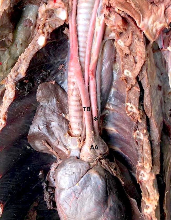
IUCN Otter Spec. Group Bull. 38(3) 2021

dioxide that results from cellular metabolites. The aortic artery is the main vessel that
distributes arterial blood to all systems. The head, neck, chest cavity, and chest organs
are supplied with blood from the arteries that emerge from the aortic arch. Knowing
the distribution and behavior of the arteries that originate from the aortic arch
contributes to the understanding of the morphology, compared anatomy, and the clinic
and surgery of wild animals.
       The origin of the collateral branches that emerge from the aortic arch assumes a
varied disposition between domestic and wild animals. These variations are already
well described (Nickel et al., 1981; Sisson et al, 1975; Barone, 2010; Dyce et al.,
2017; König and Liebich, 2020).
       Studies with several species have shown a varied disposition in distributing the
arteries that emerge from the aortic arch in wild animals. Among the species studied,
Agouti paca (Oliveira et al., 2001), Didelphis albiventris (Reckziegel et al., 2003,
Schimming et al., 2016), Chinchilla lanigera (Araújo et al., 2004), Hydrochoerus
hydrochaeris (Culau et al., 2007), Kerodon rupestris (Magalhães et al., 2007), Lepus
europaeus (Brudnicki et al., 2007), Myocastor coypus (Campos et al., 2010), Mazama
gouazoubira (Schimming et al., 2012), Tamandua tetradactyla (Pinheiro et al., 2012),
Ozotoceros bezoarticus, Mazama gouazoubira and Axis axis (Perez and Erdogan,
2014), Galea spixii (Oliveira et al., 2015), and Callithrix penicillata (Souza Neto et
al., 2019).
       Publications with Procyon cancrivorus (Santos et al. 2004), Leopardus pardalis
(Martins et al., 2010), Mustela sp. (Quesenberry and Carpenter, 2012; Fox and
Marini, 2014), Cerdocyon thous (Engel et al., 2013; Lima et al., 2016), and Lontra
canadensis (Baitchman and Kollias, 2020), demonstrate that the origin of the
collateral branches of the aortic arch maintains the same pattern observed for
domestic carnivores. However, there are some variations between species, as Engel et
al. (2013) mentioned for Cerdocyon thous.

METHODS
       This study was conducted at the Animal Refuge Research Laboratory of the
Instituto Ekko Brasil in partnership with the Wild Animals Research Laboratory at the
Federal University of Santa Catarina (LAPAS/UFSC). The project's methodology for
processing otters was authorized by the Animal Use Ethics Committee
(CEUA/UFSC), process 23091.001975/10-24. Eight Lontra longicaudis were used,
three female offspring, one adult male, and four adult females. All animals are part of
the scientific collection of Instituto Ekko Brasil and were found dead by residents, run
over by cars, drowned in fishing traps, or retaliation by fishermen, and stored at -20
°C until necropsies were performed.
       The animals were thawed, and the left common carotid artery was cannulated to
fill the arterial vascular system with Latex Neoprene 450, stained in red.
Subsequently, the animals were fixed in a 10% aqueous formaldehyde solution. After
fixation, specimens were dissected, and access to the thoracic cavity was done
through the lateral opening of the ribs and the sternum's removal.
       The adjacent structures were dissected, and the vessels were identified.
Schematic drawings were made for each vascular system, and the most representative
specimen photographed for documentation. The study was based on the nomenclature
adopted by the International Committee on Veterinary Gross Anatomical
Nomenclature (2017).

                                             - 174 -
IUCN Otter Spec. Group Bull. 38(3) 2021
RESULTS
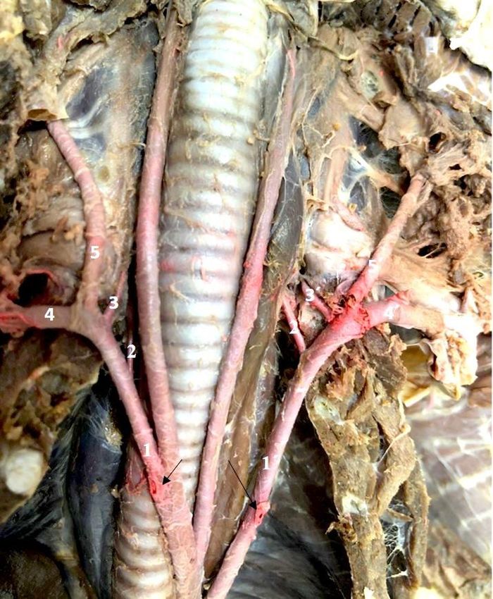
In the eight animals studied, the brachiocephalic trunk represented the first
branch of the aortic arch, followed by the left subclavian artery as independent
branches (Fig. 1). The branches originating from these arteries also did not show
variations between the analyzed specimens.
Figure 1. View of the aortic arch and its branches. AA - Aortic arch; TB - Brachiocephalic trunk; *Aa.
Subclavian artery
The internal thoracic artery appeared at the level of the first intercostal space,
inside the thoracic cavity, and advanced in the caudoventral direction towards the
sternum and transverse muscle of the chest. At the chest entrance, the right and left
subclavian arteries branched into two arteries, the superficial cervical artery that
supplies the cervical musculature's ventrolateral surface, and the axillary artery that
supplies the muscles of the thoracic limbs.
- 175 -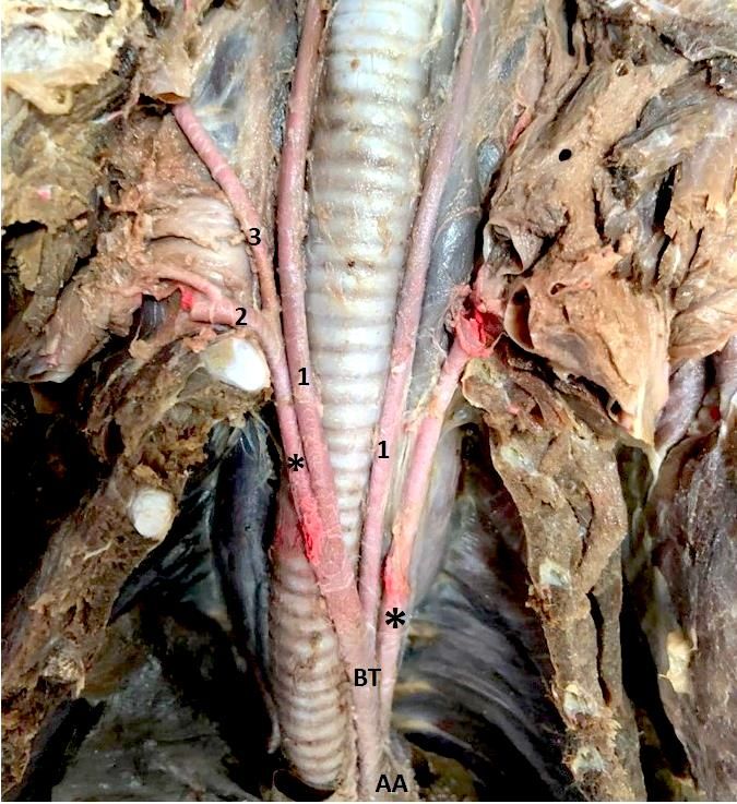
IUCN Otter Spec. Group Bull. 38(3) 2021

      The brachiocephalic trunk followed cranially over the trachea's ventral face and
emitted three branches, the left common carotid artery, the right common carotid
artery, and the right subclavian artery.
      In all specimens studied, the left common carotid artery was the first branch
emitted by the brachiocephalic trunk, followed by the formation of a common trunk
that originated the right common carotid artery and the right subclavian artery at the
level of the first intercostal space, in the right antimere of the thoracic cavity (Fig. 2).

Figure 2. View of the brachiocephalic trunk and its branches. 1- common carotid artery; 2- axillary
artery; 3 - superficial cervical artery; AA - aortic arch; BT - brachiocephalic trunk; * subclavian artery.

                                                   - 176 -
IUCN Otter Spec. Group Bull. 38(3) 2021

      In the eight animals analyzed the right and left subclavian arteries emitted five
branches: the vertebral artery, the costocervical trunk, the internal thoracic artery, the
superficial cervical artery, and the axillary artery (Fig. 3).

Figure 3. View of the subclavian arteries and their branches. 1 - right subclavian artery; 1’ - left
subclavian artery; arrows - right internal thoracic artery; 2 - right vertebral artery; 2’ - left vertebral
artery; 3 - right costocervical trunk; 3’ - left costocervical trunk; 4 - right axillary artery; 4’ - left
axillary artery; 5 - right superficial cervical artery; 5’ - left superficial cervical artery.

                                                   - 177 -
IUCN Otter Spec. Group Bull. 38(3) 2021

      The vertebral artery went craniodorsally towards the cervical vertebrae. The
costocervical trunk originated from the subclavian right after the vertebral artery and
proceeded to the cervical region's dorsal muscles and the chest (Fig. 4).

Figure 4. View of the right subclavian artery and their branches in the cranial part of the thoracic
cavity. *1 - right subclavian artery; arrow - right internal thoracic artery; 2 - right vertebral artery; 3 -
right costocervical trunk; 4 - right axillary artery; 5 - right superficial cervical artery.

DISCUSSION
      The study of anatomy in domestic animals shows that the origin of the vessels
that emerge from the aortic arch varies according to species, and the same occurs in
wild animals. Considering the carnivorous order, the treatises on animal anatomy
(Nickel and Seiferle, 1981; Sisson et al, 1975; Barone, 1996; Dyce et al., 2019; König
and Liebich, 2020), describe that the aortic arch gives rise to the brachiocephalic
trunk and then to the left subclavian artery. This same disposition was verified in this
study, as well as in Lontra canadensis (Baitchman and Kolle, 2000), in Procyon
cancrivorus (Santos et al., 2004), in Mustela sp. (Fox and Marini, 2014; Quesenberry
and Carpenter, 2020) Leopardus pardalis (Martins et al., 2010), and in Cerdocyon
thous (Lima et al., 2016).
      With the results obtained in this work, it is possible to suggest a pattern in the
origin of the vessels that emerge from the aortic arch in carnivores, whether wild or
domestic, although variations may occur. Example for such variation are cases
mentioned by Culau et al. (2004), when they reported the ectopic origin of the right
subclavian artery and the bicarotid trunk in a dog, and by Engel et al. (2013) studying
Cerdocyon thous, originating from a bicarotid trunk in the aortic arch.

                                                   - 178 -
IUCN Otter Spec. Group Bull. 38(3) 2021

       In the neotropical otter, the origin and disposition of the arteries in the
brachiocephalic trunk is following that described for domestic carnivores (Sisson et
al, 1985; Nickel et al., 2010; Dyce, 2017; König and Liebich, 2020), where the
brachiocephalic trunk first originates the common carotid artery left and posteriorly
the right common carotid arteries and the right subclavian artery.
       A study carried out with Leopardus pardalis (Martins et al., 2010) presented
these same results. However, Santos et al. (2004), studying a specimen of Procyon
cancrivorus, report that the brachiocephalic trunk of this species undergoes a
trifurcation at the level of the first intercostal space, in the left antimere of the thoracic
cavity, originating the left and right common carotid arteries, and the right subclavian
artery. Indeed, Sisson et al (1975) mentions that the brachiocephalic trunk may
present this trifurcation in domestic carnivores.
       In Cerdocyon thous (Lima et al. 2016), the brachiocephalic trunk first originated
the right subclavian artery and followed in a bicarotid trunk typical right and left
carotid arteries. Filho and Borelli (1970), in a study with 240 cats (Felis catus
domestica), reported that 75 animals showed the formation of the bicarotid trunk.
       Wild carnivores work evidenced the dispositions described in the treatises of
animal anatomy and comparative anatomy for the origin of the vessels'
brachiocephalic trunk in domestic carnivores. The arrangement found in Lontra
longicaudis and Leopardus pardalis were the same as those mentioned for domestic
animals. These animals have a standard trunk between the right common carotid and
right subclavian arteries after the left common carotid artery's origin.
       In the carnivorous order such as Lontra canadensis (Baitchman and Kollias,
2020), Leopardus pardalis (Martins et al., 2010) and Cerdocyon thous (Lima et al.,
2016), the subclavian arteries gave rise to the collateral branches: vertebral artery,
costocervical trunk, internal thoracic artery, superficial cervical artery and axillary
artery, the same branches found in our study. This same origin was also mentioned in
Mustela sp. (Fox and Marini, 2014; Quesenberry et al., 2020). However, Santos et al.
(2004) did not mention the vertebral artery and the axillary artery as branches
originating from the subclavias in Procyon cancrivorus. Our results align with what is
described in the literature for dogs and cats (Filho and Borelli, 1970; Sisson et al,
1975; Dyce et al., 2019; König and Liebich, 2020).
       This work describes the organization of the branches that originate from the
aortic arch in a neotropical otter and notes that the origin of these vessels is similar to
that described for domestic carnivores, and for wild carnivores. In carnivores in
general, the brachiocephalic trunk and the left subclavian artery emerge directly from
the aortic arch. Thus, we can conclude that this pattern of origin of vascularization of
the aortic arch is shared, both for domestic carnivores and wild carnivores.

REFERENCES
Araújo, A.C.P., Oliveira, J.C.D., Campos, R. (2004). Collaterals branches of the aortic arch and its
      main rami in chinchila (Chinchilla lanígera). Revista Portuguesa de Ciências Veterinárias, 99:
      53-58.
Baitchman, E.J., Kollias, G.V. (2000). Clinical anatomy of the North American river otter (Lontra
      canadensis). Journal of Zoo and Wildlife Medicine: Official Publication of the American
      Association of Zoo Veterinarians, 31: 473-483.
Barone, R. (1996). Anatomie comparée des mammifères domestiques. Tome 5: Angiologie. Paris:
      Vigot, 1996.
Brudnicki, W., Macherzynska, A., Nowicki, W. (2007). Variation in the arteries of the aortic arch in
      European Brown Hare (Lepus europaeus). Electronic Journal Polish Agricultural Universities,
      10(1). http://www.ejpau.media.pl/volume10/issue1/art-03.html. Last accessed: 1.5.2020.

                                                - 179 -
IUCN Otter Spec. Group Bull. 38(3) 2021
Campos, R., Araújo, A.C.P., Azambuja, R.C. (2010). Ramos colaterais do arco aórtico e suas
principais ramificações em nutria (Myocastor coypus). Acta Sientiae Veterinariae, 38: 139-146.
Carvalho-Junior, O. (2016). Emergy Analysis of the Peri Lake System and the Role of the
Neotropical Otter. IJRRAS, 29: 31-54.
Carvalho-Junior, O., Birolo, A.B., Macedo-Soares, L. (2010). Ecological Aspects of Neotropical
Otter (Lontra longicaudis) in Peri Lagoon, South Brazil. IUCN Otter Spec. Group Bull., 27:
105-115.
Carvalho-Junior, O., Macedo-Soares, L., Birolo, A.B. (2010). Annual and interannual food habits
variability of a Neotropical otter (Lontra longicaudis) population in Conceição Lagoon, South
of Brazil. IUCN Otter Spec. Group Bull., 27: 24-32.
Carvalho-Junior, O., Filippini, A., Salvador, C.H. (2012). Distribution of neotropical otter, Lontra
longicaudis (Olfers, 1818) (Mustelidae) in coastal islands of Santa Catarina, Southern Brazil.
IUCN Otter Spec. Group Bull., 29: 95-108.
Culau, P.O.V., Oliveira, J.C.D., Reckziegel, S.H., Lindemann, T. (2004). Origem ectópica da artéria
subclávia direita e do tronco bicarotídeo no cão. Ciência Rural, 34: 1615-1618.
Culau, P.O.V., Reckziegel, S.H., Lindemann, T., Araújo, a. C. P.; Balzaretti, F. (2007). Colaterais
do arco aórtico da capivara (Hydrochoerus hydrochaeris). Acta Scientiae Veterinariae, 35: 89-
92.
Dyce, K.M., Sack, W.O., Wensing, C.J.G. (2009). Textbook of Veterinary Anatomy. [s.l.]: Elsevier
Science Health Science Division.
Engel, S., Maiochi, R.R., Birck, A.J., Filadelpho A.L., Guimarães, G.C. (2013). Origem e
distribuição do arco aórtico do cachorro-do mato (Cerdocyon thous). Revista Científica
Eletrônica de Medicina Veterinária, 11(21).
Filho, A.F., Borelli, V. (1970). Contribuição ao estudo dos colaterais calibrosos do arco aórtico no
gato. Revista da Faculdade de Medicina Veterinária, 8: 385-388.
Fox, J., Marini, R.P. (2014). Biology and Diseases of the Ferret. 3.ed. [s.l.]: Wiley–Blackwell.
International Committee on Veterinary Gross Anatomical Nomenclature. (2017). Nomina
Anatomica Veterinaria. 6. ed. [s.l.]: Editorial Committee Hanover (Germany), Ghent (Belgium),
Columbia, MO (U.S.A.), Rio de Janeiro (Brazil).
000König, H.E., Liebich, H. (Orgs.). (2020). Veterinary Anatomy of Domestic Animals: Textbook
and Colour Atlas. 7th Updated and Extended ed. Edição. Stuttgart; New York: Thieme Medical
Publishers.
Lima, A.R.; Souza, D.C.; Carmo, D.C. do; et al. (2016). Ramos colaterais do arco aórtico e suas
principais ramificações no cachorro-do-mato (Cerdocyon thous). Pesquisa Veterinária
Brasileira, 36: 647-651.
Magalhães, M.D.S., Albuquerque, J.F.G., Oliveira, M.F., Papa, P.D.C., Moura, C.E.B. (2007).
Ramos do arco aórtico no mocó (Kerodon rupestris). Revista Portuguesa de Ciências
Veterinárias, 102: 49-52.
Martins, D.M., Lima, A.R., Pinheiro, L.L., Brígida, S.S.S., Araújo, E.B., Melul, R., Lacreta
Júnior, A.C.C., Meneses, A.M.C., Souza, A.C.B., Pereira, L.C. Fioretto, E.T., Branco, E.
(2010). Descrição Morfológica dos Ramos Colaterais do Arco Aórtico e suas Principais
Ramificações em Leopardus pardalis. Acta Veterinaria Brasilica, 4: 74-77.
Nickel, R., Schummer, A., Seiferle, E. (1981). Anatomy of the Domestic Animals. Berlin: Verlag
Paul Parey.
Oliveira, F.S., Machado, M.R.F., Miglino, M.A., Nogueira, T.M. (2001). Gross anatomical study of
the aortic ARC branches of the paca (Agouti paca, Linnaeus, 1766). Brazilian Journal of
Veterinary Research and Animal Science, 38: 103-105.
Oliveira, R.E.M., Oliveira, G.B., Barbosa, P.M.L., Bezerra, F.V.F., Albuquerque, J.F.G.,
Ambrósio, C.E., Miglino, M.A., Oliveira, M.F. (2015). Ramos colaterais do Arco aórtico do
preá (Galea spixii Wagler, 1831). Pesquisa Veterinária Brasileira, 35: 762-766.
Pérez, W., Erdogan, S. (2014). Arterial thoracic vascularization in some deer species: pampas deer
(Ozotoceros bezoarticus), brown brocket deer (Mazama gouazoubira) and axis deer (Axis axis).
Anatomia, Histologia, Embryologia, 43: 490-494.
Pinheiro, V.L.C., Lima, A.R., Pereira, L.C., Gomes, B.D., Branco, E. (2012). Descrição anatômica
dos ramos colaterais do arco aórtico do tamanduá-mirim (Tamandua tetradactyla). Biotemas, 25:
133-137.
Quadros, J., Monteiro-Filho, E.L.A. (2001). Diet of the Neotropical Otter, Lontra longicaudis, in an
Atlantic Forest Area, Santa Catarina State, Southern Brazil. Studies on Neotropical Fauna and
Environment, 36: 15-21.
- 180 -IUCN Otter Spec. Group Bull. 38(3) 2021
Quesenberry, K., Carpenter, J.W. (2011). Ferrets, Rabbits and Rodents - E-Book: Clinical Medicine
and Surgery. [s.l.]: Elsevier Health Sciences.
Quintela, F.M., Porciuncula, R.A., Colares, E. (2008). Diet of Lontra longicaudis (Olfers) in a
coastal stream in southern Rio Grande do Sul State, Brazil. Neotrop. Biol. Cons., 3: 119-125.
Reckziegel, S.H., Lindemann, T., Culau, P.O.V. (2003). Collaterals of the aortic arch in opossun
(Didelphis albiventris). Ciência Rural, 33: 507-511.
Rheingantz, M.L., Waldemarin, H.F., Rodrigues, L., Moulton, T.P. (2011). Seasonal and spatial
differences in feeding habits of the Neotropical otter Lontra longicaudis (Carnivora:
Mustelidae) in a coastal catchment of southeastern Brazil. Zoologia (Curitiba), 28: 37-44.
Rodrigues, L.A. (2013). Avaliação do risco de extinção da Lontra neotropical Lontra longicaudis
(Olfers, 1818) no Brasil. Biodiversidade Brasileira, 3: 216-227.
Santos, A.L.Q., Moraes, F.M., Malta, T.S., Carvalho S.F.M., Alves Júnior, J.R.F. (2004).
Topografia dos Colaterais Calibrosos do Arco Aórtico de um Mão - Pelada (Procyon
cancrivorus GRAY, 1865) (Carnivora Procyonidae). Archives of Veterinary Science, 9: 67-72
Schimming, B.C., Matteis, R., Silva, J.R.C.P., Guazzelli Filho, J. (2012). Ramos do arco aórtico no
veado-catingueiro. Revista Científica Eletrônica de Medicina Veterinária, 10: 1-8.
Schimming, B.C., Jesus, L.S.B.S., Filadelpho, A.L. (2016). Branching pattern of aortic arch in the
white-eared opossum (Didelphis albiventris). Braz. J. Vet. Res. Anim. Sci. (Online), 53: 235-
242.
Sisson, S., Grossman, J.D., Getty, R. (1975). Anatomy of the Domestic Animals. 5a Edição.
Philadelphia: W B Saunders Co.
Souza Neto, J.R.N. de, Lima, A.R., Branco, É. (2019). Ramos colaterais do arco aórtico de Callithrix
penicillata (É. Geoffroy, 1812) (Primates, Cebidae). Biotemas, 32: 85-91.
Uchôa, T., Vidolin, G.P., Fernandes, T.M., Velastin, G.O., Mangini, P.R. (2004). Aspectos
ecológicos e sanitários da lontra (Lontra longicaudis OLFERS, 1818) na Reserva Natural Salto
Morato, Guaraqueçaba, Paraná, Brasil. Cad. Biodivers., 4: 19-28.
RÉSUMÉ
VASCULARISATION DE L’ARC AORTIQUE CHEZ LA LOUTRE À
LONGUE QUEUE (Lontra longicaudis, OLFERS 1818)
Lontra longicaudis est un mammifère carnivore semi-aquatique appartenant à la
famille des Mustelidae et à la sous-famille des Lutrinae. On la trouve en Amérique du
Sud et en Amérique centrale, du sud du Mexique à l'Uruguay, jusqu'à 3000 m
d'altitude. Dans cette étude, la vascularisation de l'arc aortique est décrite chez la
loutre à longue queue. Huit animaux ont été étudiés, trois loutrons et cinq adultes du
Projet Loutre de l'Institut Ekko Brasil. Les résultats indiquent que le tronc
brachiocéphalique et l'artère sous-clavière gauche provenaient de l'arc aortique chez
tous les animaux étudiés. Cet agencement est similaire à celui trouvé chez les
carnivores sauvages et domestiques. Le tronc brachiocéphalique provient de trois
artères: l'artère carotide commune gauche, l'artère carotide commune droite et l'artère
sous-clavière droite, les deux dernières étant émises par un tronc commun. Les artères
sous-clavières droite et gauche donnent naissance à cinq vaisseaux représentés par
l'artère vertébrale, l'artère cervicale superficielle, l'artère thoracique interne, le tronc
costocervical et l'artère axillaire.
RESUMEN
VASCULARIZACIÓN DEL ARCO AÓRTICO EN LA NUTRIA
NEOTROPICAL (Lontra longicaudis, OLFERS 1818)
Lontra longicaudis es un mamífero carnívoro semi-acuático que pertenece a la familia
Mustelidae y la subfamilia Lutrinae. Se encuentra en América del Sur y Central,
desde Mexico hasta Uruguay y hasta 3.000 m de altitud. En este estudio, se describe
la vascularización del arco aórtico en la nutria neotropical. Se estudiaron ocho
- 181 -IUCN Otter Spec. Group Bull. 38(3) 2021
animales, tres crías y cinco adultos, del Proyecto Nutria del Instituto Ekko Brasil. Los
resultados indican que el tronco braquiocefálico y la arteria subclavia izquierda se
originaron en el arco aórtico en todos los animales estudiados. Este patrón es similar
al encontrado en carnívoros silvestres y domésticos. El tronco braquiocefálico origina
tres arterias, la arteria carótida izquierda común, la arteria carótida derecha común, y
la arteria subclavia derecha, siendo las dos últimas emitidas a partir de un tronco
común. Las arterias subclavia derecha e izquierda dan origen a cinco vasos
respresentados por la arteria vertebral, la arteria cervical superficial, la arteria torácica
interna, el tronco costocervical, y la arteria axilar.
RESUMO
VASCULARIZAÇÃO DO ARCO AÓRTICO EM LONTRA NEOTROPICAL
(Lontra longicaudis, OLFERS 1818)
Lontra longicaudis é um mamífero carnívoro semiaquático que pertence à família
Mustelidae e à subfamília Lutrinae. É encontrada na América do Sul e Central, do sul
do México ao Uruguai e até 3.000 m de altitude. Neste estudo, a vascularização do
arco aórtico é descrita na lontra neotropical. Foram estudados oito animais do Projeto
Lontra do Instituto Ekko Brasil, sendo três filhotes e cinco adultos. Os resultados
indicam que, em todos os animais estudados, o tronco braquiocefálico e a artéria
subclávia esquerda originam-se do arco aórtico. Este padrão é semelhante ao
encontrado em carnívoros selvagens e domésticos. O tronco braquiocefálico origina
três artérias, a artéria carótida comum esquerda, a artéria carótida comum direita, e a
artéria subclávia direita, sendo as duas últimas emitidas a partir de um tronco comum.
As artérias subclávias direita e esquerda dão origem a cinco vasos representados pela
artéria vertebral, a artéria cervical superficial, a artéria torácica interna, o tronco
costocervical e a artéria axilar.
- 182 -You can also read