Domestic cat's internal carotid artery in ontogenesis
←
→
Page content transcription
If your browser does not render page correctly, please read the page content below
Original Paper                                                  Veterinarni Medicina, 66, 2021 (07): 292–297

                                                                    https://doi.org/10.17221/116/2020-VETMED

Domestic cat’s internal carotid artery in ontogenesis
Hanna Ziemak, Hieronim Frackowiak, Maciej Zdun*

Institute of Veterinary Medicine, Nicolaus Copernicus University in Toruń,
Toruń, Poland
*Corresponding author: maciejzdun@onet.eu

Citation: Ziemak H, Frackowiak H, Zdun M (2021): Domestic cat’s internal carotid artery in ontogenesis. Vet Med-
Czech 66, 292–297.

Abstract: The aim of the study was to trace the presence of the internal carotid artery in the system of cerebral
arteries of the domestic cat and to determine the role of this artery in supplying blood to the brain in ontogenesis.
The available publications provide ambiguous or even contradictory information. The authors of some studies
claim that there is no extracranial segment in the domestic cat’s internal carotid artery. Other authors reported
the internal carotid artery in the arterial pattern of the encephalon base. The study was conducted on sixty-one
domestic cats: fifteen foetuses, sixteen juvenile cats, and thirty adult cats were analysed. The internal carotid
artery – a vessel with a relatively large lumen – was fully preserved in all the foetuses and most of the juvenile
animals. This artery was not complete with regard to the adults and some juvenile individuals, because it had
lost the extracranial segment as a result of the obliteration process. A precise description of this area is not only
of biological, but also of clinical, significance. The knowledge of the anatomical structure of cerebral vessels
is particularly important to correctly interpret images obtained during diagnostic tests and to conduct surgical
procedures correctly.

Keywords: anatomy; brain arteries; brain supply; morphology

  The available publications provide ambiguous              knowledge of the anatomical structure of cerebral
or even contradictory information on the internal           vessels is particularly important to correctly in-
carotid artery in the arterial system of a cat’s head.      terpret images obtained in diagnostic tests, both
The authors of some studies state that there is no ex-      by computed tomography (CT) and magnetic reso-
tracranial segment in the domestic cat’s internal           nance imaging (MRI).
carotid artery (Kamijyo and Garcia 1975; Bugge                Unlike classic X-ray images, CTs and MRIs give
1978; Klein 1980; Simoens et al. 1987; Frackowiak           images with good tissue contrast in places with
2003; Frackowiak and Godynicki 2003; Kier et al.            high-complexity tissue structures such as the cen-
2019). Some authors (Nickiel and Schwarz 1963;              tral nervous system (CNS), chest, and abdominal
McClure et al. 1973; Nickel et al. 1996) did, how-          cavity (Caine et al. 2019). Information about arte-
ever, find an extracranial segment of the internal          rial vessels is particularly important during brain
carotid artery in cats. The authors of some studies         surgery as it enables the precise location and liga-
analysed very few specimens, for example, Kamijyo           tion of the vessels supplying blood to the brain (Ijiri
and Garcia (1975) analysed seven adults, while              et al. 2014).
Simoens et al. (1987) analysed a total of five cats.          The aim of the study was to trace the presence
These facts inspired our research in an attempt             of the internal carotid artery in the cerebral arterial
to verify this ambiguous information.                       system of the domestic cat and to determine the
  A precise description of this area is not only            role of this artery in supplying blood to the brain
of biological, but also of clinical, significance. The      in ontogenesis.

292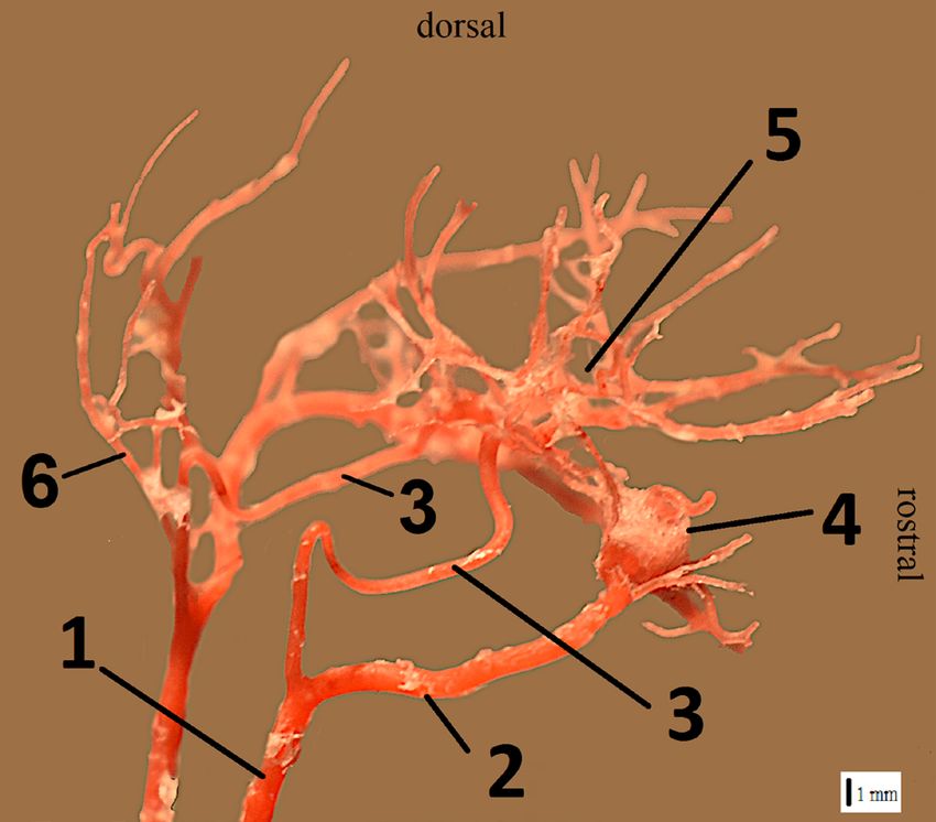
Original Paper Veterinarni Medicina, 66, 2021 (07): 292–297
https://doi.org/10.17221/116/2020-VETMED
MATERIAL AND METHODS arterial circle. The extracranial segment of the in-
ternal carotid artery was a branch of the final divi-
The study was conducted on sixty-one domestic sion of the common carotid artery.
cats of both sexes (thirty-nine females and twenty- The common carotid artery was divided into the
two males) divided into three groups: fifteen foetus- external carotid artery and internal carotid artery
es with a crown-rump length of 84–126 mm, sixteen bilaterally in all the foetuses and in twelve out of the
juvenile cats aged 4–8 weeks, and thirty adult sixteen juvenile animals. The external carotid artery
cats. The cadavers were delivered from veterinary continued as the main arterial vessel in the head. The
clinics. These animals had been euthanised [using internal carotid artery supplies blood to the enceph-
xylazine (intramuscular; i.m.), ketamine (i.m.), and alon and is first directed dorsally, entering the jugu-
pentobarbital (intravenous; i.v.)] for medical reasons lar foramen without passing through it, and forming
other than circulatory or neurological disorders. an arc of about 180°. It is shaped like an inverted let-
Forty-two randomly selected cadavers were pro- ter U (Figure 1). In its further course, it is directed
cessed by injecting a COLOREX ® (Śnieżka, War- towards the cranial cavity, in which it enters through
szawa, Poland) stained solution of the chemo-setting the carotid artery canal. The vessel has a relatively
acrylic material Duracryl® Plus (SpofaDental, Jičín, large lumen throughout its course.
Czech Republic) into both the common carotid ar- The internal carotid artery in all the adult cat
teries. After a short time (15–20 min) necessary for specimens and four juveniles was incomplete by vir-
setting, the specimens were enzymatically macerated tue of the extracranial segment being missing. All
with Persil® powder (Henkel, Düsseldorf, Germany) the adult cats had the carotid sinus at the point
diluted in water at 42 °C for about a month. This where the obliterated extracranial segment of the
procedure resulted in corrosion castings of the internal carotid artery diverged from the common
vessels on a bone scaffold (without the animal’s carotid artery. The carotid sinus is a very short sec-
tissues, except the bones). The second method, ap- tion of the extracranial part of the internal carotid
plied to nineteen specimens, consisted of passing artery and it was not found in the foetuses or juve-
the liquid-stained latex LBS 3060 into both common nile cats. The occipital artery branched from the
carotid arteries, leaving it to set in a 5% formalin so- carotid sinus; however, in the foetuses and juvenile
lution for 2 weeks, then preparing the blood vessels animals, it branched directly from the common ca-
manually using surgical instruments during dissec- rotid artery, bilaterally in nineteen cats and unilater-
tion, in order to view them within the tissue.
No ethics committee approvals were required Figure 1
to conduct the experiments on the cadavers. An eth-
ical commission approval is only needed to use live
animals. The transport and collection of the cadav-
ers were carried out in accordance with the standard
operating procedure: SOP-17-CW-03.
The names of the anatomical structures were
standardised according to Nomina Anatomica Vet-
erinaria (International Committee on Veterinary
Gross Anatomical Nomenclature 2017).
RESULTS
The analysis of the course of the internal carotid
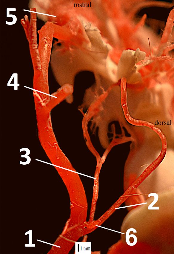
artery in the cat cadavers revealed an intracranial Figure 1. Corrosion cast of the common carotid artery
segment located in the cranial cavity and an ex- and its branches in a cat foetus. The internal carotid
tracranial segment located outside the skull. The artery is fully functional
intracranial segment finally branched into the ros- 1 – common carotid artery; 2 – external carotid artery; 3 –
tral cerebral artery and the caudal communicating internal carotid artery; 4 – rete mirabile of the maxillary
artery, i.e., the main components of the cerebral artery; 5 – arterial circle of the brain; 6 – occipital artery
293Original Paper Veterinarni Medicina, 66, 2021 (07): 292–297
https://doi.org/10.17221/116/2020-VETMED
ally in three. In five foetuses and the juvenile cats,
Figure 2
the occipital artery branched bilaterally from the ex-
ternal carotid artery, and unilaterally in three. The
ascending pharyngeal artery is a branch of the oc-
cipital artery running along the lateral surface of the
bulla tympanica and dividing into two branches
in the middle of its course. One of these, located
medially, was directed towards the pharyngeal wall,
and the branch with a lateral course was directed
towards the carotid artery canal. The lateral branch
of the ascending pharyngeal artery was connected
with the internal carotid artery in the carotid artery
canal in ten foetal specimens. A similar connection
of the arteries was found in the specimens of eleven
juvenile cats (Figure 2). No connection, of these ar-
teries, was observed in the remaining specimens
from these groups. There was also no extracranial
segment of the internal carotid artery found in the
specimens of five juvenile animals.
No extracranial segment of the internal carotid
artery was observed in any specimen from the group
of thirty adult animals, with only the intracranial
segment of this vessel apparent. The connection
of the lateral branch of the ascending pharyngeal
artery with the intracranial segment of the internal
carotid artery was observed in nineteen specimens.
There was a unilateral connection of these arter- Figure 2. Corrosion cast of the common carotid artery
ies in three specimens. These arteries did not con- and its branches in a juvenile cat. The ascending pharyn-
nect in the remaining eight specimens because the geal artery is a branch of the occipital artery. The lateral
final fragment of the ascending pharyngeal artery branch of the ascending pharyngeal artery is connected
lost its lumen and did not reach the intracranial to the internal carotid artery
segment of the internal carotid artery. Regardless 1 – common carotid artery; 2 – internal carotid artery; 3 –
of the presence or absence of this connection be- ascending pharyngeal artery; 4 – caudal auricular artery;
tween these vessels, the ascending pharyngeal ar- 5 – rete mirabile of the maxillary artery; 6 – beginning
tery was always a vessel with a very small lumen. of the occipital artery (the remainder of the occipital artery
There was a permanent connection between the has been removed to allow better visibility of the internal
intracranial segment of the internal carotid artery carotid artery)
Table 1. Summary of the blood vessels in the analysed cats
Name of vessel From In whom it occurs
The internal carotid artery in all the foetuses and in 12 out
the common carotid artery
(extracranial and intracranial segments) of the 16 juvenile cats
The occipital artery the carotid sinus in the adult cats
in the foetuses and juvenile cats:
The occipital artery the common carotid artery
19 bilaterally, 3 unilaterally
in the foetuses and juvenile cats:
The occipital artery the external carotid artery
5 bilaterally, 3 unilaterally
Lateral branch of the ascending bilaterally in 10 foetuses, 11 juvenile cats,
the occipital artery
pharyngeal artery 19 adult cats, unilaterally in 3 adult cats
Rami retis the maxillary artery in all the cats
294Original Paper Veterinarni Medicina, 66, 2021 (07): 292–297
https://doi.org/10.17221/116/2020-VETMED
and the rami retis passing through the orbital fis- following studies stated that they observed the ex-
sure in all the cat specimens of all age groups. The tracranial segment of the internal carotid artery
branches emerged from the rete mirabile of the max- in their cat specimens (Nickiel and Schwarz 1963;
illary artery. McClure et al. 1973; Nickel et al. 1996).
It is also possible that the ascending pharyngeal The specific arterial pattern of the domestic
artery provides blood flow to the intracranial seg- cat’s encephalon base was confirmed by the phy-
ment of the internal carotid artery (Table 1). logenesis of the species from the Felidae family.
In the adult specimens, the lumen of the intracra- Obliterations of the extracranial segment of the in-
nial segment of the internal carotid artery was larger ternal carotid artery and the participation of the
from the connection with the rami retis to the site rete mirabile of the maxillary artery in the distribu-
of the final division into the vessels of the arterial tion of blood to the encephalon were demonstrat-
circle of the brain. However, the caudal fragment ed in the African lion (Hsieh and Takemura 1994;
of this artery was characterised by a very small lu- Frackowiak and Godynicki 2003), serval, Eurasian
men, which was comparable to the cross-section lynx, Bengal cat, jungle cat, puma, leopard, jaguar,
of the ascending pharyngeal artery with which and tiger (Frackowiak and Godynicki 2003). Apart
it connects. from those, such an arterial pattern of the encepha-
lon base was also observed in other representa-
tives of the Carnivora order, e.g., the large Indian
DISCUSSION civet of the Viverridae family (Frackowiak 2003) and
the hyena of the Hyaenidae family (Bugge 1978).
Our study showed that the internal carotid ar- Interestingly, the extracranial segment of the in-
tery in the studied domestic cats underwent funda- ternal carotid artery is not obliterated in the rep-
mental changes at different periods of ontogenesis. resentatives of other families and species of the
In the foetuses and the majority of the juvenile cats, Carnivora order. During the entire ontogenesis,
it was fully developed (contained extracranial and it is a source of blood for the brain, as was demon-
intracranial segments) and connected to the com- strated in studies on the dog, fox, raccoon, and rep-
mon carotid artery with the arterial circle of the resentatives of the Phocidae, Mustelidae, Ursidae,
brain. However, the initial segment, i.e., the extrac- and Procyonidae families (Nickiel and Schwarz
ranial segment of the internal carotid artery, became 1963; Frackowiak 2003; Skoczylas et al. 2016).
obliterated in the older and adult animals. The only As the internal carotid artery in the dog and other
trace of its earlier presence was the carotid sinus – canines (Caniformia) is active throughout their
the bulb remaining on the common carotid artery. lifetime (Nickiel and Schwarz 1963), the arterial
As a result of the obliteration of the extracranial patterns of the encephalon base are fundamentally
segment of the internal carotid artery, the connec- different in Feliformia. In the available literature,
tion of the common carotid artery with the arterial speculation can be found on the advisability of a loss
circle of the brain was broken. In the foetuses and of the internal carotid artery in some animal spe-
juvenile cats, the blood supply to the brain came cies. According to Zedenov (1937), the obliteration
from two sources: the complete internal carotid of a ruminants’ extracranial segment of the inter-
artery and the maxillary artery. Blood was distrib- nal carotid artery takes place to eliminate factors
uted from the maxillary artery to the arterial circle emitting interference with low-frequency sounds.
of the brain through the vessels of the rete mira- This process consists of the displacement of the
bile of the maxillary artery. The rami retis passed tympanic part of the temporal bone.
through the orbital fissure and anastomosed with The internal carotid artery is also the main source
the intracranial segment of the internal carotid ar- of blood in animals belonging to various other taxa,
tery. Some researchers did not find the extracranial e.g., the domestic horse and donkey (Nickiel and
segment of the internal carotid artery (Kamijyo and Schwarz 1963; Aly et al. 1981) as well as the European
Garcia 1975; Bugge 1978; Klein 1980; Simoens et al. rabbit and European hare (Brudnicki et al. 2012;
1987; Frackowiak 2003; Frackowiak and Godynicki Brudnicki et al. 2015). The connection of the arte-
2003; Kier et al. 2019). The results of their studies, rial circle of the brain with the internal carotid ar-
in line with ours, found it only in adult cats as well tery was found in some rodents (Rodentia), e.g., the
as in some juvenile individuals. The authors of the Cairo spiny mouse, muskrat, brown rat, and North
295Original Paper Veterinarni Medicina, 66, 2021 (07): 292–297
https://doi.org/10.17221/116/2020-VETMED
American porcupine (Bugge 1978; Frackowiak 2003; Aydin A. The morphology of circulus arteriosus cerebri
Szczurkowski et al. 2007; Esteves et al. 2013). The in the red squirrel (Sciurus vulgaris). Vet Med-Czech.
obliteration of the extracranial segment of the inter- 2008 May;53(5):272-6.
nal carotid artery, which was demonstrated in our Brudnicki W, Kirkillo-Stacewicz K, Skoczylas B, Nowicki W,
study and described in other representatives of the Jablonski R, Brudnicki A, Wach J. The arteries of the brain
Felidae family, was also observed in animals of other in hare (Lepus europaeus Pallas, 1778). Anat Rec (Hobo-
orders. The phenomenon of the obliteration of the ken). 2015 Oct;298(10):1774-9.
extracranial segment of the internal carotid artery Brudnicki W, Nowicki W, Skoczylas B, Brudnicki A, Kirkillo-
or the presence of a vessel with a very narrow lumen, Stacewicz K, Wach J. Arteries of the brain in wild Euro-
without significance to the supply of blood to the pean rabbit Oryctolagus cuniculus (Linnaeus, 1758).
brain, was found in some animals of the Rodentia Folia Biol (Krakow). 2012;60(3-4):189-94.
order, such as the African crested porcupine, guinea Brudnicki W, Skoczylas B, Jablonski R, Nowicki W, Brud-
pig, common degu, long-tailed chinchilla, and red nicki A, Kirkillo-Stacewicz K, Wach J. The arteries of the
squirrel (Ocal and Ozer 1992; Aydin et al. 2005; brain base in the degu (Octodon degus Molina 1782). Vet
Aydin 2008; Brudnicki et al. 2014; Kuchinka 2017), Med-Czech. 2014 Jul;59(7):343-8.
and in ruminants (Wible 1986). Bugge J. The cephalic arterial system in carnivores, with
Our study showed that the weak ascending pha- special reference to the systematic classification. Acta
ryngeal artery in the cat may connect the common Anat (Basel). 1978;101(1):45-61.
carotid artery with the intracranial segment of the in- Caine A, Brash R, De Risio L, Van Dijk J, Cherubini GB,
ternal carotid artery. There were similar descriptions Dennis R. MRI in 30 cats with traumatic brain injury.
of the domestic cat’s lateral branch of the ascending J Feline Med Surg. 2019 Dec;21(12):1111-9.
pharyngeal artery in other publications (Kamijyo and Esteves A, Freitas A, Rossi-Junior W, Fernandes G. Ana-
Garcia 1975; Klein 1980; Kier et al. 2019). tomical arrangement and distribution of the cerebral
In conclusion, it can be stated, that: arterial circle in rats. J Morphol Sci. 2013 Jul;30(2):
1. The internal carotid artery is fully preserved 132-9.
in the foetuses only and, in most cases, juve- Frackowiak H, Godynicki S. Brain basal arteries in various
nile cats up to about 8 weeks of age. species of Felidae. Pol J Vet Sci. 2003;6(3):195-200.
2. After the obliteration of the extracranial seg- Frackowiak H. Magistrale tetnicze glowy u niektorych rze-
ment of the cat’s internal carotid artery, the ar- dow ssakow [Arterial patterns of the head in selected
terial circle of the brain connects with the mammalian orders]. Poznań: Akademii Rolniczej im.
common carotid artery only through the thin Augusta Cieszkowskiego; 2003. p. 5-80. Polish.
(weak) ascending pharyngeal artery. Hsieh HM, Takemura A. The rete mirabile of the maxillary
3. After the obliteration of the extracranial seg- artery in the lion (Panthera leo). Okajimas Folia Anat Jpn.
ment of the cat’s internal carotid artery, the 1994 May;71(1):1-11.
maxillary artery becomes the main source Ijiri A, Yoshiki K, Tsuboi S, Shimazaki H, Akiyoshi H, Na-
of blood for the encephalon. kade T. Surgical resection of twenty-three cases of brain
meningioma. J Vet Med Sci. 2014 Mar;76(3):331-8.
International Committee on Veterinary Gross Anatomical
Conflict of interest Nomenclature. Nomina anatomica veterinaria. 6 th ed.
Hannover: Editorial Committee Germany (Hanover),
The authors declare no conflict of interest. Belgium (Ghent), U.S.A. (Columbia), Brazil (Rio de Ja-
neiro; 2017. 160 p.
Kamijyo Y, Garcia JH. Carotid arterial supply of the feline
REFERENCES brain. Applications to the study of regional cerebral isch-
emia. Stroke. 1975 Jul-Aug;6(4):361-9.
Aly MA, Anis H, Moustafa SM. Morphological studies Kier EL, Conlogue GJ, Zhuang Z. High-resolution computed
on the arterial supply of the brain of the donkey in Egypt. tomography imaging of the cranial arterial system and
Assuit Vet Med J. 1981 Jan;8(15-16):2-6. rete mirabile of the cat (Felis catus). Anat Rec (Hoboken).
Aydin A, Yilmaz S, Dinc G, Ozdemir G, Karan M. The mor- 2019 Nov;302(11):1958-67.
phology of circulus arteriosus cerebri in the porcupine Klein T. Korrosionsanatomische Untersuchungen am Blut-
(Hystrixcristata). Vet Med-Czech. 2005 Mar;50(3):131-5. gefaßsystem des Encephalon und der Meninges bei Felis
296Original Paper Veterinarni Medicina, 66, 2021 (07): 292–297
https://doi.org/10.17221/116/2020-VETMED
domestica [Corrosion-anatomical study of the blood ves- in the domestic mammals. Schweiz Arch Tierheilkd. 1987
sels of the encephalon and meninges of Felis domestica]. Jun;129(6):295-307.
Anat Histol Embryol. 1980 Sep;9(3):236-79. German. Skoczylas B, Brudnicki W, Kirkillo-Stacewicz K, Nowicki W,
Kuchinka J. Morphometry and variability of the brain arte- Wach J. Cortical branches of the middle cerebral artery
rial circle in chinchilla (Chinchilla laniger, Molina). Anat in silver fox (Vulpes vulpes). Pesq Vet Bras. 2016 Oct;
Rec (Hoboken). 2017 Aug;300(8):1472-80. 36(10):1053-7.
McClure RC, Dallman MJ, Garret PG. Cat anatomy. Phila- Szczurkowski A, Kuchinka J, Nowak E, Kuder T. Topogra-
delphia: Lea and Febiger; 1973. 240 p. phy of arterial circle of the brain in Egyptian spiny mouse
Nickel R, Schummer A, Seiferle E. Lehrbuch der Anatomie (Acomys cahirinus, Desmarest). Anat Histol Embryol.
der Haustiere [Textbook of the anatomy of domestic an- 2007 Apr;36(2):147-50.
imals]. Band III. Berlin: Parey Buchverlag; 1996. 664 p. Wible JR. Transformations in the extracranial course of the
German. internal carotid artery in mammalian phylogeny. J Vertebr
Nickiel R, Schwarz R. Vergleichende Betrachtung der Kopf- Paleontol. 1986 Dec;6(4):313-25.
arterien der Haussaugetiere (Katze, Hund, Schwein, Rind, Zedenov W. Sosudistaja sistema Bovinae w sravnitelno-
Schaf, Ziege, Pferd) [Comparative analysis of the head anatomioceskom izuceni i voprosy specyficnosti jeje
arteries of domestic mammals (cat, dog, pig, cattle, sheep, morfołogii. IV. K voprosu obliteracji vnutrennoj sonnoj
goat, horse)]. Zbl Vet Med A. 1963 Mar;10(2):89-120. arterii u krupnogo rogatego skota [The Bovinae vascular
German. system in comparative anatomical studies and questions
Ocal MK, Ozer M. The circulus arteriosus cerebri in the about the specificity of its morphology. IV. To the ques-
guinea pig. Ann Anat. 1992 Jun;174(3):259-60. tion of obliteration of the internal carotid artery in cattle].
Simoens P, Lauwers H, De Geest JP, De Schaepdrijver L. Arch Anat Gist Embriol. 1937;16:490-508. Russian.
Functional morphology of the cranial retia mirabilia
Received: May 27, 2020
Accepted: February 26, 2021
297You can also read