First case report of dermatitis associated with Leporacarus gibbus in cat
←
→
Page content transcription
If your browser does not render page correctly, please read the page content below
Dumitrache et al. BMC Veterinary Research              (2021) 17:4
https://doi.org/10.1186/s12917-020-02681-0

 CASE REPORT                                                                                                                                         Open Access

First case report of dermatitis associated
with Leporacarus gibbus in cat
Mirabela Oana Dumitrache* , Adriana Györke, Gianluca D’Amico and Viorica Mircean

  Abstract
  Background: Leporacarus gibbus is a highly specific acarian parasitizing in rabbits, with a proven zoonotic potential.
  While the majority of cases of L. gibbus infestation are asymptomatic, several cases of pruritic cutaneous condition
  in both laboratory and pet rabbits were reported. Up to date, L. gibbus has not been linked with clinical signs in
  any other species than rabbits and humans.
  Case presentation: This case report described the clinical case of a 14-month-old cat with a dermatitis linked to L.
  gibbus. Mites specimens were collected by brushing, followed by light microscopy examination and species
  identification. To the best of our knowledge, this is the first report of L. gibbus-related dermatitis in cat.
  Conclusions: L. gibbus infestation should be considered as a possible differential diagnosis of pruritic skin
  conditions in cat.
  Keywords: Leporacarus gibbus, Cat, Dermatitis

Background                                                                              a hypersensitivity reaction could determine the occur-
Leporacarus gibbus (formerly Listrophorus gibbus), the                                  rence of clinical signs [4]. L. gibbus has been occasionally
rabbit fur mite, belongs to the family Listrophoridae,                                  reported as the aetiological agent of a pruritic cutaneous
division Psoroptida, order Astigmata [1]. The life cycle                                condition in both laboratory and pet rabbits. Clinically,
of the parasite is characterized by a complete metamor-                                 this ectoparasitosis is characterized by pruritus, moist
phosis and occurs entirely on the rabbit coat. L. gibbus                                dermatitis, poorly demarcate alopecia, erythema and
feeds on sebum and skin epithelial cells. Different stud-                               scaling on the dorsum and hindlimbs [3, 5]. Although it
ies questioning its pathogenicity. Due to the asymptom-                                 is considered as uncommon in humans, several case re-
atic evolution of the majority of cases, L. gibbus is also                              ports confirmed its zoonotic potential [5]. Up to date, L.
considered as commensal, which can be commonly                                          gibbus has not been linked with clinical signs in any
found on the rabbits’ skin [2, 3]. Despite of being consid-                             other species than rabbits and humans [1, 5].
ered as a cosmopolitan species for both wild and domes-
tic rabbits in Europe, only few studies are available on                                Case presentation
this topic. There is a lack of epidemiological studies and                              A 14-month-old, sterilised female Ragdoll cat, only pet,
reports of L. gibbus in many countries. In addition, the                                living indoor, was presented with a history of one year of
description of the life cycle and contamination method                                  pruritus. The anamnesis revealed that the cat was pur-
remained unclear for many years [3]. It is though that L.                               chased online a year ago and the owner observed the
gibbus is generally well tolerated by the rabbit host, but                              presence of generalised pruritus, the lack of hair on the
                                                                                        tail and face, and black cerumen in the both ears during
* Correspondence: mirabela.dumitrache@usamvcluj.ro                                      the first few days following acquisition. At that time, the
Department of Parasitology and Parasitic Diseases, University of Agricultural
Sciences and Veterinary Medicine Cluj-Napoca, Calea Mănăştur 3-5,                       patient was diagnosed by a primary care veterinarian
Cluj-Napoca 400372, Cluj, Romania                                                       with otodectic mange. The treatment applied was a
                                         © The Author(s). 2020 Open Access This article is licensed under a Creative Commons Attribution 4.0 International License,
                                         which permits use, sharing, adaptation, distribution and reproduction in any medium or format, as long as you give
                                         appropriate credit to the original author(s) and the source, provide a link to the Creative Commons licence, and indicate if
                                         changes were made. The images or other third party material in this article are included in the article's Creative Commons
                                         licence, unless indicated otherwise in a credit line to the material. If material is not included in the article's Creative Commons
                                         licence and your intended use is not permitted by statutory regulation or exceeds the permitted use, you will need to obtain
                                         permission directly from the copyright holder. To view a copy of this licence, visit http://creativecommons.org/licenses/by/4.0/.
                                         The Creative Commons Public Domain Dedication waiver (http://creativecommons.org/publicdomain/zero/1.0/) applies to the
                                         data made available in this article, unless otherwise stated in a credit line to the data.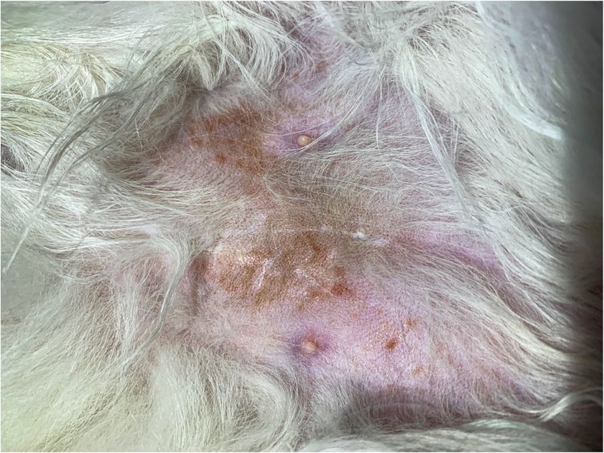
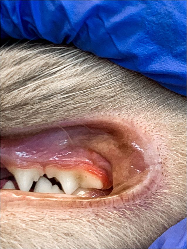
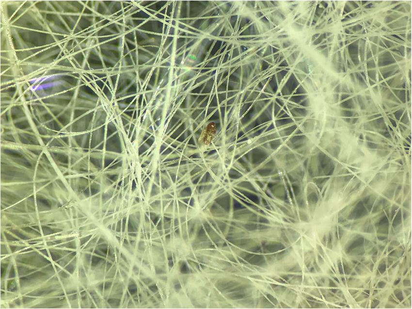
Dumitrache et al. BMC Veterinary Research          (2021) 17:4                                                                  Page 2 of 5

single spot-on application of 45 mg selamectin (Strong-
hold ®, Zoetis) that lead to clinical resolution of the oto-
dectic mange and skin lesions, and a substantial
improvement of the generalised pruritus. The later
symptom persisted at a low level of intensity during the
following months. Furthermore, transitory digestive
signs (vomiting, diarrhoea) occurred. At the age of 13
months, an intensification of the pruritus was observed.
The owner linked this event with the antirabic vaccin-
ation. The same practitioner recommended an exclusion
diet with an anallergenic food and two doses of dexa-
methasone (Dexamethasone ®, Kepro) administrated at
3 days interval. After a month of therapy, the case was
referred to our clinic because only a slightly improve-
ment of the symptoms has been noticed.
  At the moment of presentation, the clinical examin-
ation revealed a self-induced alopecia on the abdomen,
the presence of papulosquamous lesions in the same re-
gion (Fig. 1), and gingivitis (Fig. 2).
  Both skin scrapings and scotch test proved negative.
However, stereomicroscope examination of the coat
brushing revealed the presence of several mites. The
image of a female mite at a low power objective (3x) is
provided (Fig. 3). The mite specimens were collected
and the microscopic examination using the 10x objective
confirmed the presence of L. gibbus based on the follow-          Fig. 2 Gingivitis associated with L. gibbus infestation in a cat
ing morphological characteristics: subcylindrical body,
finely striated cuticle, sclerotised gnathostoma, two stri-
ated membranous flaps at each of the coxae of the first          Genomic DNA Kit, Bioline, London, UK) following the
pair of legs, and presence of two elongated adanal pro-          manufacturer’s instructions. The COI gene fragment was
cesses and distinct adanal suckers in males (Fig. 4a) [3].       PCR-amplified using primer pairs bcdF05/bcdR04 [6, 7].
No other mite species were visualized. For comparison,           The PCR products were purified using FavorPrep GEL/
a L. gibbus male collected from a rabbit is provided             PCR Purification Mini Kit (Favorgen Biotech Corp.,
(Fig. 4b). In order to confirm the morphological species         Taiwan) and further sequenced (Macrogen Europe). The
identification, the genomic DNA was extracted from               obtained sequences were compared with the available se-
specimens collected from the cat (n = 2) and from the            quence in GenBank (acc. no. GQ864335.1). The BLAST
rabbit (n = 10), by using a commercial kit (Isolate II           analysis showed 92.39%, and 92.21% similar degree

 Fig. 1 Ventral aspect of a cat infested with L. gibbus           Fig. 3 Stereomicroscope examination of the coat brushing
Dumitrache et al. BMC Veterinary Research         (2021) 17:4                                                                                Page 3 of 5

 Fig. 4 Microscopic image of L. gibbus male, from cat (a) and from rabbit (b), showing finely striated cuticle (black arrows), sclerotised “dark
 hooded” gnathostoma (red arrow), two elongated adanal processes (blue arrows)

identity with L. gibbus for the cat, and rabbit strains re-                   clinical patterns are: eosinophilic syndrome, miliary
spectively. The sequences obtained in this study from                         dermatitis, head and/or neck excoriation, regional or
the parasites collected from the cat and from rabbits                         generalized scaling and/or crusting dermatoses, and
were submitted to the GenBank database under the fol-                         symmetrical self-induced alopecia [10]. Nevertheless, for
lowing accession numbers: MW286180 and MW286214,                              the development of an efficient treatment approach of
respectively.                                                                 feline pruritus, the case history might be of a great
  To evaluate one of the most common causes of im-                            importance.
munosuppression in cats [8], a rapid test for FIV/FeLV                           In this case report, the history of the cat, its origin, the
infections (SNAP FIV/FeLV Combo Test, IDEXX) was                              young age, the lack of anti-parasitic treatments, and the
carried out, but tested negative. The cat was treated with                    clinical presentation with the self-induced alopecia clin-
two spot-on applications of 45 mg selamectin (Strong-                         ical pattern suggested a parasitic disease as first hypo-
hold ®, Zoetis) at two weeks interval, a protocol proved                      thetical diagnosis, followed by hypersensitivity dermatitis
to be effective by Birke et al. [1]. Coat brushing tested                     that could have been caused by flea bite, cutaneous ad-
negative for mites at 7 days after the second dose of sela-                   verse food reactions, and/or atopic dermatitis [10]. Cats
mectin. The skin lesions and the pruritus disappeared                         with nonseasonal pruritus and suggestive clinical pat-
within the first 14 days of treatment. The owner did not                      terns should undergo a dietary elimination-challenge
show any lesions on the course of the disease evolution                       trial to determine the importance of food allergen in the
nor at the time of rabbit examination.                                        aetiology of a condition [10]. A restriction diet of 6 to 8
                                                                              weeks is recommended to be followed. In our case, the
Discussions and conclusion                                                    cat was already following a strict diet with anallergenic
Although pruritus is a frequent sign of dermatological                        food at the time of the initial examination. The lack in
conditions in cats, this species develops less relevant                       clinical signs improvement following the exclusion diet
diagnostic lesions than those observed in dogs. More-                         and anti-pruritic treatment with corticosteroids allowed
over, the primary inflammatory lesions that may be                            us to eliminate food allergy as a hypothetical diagnosis.
present and could be relevant for identification of the                       It is well known that in cats with food allergy, pruritus,
pruritus cause are frequently transformed in secondary                        which is the main complain of owners, is corticoid-
lesions due to the different behaviours triggered by itch                     resistant in the majority of cases [11]. Altogether, the
(e.g. scratching, licking) [9]. Given this, the diagnosis ap-                 parasitic aetiology remained a possible cause and add-
proach of a pruritic skin condition should be made ac-                        itional diagnostic test were performed to confirm or to
cording to the cutaneous reaction patterns [9]. The                           exclude fleas’ infestation, cheyletiellosis, demodicosis,
causes of pruritus in cats are various, such as hypersensi-                   pediculosis and/or Lynxacarus radovskyi infestation.
tivity dermatitis, ectoparasites, fungal infections, bacter-                     While skin scrapings and scotch test yielded negative
ial infections, or cutaneous reaction to systemic diseases                    results, the stereomicroscope examination of the coat
[9, 10]. As diverse as the causes are, as various the                         brushing confirmed our hypothesis of a parasitic skin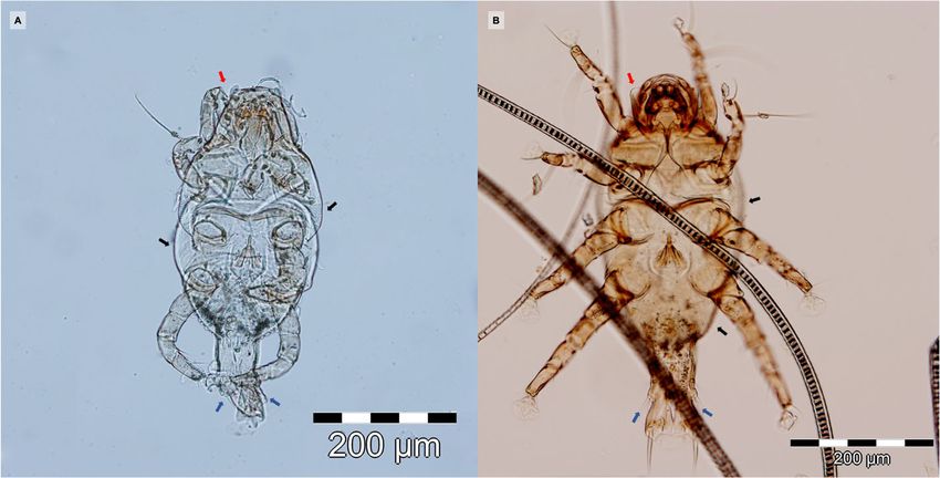
Dumitrache et al. BMC Veterinary Research   (2021) 17:4                                                                                Page 4 of 5

condition by revealing the mites. Initially, we suspected         Infestation with other mites such as L. radovskyi in
an infestation with L. radovskyi, due to the similar            cats, were previously associated with gastrointestinal
morphology of the mites and classification in the same          signs, gingivitis, fever, vulvitis/proctitis, weight loss, etc.
family [12]. The presence of some uncharacteristic mor-         [12]. However, as now other similar cases are reported,
phological features, as well as the few cases reported in       it is difficult to correlate the presence of the non-
the scientific literature [13] and L. radovskyi distribution    dermatological signs identified in our patient, with L.
in tropical regions [14] leaded us to perform a more de-        gibbus infestation. However,
tailed examination. Surprisingly, the microscopic identi-         The findings of this case report suggest that L. gibbus
fication based on the morphological characteristics             infestation, although uncommon, should be considered
revealed L. gibbus as the aetiological agent. Given that        as a possible differential diagnosis of pruritic skin condi-
the cat was living exclusively indoor and considering its       tions in cat.AbbreviationPCR: polymerase chain reac-
origin, we can assume that the contamination occurred           tions; FIV: feline immunodeficiency virus; FeLV: feline
through the close contact with infested rabbits from the        leukemia virus.
pet shop. Diagnosis of L. gibbus in rabbits is challenging
                                                                Acknowledgements
because the tape test could be inefficient due to the           Not applicable.
localization of the parasite on the first third of the hair
shaft. This seems to be the principal cause of underdiag-       Authors’ contributions
                                                                MOD wrote the manuscript. AG and VM performed the clinical examination
nosis in rabbits [1] and might explain why the scotch           of the cat. GD and MOD collected the parasites and performed the
test we performed was negative in cat.                          morphological identification. VM designed the manuscript. All authors have
   L. gibbus has a zoonotic potential and it has been           read and approved the manuscript.
linked twice with papular eruptions in humans [5, 15].
                                                                Funding
Both reports highlight the difficulty in demonstrating          The research was funded by USAMV Cluj-Napoca Internal Grant number
the aetiology of the lesions in rabbit’s owners because no      6270/2017. and by the Ministry of Research and Innovation of Romania, Pro-
mites were detected on their skin. In one report, the           jects for Financing the Excellence in CDI, Contract no. 37PFE/06.11.2018.

owners’ dermatologist admitted a possible parasitic aeti-       Availability of data and materials
ology and suggested that the pruritic erythematous pap-         All relevant data are within this paper.
ules were most likely the result of a hypersensitivity
                                                                Ethics approval and consent to participate
reaction to mites’ biting [5]. Morover, the resolution of       Not applicable.
the owners’ skin lesions following the specific treatment
of rabbits is an indirect, but substantial proof for that in-   Consent for publication
                                                                The cat owner provided written consent for publication.
criminates L. gibbus [5]. Despite of being considered as a
common parasite of rabbits or even a commensal of this          Competing interests
species, only few reports on the zoonotic character of L.       The authors declare that they have no competing interests.
gibbus are available. Hence, we can suspect that that the
                                                                Received: 6 May 2020 Accepted: 18 November 2020
zoonotic transmission is linked to a certain immunologic
status of the owner, like in the case of other mites [16],
or uncommon.                                                    References
                                                                1. Birke LL, Molina PE, Baker DG, Leonard ST, Marrero LJ, Johnson M, Simkin J.
   Several treatment methods against L. gibbus have been            Comparison of selamectin and imidacloprid plus permethrin in eliminating
proposed [1, 2, 17]. Although both imidocloprid plus                Leporacarus gibbus infestation in laboratory rabbits (Oryctolagus cuniculus). J
permethrin and selamectin proved to be effective against            Am Assoc Lab Anim Sci. 2009;48(6):757–62.
                                                                2. Hansen O, Mencke N, Pfister K, Beck W. Efficacy of a formulation containing
this parasite, selamectin seems to eliminate the infest-            imidacloprid and permethrin against naturally acquired ectoparasite
ation more rapidly and is considered that a single sela-            infestations (Ctenocephalides felis, Cheyletiella parasitovorax, and Listrophorus
mectin application has a 100% efficacy against L. gibbus.           gibbus) in rabbits. Int J Appl Res Vet M. 2006;4:4: 320.
                                                                3. Kirwan AP, Middleton B, Mcgarry JW. Diagnosis and prevalence of
However, some authors recommend monthly treatment                   Leporacarus gibbus in the fur of domestic rabbits in the UK. Vet rec. 1998;
until no live mites are detected [16]. These reports could          142:20–1.
explain the results obtained in our case. The first spot-       4. Patel A, Robinson KJE. Dermatosis associated with Listrophorus gibbus in the
                                                                    rabbit. J Small Anim Pract. 1993;34.8:409–11.
on treatment most likely has eliminated most of the par-        5. d’Ovidio D, Santoro D. Leporacarus gibbus infestation in client-owned
asites and allowed a temporary improvement of cat’s                 rabbits and their owner. Vet Dermatol. 2014;25(1):46-e17.
condition. As no other treatment followed this first ad-        6. Dabert J, Ehrnsberger R, Dabert M. Glaucalges tytonis sp. n. (Analgoidea:
                                                                    Xolalgidae) from the barn owl Tyto alba (Strigiformes: Tytonidae): compiling
ministration, the few parasites remaining on the skin               morphology with DNA barcode data for taxon descriptions in mites (Acari).
continued their lifecycle and determined a low intensity            Zootaxa. 2008;1719:41–52.
but permanent pruritus during the following months.             7. Dabert M, Witali nski W, Kazmierski A, Olszanowski Z, Dabert J. Molecular
                                                                    phylogeny of acariform mites (Acari, Arachnida): strong conflict between
The exacerbation of pruritus following vaccination could            phylogenetic signal and long-branch attraction artifacts. Mol Phylogenet
be related to the stress associated with this procedure.            Evol. 2010;56:222–241.
Dumitrache et al. BMC Veterinary Research (2021) 17:4 Page 5 of 5
8. Hartmann K. Clinical aspects of feline retroviruses: a review. Viruses. 2012;
4(11):2684–710.
9. Foil CS. Differential diagnosis of feline pruritus. Vet Clin North Am Small
Anim Pract. 1988;18(5):999–1011.
10. Hobi S, Linek M, Marignac G, et al. Clinical characteristics and causes of
pruritus in cats: a multicentre study on feline hypersensitivity-associated
dermatoses. Vet Dermatol. 2011;22(5):406–13.
11. Verlinden A, Hesta M, Millet S, Janssens GPJ. Food allergy in dogs and cats:
a review. Crit Rev Food Sci Nutr. 2006;46(3):259–73.
12. Foley RH. An epizootic of a rare fur mite in an island’s cat population. Feline
Pract. 1991;19.3:17–9.
13. Jayanthy C, Nagarajan B, Latha BR. Cat fur mite Lynxacarus radovskyi in India.
J Parasit Dis. 2017;41(4):1102–4.
14. Ketzis JK, Dundas J, Shell LG. Lynxacarus radovskyi mites in feral cats: a study
of diagnostic methods, preferential body locations, co-infestations and
prevalence. Vet Dermatol. 2016;27(5):425-e108.
15. Burns DA. Papular urticaria produced by the mite Listrophorus gibbus. Clin
Exp Dermatol. 1987;12:200–1.
16. Jefferson RV, Fusaro AE, da Silva Duarte AJ, Sato MN. Preconception
maternal immunization to dust mite inhibits the type I hypersensitivity
response of offspring. J Allergy Clin Immunol. 2003;111(2):269–77.
17. Hughs JE. Diagnosis and treatment of selected rabbit dermatologic
disorders. Exotic DVM. 2004;5.6:18–20.
Publisher’s Note
Springer Nature remains neutral with regard to jurisdictional claims in
published maps and institutional affiliations.You can also read