Demodectic Mange Associated With Lymphoma in a Ferret
←
→
Page content transcription
If your browser does not render page correctly, please read the page content below

AEMV Forum

Demodectic Mange Associated With
Lymphoma in a Ferret
Hugues Beaufrere, Dr.Med.Vet.,
Michal Neta, DVM,
Dale A. Smith, DVM, DVSc,
and W. Michael Taylor, DVM

              Abstract
              Clinical demodicosis is rare in the ferret, and in other species this condition is
              commonly associated with concurrent immunosuppression. A 4-year-old ferret was
              examined for alopecia of the face and tail. Skin scrapings of the face and otic
              cerumen preparations revealed the presence of a large number of Demodex species.
              The ferret was also diagnosed with adrenal cortical disease based on clinical signs
              and elevated blood estradiol. Dermatologic lesions improved with oral ivermectin
              treatment, but the ferret re-presented for lethargy, weight loss, and anorexia 3
              weeks later. Ultrasound-guided fine-needle aspirates from an enlarged mesenteric
              lymph node and the spleen revealed cytologic abnormalities consistent with high-
              grade lymphoma. This case description is the first, to our knowledge, of sponta-
              neous clinical demodicosis in a ferret. Copyright 2009 Elsevier Inc. All rights
              reserved.

              Key words: demodicosis; Demodex species; ferret; immunosuppression; lymphoma

A
        924-gram, 4-year-old neutered male ferret was         logic examination revealed periorbital, peribuccal,
        presented to the Veterinary Teaching Hospi-           and tail hair loss (Fig 1). Affected facial skin was also
        tal, Ontario Veterinary College, University of        erythematous and thickened with small follicular
Guelph for assessment of hair loss and pruritus               papules. Examination of the ears showed increased
around the eyes, mouth, and tail. The diet consisted
of pelletized ferret food (Totally Ferret; Perfor-
mance Foods Inc., Broomfield, CO USA). The
owner reported that the hair loss and pruritus had               From the Department of Veterinary Clinical Sciences, Louisiana
started 9 months previously and was initially                 State University–School of Veterinary Medicine, Baton Rouge,
treated by another veterinarian with imidacloprid-            LA USA, Department of Pathobiology, Ontario Veterinary College,
moxidectin spot-on (Advantage Multi; Bayer Health             University of Guelph, Guelph, Ontario, Canada, and the Avian
Care, Toronto, Canada) once monthly for 6 months.             and Exotic Service, Veterinary Teaching Hospital, Ontario Veter-
                                                              inary College, University of Guelph, Guelph, Ontario, Canada.
The lesions resolved after the 6-month treatment but
                                                                 Address correspondence to: Hugues Beaufrere, Dr.Med.Vet.,
recurred 1 month before presentation. Another fer-
                                                              Department of Veterinary Clinical Sciences, Louisiana State Uni-
ret in the household was unaffected.                          versity–School of Veterinary Medicine, Skip Bertman Dr, Baton
   On presentation, the ferret was bright and alert.          Rouge, LA 70803. E-mail: hbeaufrere@vetmed.lsu.edu.
Abnormalities noticed during the external physical               © 2009 Elsevier Inc. All rights reserved.
examination included a mildly painful cranial abdo-              1557-5063/09/1801-$30.00
men and a moderately enlarged spleen. Dermato-                   doi:10.1053/j.jepm.2008.10.007

Journal of Exotic Pet Medicine, Vol 18, No 1 ( January), 2009: pp 57– 61                                                    57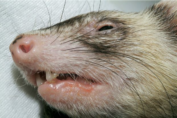
58 Beaufrere et al
underlying disease might have been the primary
cause of the patient’s immunosuppressive state.
Three weeks after the initial presentation, the
ferret was re-presented for lethargy, anorexia, and
weakness. The facial dermatologic lesions had mildly
improved with hair regrowth, decreased erythema,
and reduced amount of cerumen (Fig 2); however,
skin scrapings were not repeated. Within the cranial
abdomen, a small, apparently painful mass was noted
on palpation. The ferret had lost 18% of its body
weight since initial presentation and now weighed
755 g. Core body temperature was 36.5°C (reference
range, 37.8-40°C) and blood glucose was 4 mmol/L
Figure 1. Peribuccal and periocular alopecia, erythema, and skin (reference range, 3.4-7.4 mmol/L).2 Electrolytes and
thickening in a ferret. Demodex species were found in skin scrapings blood gas analysis revealed a moderate respiratory
from these areas. acidosis with pH: 7.13, HCO3-: 21.3 mmol/L, and
pCO2: 63.6 mm Hg (reference ranges in the cat, pH:
7.277-7.409, HCO3-: 18-23.2 mmol/L, pCO2: 32.7-
amounts of brown cerumen and erythema of the 44.7 mm Hg).3 Indirect arterial systolic blood pres-
auricular ducts. sure was normal. Supportive care was initiated with
The ferret was anesthetized with isoflurane, and a intravenous fluid administration (Lactated Ringer’s
blood sample was collected from the cranial vena solution ⫹ KCl 20 meq/L ⫹ dextrose 5%) and tri-
cava, 4 skin scrapings were taken from the face, and methoprim/sulfamethoxazole (15 mg/kg, every
several cerumen samples were collected from the 12 hours, intravenously, Tribrissen 48% injection;
ears. Microscopic examination of skin scrapings and Schering-Plough, Pointe Claire, Canada). An ab-
the cerumen preparations revealed a large number dominal ultrasound examination was performed
of adults and larvae of Demodex species that were and revealed an enlarged mesenteric lymph node
morphologically similar to the large-bodied D. canis measuring 4.2 ⫻ 2.5 ⫻ 4.2 cm with a complex echo-
in dogs. Otodectes cynotis was not present in the ceru- genicity and predominantly hypoechoic tissue. The
men preparations. Cytology of the skin surface was spleen contained 6 to 8 hypoechoic nodules within
unremarkable, thus bacterial culture was not per- its parenchyma. Adrenal gland sizes were within nor-
formed. The complete blood count and bioche- mal limits, with 0.35 ⫻ 0.29 ⫻ 0.79 cm for the left
mistry profile were unremarkable. A ferret adrenal and 0.38 ⫻ 0.26 ⫻ 0.74 cm for the right (reference
hormone panel submitted to the University of Ten- range, left 4-8.1 ⫻ 1.8-3.9 mm; right 4.9-10.6 ⫻ 1.3-
nessee Endocrinology Laboratory revealed an in- 3.7 mm).4 Fine-needle aspirates were collected from
creased estradiol level at 195.2 pmol/L (reference
the lymph node and the splenic nodules.
range provided by the University of Tennessee: 20-
The lymph node aspirates (Fig 3) consisted al-
180 pmol/L) with normal 17-hydroxyprogesterone
most exclusively of highly monomorphic lympho-
and androstenedione blood levels. Abdominal ultra-
sound examination was declined at that time.
After the physical examination and diagnostic test
results, a tentative diagnosis was facial demodectic
mange and adrenal cortical disease. The ferret was
treated with an injection of leuprolide acetate (100
g, intramuscularly, Lupron Depot; TAP Pharma-
ceuticals, Lake Forest, IL USA). He was also sent
home with oral ivermectin (50 g/kg every 24 hours,
orally, Ivomec; Merial Limited, Duluth, GA USA).
The dosage was progressively increased over the first
2 weeks to 300 g/kg, and the overall treatment was
planned to last until 1 month after negative skin
scrapings. Progressive dosage increase was elected as
Figure 2. Mild improvement of dermatologic lesions caused by
recommended in the dog.1 The owner was encour- Demodex species 3 weeks after starting oral ivermectin treatment.
aged to permit an abdominal ultrasound examina- The skin was less erythematous and thickened, and the auricular
tion as soon as possible to determine whether an ducts were less inflamed.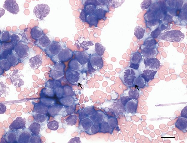
Demodicosis in a Ferret 59
alopecia and pruritus that were diagnosed with de-
modicosis based on skin scrapings and biopsies.
Demodex species are thought to live as commensals
in the hair follicles and sebaceous glands of most
mammals and may proliferate with an underlying
immunological deficiency.9 A retrospective his-
topathologic study on normal ferret skin found 9 of
25 ferrets, aged 4 to 32 months, with commensal
Demodex species present within the hair follicles and
sebaceous glands in the perianal, vulvar, preputial,
facial, and caudal abdominal regions.10 Adult onset
of demodectic mange in dogs and cats is generally
triggered by conditions altering the immune system,
most commonly Cushing’s disease, hypothyroidism,
diabetes mellitus, immunosuppressive drug therapy,
Figure 3. Cytological appearance of a fine-needle aspirate of the feline immunodeficiency virus infection, feline leu-
mesenteric lymph node from a ferret with lymphoma and concurrent kemia virus infection, or neoplasia.1,9 In the previous
demodicosis. There were large monomorphic lymphocytes, with
frequent mitotic activity (arrows). Wright’s stain, bar ⫽ 10 m. case report by Noli and coworkers, demodicosis was
induced by the use of a glucocorticoid-based oint-
ment containing triamcinolone (Tresaderm; Merial
cytes. These were large cells (approximately 30 m Limited, Iselin, NJ USA) for 3 months for the treat-
in diameter) with increased nuclear-cytoplasmic ra- ment of recurrent ear mite infestations.8
tios. Cytoplasm was grainy and stained deeply baso- The dermatologic lesions in this ferret responded
philic. There was sparse cytoplasmic vacuolation and twice to acaricidal therapy. Postmortem examination
small, indistinct Golgi zones. Nuclei were large, with and histopathology of the skin would have been very
a finely reticulated chromatin pattern and occa- informative but were declined by the owner. Skin
sional obscure nucleoli. There was minimal anisocy- scrapings were not repeated at the 3-week recheck
tosis and anisokaryosis, but mitotic activity was high because of the early stage of the therapy. The two
(2-4 mitotic figures per high power field). The aspi- ferrets described by Noli and coworkers showed der-
rate from the spleen (Fig 4) was extremely cellular matologic lesions consisting of alopecia and sebor-
and revealed extensive extramedulllary hematopoie- rhoea localized behind the ears, in the inguinal ar-
sis which is commonly seen in ferrets. However, eas, and on the ventral aspect of the tail with an
among the hematopoietic precursors were low num- increased cerumen production in the ears. Skin
bers of large lymphocytes, morphologically similar to
those noted in the mesenteric lymph node and with
high mitotic activity. These cells were scattered in a
patchy, uneven distribution and, although overall
they were not a prominent population, they were
clearly abnormal and predominated in certain areas
of the slide. Findings were interpreted as a high-
grade lymphoma in the mesenteric lymph node and
the spleen.
The ferret’s clinical condition did not improve
over 24 hours, and the owner requested euthanasia.
Postmortem examination was declined. The final
diagnosis was demodicosis secondary to a general-
ized lymphoma with concurrent adrenal cortical dis-
ease.
Discussion
Figure 4. Cytological appearance of a fine-needle aspirate of the
spleen from a ferret with lymphoma and concurrent demodicosis.
Clinical demodicosis is considered an uncommon Large lymphocytes are present among hematopoietic precursors.
dermatosis of ferrets and has rarely been reported.5-7 Megakaryocytes (MK) and a mitotic figure (arrow) were also noted.
Noli and coworkers8 described two ferrets with local Wright’s stain, bar ⫽ 10 m.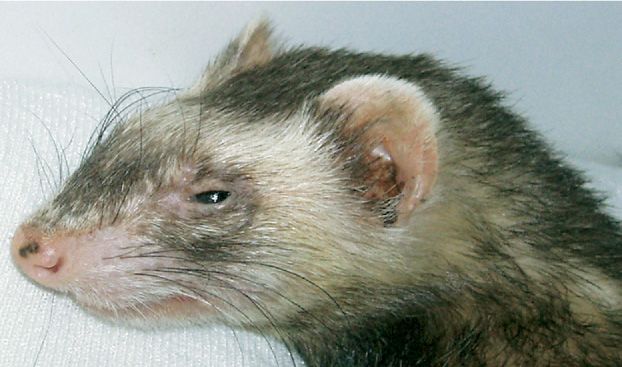
60 Beaufrere et al
scrapings, smears of exudates from the ears, and skin in association with lymphoproliferative disorders has
biopsies revealed the presence of adult and larvae of been described in dogs, cats,9,14 and humans.15-17
a short-bodied Demodex species. However, localiza- Immune depression or suppression predisposes to a
tion of skin scrapings and biopsies were not docu- variety of skin diseases, including demodicosis, in
mented. They were successfully treated with amitraz small animals.18 Deficiencies in cell-mediated im-
dips without side effects, 0.0125% at 7-day intervals mune response play an important role in the patho-
for 3 treatments followed by 0.0375% at 5-day inter- genesis of demodicosis in humans and dogs.15 Effects
vals for 3 final treatments. The ferrets had no evi- of lymphoma on the immune system have been
dence of mites 1 month after the end of the treat- poorly investigated in ferrets. Although there are
ment.8 In our case, Demodex species were similarly numerous reports of lymphoid neoplasia of ferrets
found in the skin scrapings and ear exudate but were in the literature, none describe concurrent demodi-
different in morphology and more comparable to cosis.19
the long-bodied dog D. canis and the Demodex species This report is the first, to our knowledge, to
described in normal ferret skin by Martin and co- present the spontaneous occurrence of demodectic
workers10 On skin biopsies presented by Noli and mange in a ferret. We suggest that cases of con-
coworkers, the mites were superficial and not seen in firmed demodicosis be screened carefully for the
the sebaceous glands and deeper portions of the hair presence of an underlying disease process that re-
follicles, whereas the commensal Demodex species re- sults in an immunosuppressive state.
ported by Martin and coworkers were present in the
sebaceous glands. This suggests two different species
of Demodex, the one recovered from our ferret being
more consistent with the report from Martin and
References
coworkers. On the other hand, dermatologic lesions
1. Mueller RS: Treatment protocols for demodicosis: an
were only localized on the face and were also char- evidence-based review. Vet Dermatol 15:75-89, 2004
acterized by erythema and thickening of the skin. 2. Quesenberry KE, Orcutt C: Basic approach to vet-
The dermatologic lesions on the tail were only alo- erinary care, in Quesenberry KE, Carpenter JW
pecic and very different from the facial ones. Be- (eds): Ferrets, Rabbits, and Rodents Clinical Med-
icine and Surgery (ed 2). St Louis, MO, Elsevier/
cause tail alopecia is a common sign of adrenal
Saunders, pp 13-24, 2004
cortical disease in ferrets, investigation of the skin in 3. DiBartola SP: Introduction to acid-base disorders, in
this area was not pursued. DiBartola SP (ed): Fluid Therapy in Small Animal
Treatment recommendations for dogs and cats Practice (ed 2). Philadelphia, PA, W.B. Saunders,
with demodicosis include topical amitraz (0.025- pp 189-210, 2000
4. Kuijten AM, Schoemaker NJ, Voorhout G: Ultrasono-
0.06%, every 7-14 days, Mitaban; Pfizer Animal graphic visualization of the adrenal glands of healthy
Health, New York, NY USA); ivermectin, (300 g/ ferrets and ferrets with hyperadrenocorticism. J Am
kg, every 24 hours, orally); milbemycin oxime (2 Anim Hosp Assoc 43:78-84, 2007
mg/kg, every 24 hours, orally, Interceptor; Novartis 5. Lewington JH: Parasitic diseases of ferrets, in Lew-
Animal Health, Basel, Switzerland); and moxidectin ington JH (ed): Ferret Husbandry, Medicine and
Surgery (ed 2). Philadelphia, PA, Elsevier/Saunders,
(400 g/kg, every 24 hours, orally, ProHeart 6; pp 224-257, 2007
Wyeth Animal Health, Guelph, Ontario, Canada).1 6. Orcutt C: Dermatologic diseases, in Quesenberry KE,
The ferret described here showed an improvement Carpenter JW (eds): Ferrets, Rabbits, and Rodents
of dermatologic lesions after a 3-week treatment with Clinical Medicine and Surgery (ed 2). St Louis, MO,
Elsevier/Saunders, pp 107-114, 2004
daily oral ivermectin. Nevertheless, the progression
7. Marini RP, Otto G, Erdman S, et al: Biology and
of the underlying lymphoma prevented us from fol- diseases of ferrets, in Fox JG, Anderson LC, Loew
lowing up on demodicosis. FM, et al (eds): Laboratory Animal Medicine (ed 2).
It is possible that the ferret presented here was San Diego, MO, Academic Press, pp 483-518, 2002
immunocompromised as a result of the systemic lym- 8. Noli C, VanderHorst HH, Willemse T: Demodicosis
in ferrets (Mustela putorius furo). Vet Quart 18:28-31,
phoma. In humans, dogs, and cats, immunosuppres- 1996
sion secondary to lymphoma and other malignancies 9. Campbell KL: Other external parasites, in Ettinger
is well documented, and the pathogenesis includes SJ, Feldman EC (eds): Textbook of Veterinary Inter-
loss of lymphoid tissues, overexpression of immuno- nal Medicine (ed 6). St Louis, MO, Elsevier/Saun-
modulating cytokines such as interleukin-10 and ders, pp 66-69, 2005
10. Martin AL, Irizarry-Rovira AR, Bevier DE, et al: His-
transforming growth factor , downregulation of im- tology of ferret skin: preweaning to adulthood. Vet
munoglobulin production, and induction of regula- Dermatol 18:401-411, 2007
tory T-cells.11-13 The proliferation of Demodex species 11. Biller BJ, Dow S: Immunotherapy of cancer, in With-Demodicosis in a Ferret 61
row SJ, Vail DM (eds): Small Animal Clinical demodicosis. J Eur Acad Dermatol Venereol
Oncology (ed 4). St Louis, MO, Elsevier/Saunders, 18:440-444, 2004
pp 211-235, 2007 16. Rawal M, Tiliakos A, Hostoffer RW: Demodicosis: an
12. Re D, Küppers R, Diehl V: Molecular pathogenesis of unusual presentation of small lymphocytic lym-
Hodgkin’s lymphoma. J Clin Oncol 23:6379-6386, phoma. J Allergy Clin Immunol 113(suppl 1):S121,
2005 1996
13. Beverley PCL: The immunology of cancer, in 17. Seyhan ME, Karincaoglu Y, Bayram N, et al: Density
Knowles M, Selby P (eds): Introduction to the Cellu- of Demodex folliculorum in haematological malignan-
lar and Molecular Biology of Cancer (ed 4). Oxford, cies. J Int Med Res 32:411-415, 2004
Oxford University Press, pp 337-355, 2005 18. Foster AP: Immunomodulation and immunodefi-
14. Scott DW, Miller WH, Griffin CE: Parasitic skin dis- ciency. Vet Dermatol 15:115-126, 2004
ease, in Scott DW, Miller WH, Griffin CE (eds): 19. Erdman SE, Brown SA, Kawasaki TA, et al: Clinical
Muller and Kirk’s Small Animal Dermatology (ed 6). and pathologic findings in ferrets with lymphoma: 60
Philadelphia, PA, W.B. Saunders, pp 423-516, 2001 cases (1982-1994). J Am Vet Med Assoc 208:1285-
15. Akilov OE, Mumcuoglu KY: Immune response in 1289, 1996You can also read