Fact sheet - Wildlife Health Australia
←
→
Page content transcription
If your browser does not render page correctly, please read the page content below
Fact sheet

Yellow fungus disease in lizards occurs as a result of infection with Nannizziopsis guarroi. Similar disease
conditions identified in Australian reptiles are linked to related fungi in the Nannizziopsis and
Paranannizziopsis genera (Sigler et al. 2013; Paré and Sigler 2016). Ophidiomyces ophiodiicola is the causative
agent of snake fungal disease, an emerging disease of free-ranging snakes in North America which is also
occasionally identified globally in captive snakes, including in Australia (Sigler et al. 2013; Stchigel et al. 2013;
Lorch et al. 2016; Paré and Sigler 2016). It has not been reported in wild Australian snakes.

Yellow fungus disease in inland bearded dragons (Pogona vitticeps) and green iguanas (Iguana iguana) is
caused by N. guarroi. Similar dermatomycoses in a variety of reptilian species are caused by other species in
the Nannizziopsis and Paranannizziopsis genera (Paré and Sigler 2016). The aetiologic agent of snake fungal
disease (previously thought to be Chrysosporium anamorph of Nannizziopsis vriessi; CANV), has now been
identified as O. ophiodiicola, the only known species of the Ophidiomyces genus (Sigler et al. 2013; Stchigel et
al. 2013)1. These fungal genera fall into the family Onygenaceae of the order Onygenales (Sigler et al. 2013;
Stchigel et al. 2013).

Nannizziopsis spp. have been reported in a wide range of lizard species, crocodiles and humans (Paré and
Sigler 2016). Paranannizziopsis spp. have been reported in lizards, snakes and tuatara (Sphenodon punctatus)

1
    The fungal agent formerly known as the Chrysosporium anamorph of Nannizziopsis vriessi (CANV) has undergone
extensive taxonomic reclassification, resulting in its division into a number of different species within three genera namely
Nannizziopsis, Paranannizziopsis and Ophidiomyces (Sigler et al. 2013; Stchigel et al. 2013).
(Paré and Sigler 2016). O. ophiodiicola has been identified in a wide range of snakes (Lorch et al. 2016; Paré and Sigler 2016). Cases of Nannizziopsis spp. and Paranannizziopsis spp. infection in reptiles have been reported in Africa, Asia, Europe, North America and Oceania (Sigler et al. 2013; Stchigel et al. 2013; Paré and Sigler 2016). O. ophiodiicola is considered a recently emerging disease of wild snakes in North America, but has been identified in captive snakes since the 1980’s in Europe, North America and Australia (Sigler et al. 2013; Stchigel et al. 2013; Paré and Sigler 2016). Species identified in Australia include N. barbata, N. crocodile, P. australasiensis and O. ophiodiicola (Thomas et al. 2002; Johnson et al. 2011; Sigler et al. 2013; Paré and Sigler 2016). Outbreaks of N. crocodili occurred on two separate occasions in 1994 and 1997 in saltwater crocodiles (Crocodylus porosus) sourced from the same crocodile farm in northern Queensland (Thomas et al. 2002; Paré and Sigler 2016). In 2008-2009 an outbreak of what is now identified as N. barbata was reported in captive coastal bearded dragons (Pogona barbata) in New South Wales (Johnson et al. 2011; Paré and Sigler 2016). A very similar isolate to N. barbata has been obtained from a free-ranging eastern water dragon (Physignathus lesueurii) with dermal lesions (Paré and Sigler 2016). A separate case of yellow fungus-like disease has been diagnosed in a “wild caught” pet bearded dragon (R. Johnson 2009, personal communication). P. australasiensis was cultured from lesions on two file snakes (Acrochordus arafura) housed in captivity in Victoria (Paré and Jacobson 2007; Sigler et al. 2013; Paré and Sigler 2016). O. ophiodiicola has been diagnosed in captive snakes in Australia, including a file snake in Queensland and a broad-headed snake in South Australia (McLelland et al. 2010; Sigler et al. 2013; Paré and Sigler 2016). There is also suggestion that mycotic dermatitis attributed to Geotrichum candidum in three captive carpet snakes in Queensland may have in fact been caused by O. ophiodiicola (McKenzie and Green 1976; Paré and Sigler 2016). Anecdotal evidence suggests that clinical cases consistent with yellow fungus disease and snake fungal disease may have been occurring in captive snakes and lizards in Australia for several decades (R Woods, A Reiss pers comm Aug 2018). N. guarroi as well as other members of the Nannizziopsis and Paranannizziopsis genera appear to be primary, obligate pathogens, rather than opportunists (as fungal pathogens have been traditionally considered) (Mitchell and Walden 2013). Koch’s postulates have been fulfilled in experimental infection of veiled chameleons (Chamaeleo calyptratus) with N. guarroi (Paré et al. 2006). In captivity, suboptimal environmental factors may make reptiles more susceptible to these infections and the pathogens are contagious between animals in close proximity (Thomas et al. 2002; Paré and Sigler 2016). Although cases
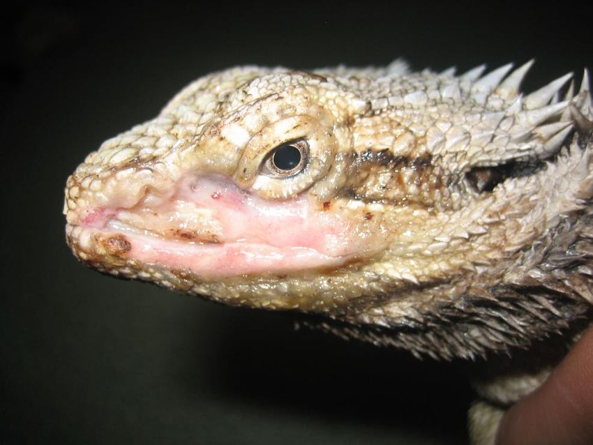
have been identified in free-ranging reptiles, there is little available information on the epidemiology in the
wild (Paré and Sigler 2016).

O. ophiodiicola is now well recognised as a pathogen of free-ranging snakes, particularly in North America.
Experimental infections have fulfilled Koch’s postulates and have demonstrated action as a primary pathogen
(Allender et al. 2015a; Lorch et al. 2015). The fungus has not been identified on the skin of healthy snakes, but
is found in copious amounts in the lesions of affected snakes, indicating that it is not part of the cutaneous
mycobiota (Allender et al. 2011; Allender et al. 2015b; Paré and Sigler 2016). Although the fungus does not
grow at temperatures less than 15 C, snakes undergoing brumation may be susceptible during the autumnal
decline and spring rise in temperatures, when the snakes’ immune systems are depressed (Lorch et al. 2016;
Paré and Sigler 2016). Small skin lesions colloquially termed “hibernation” sores or blisters that have been
recognised in snakes for many years also test positive for the fungus, suggesting infection has been occurring
historically, but severity of clinical disease (at least in some incidences) may have been increasing in recent
years. The cause of this increased severity (from small healing sores to a fatal disease) is unclear (Lorch et al.
2016; Paré and Sigler 2016).

Lesions are similar for all pathogens, although appear to vary in severity depending on the host species and
likely other environmental factors. Grossly, lesions are initially yellow, and then thicken to form brown,
hyperkeratotic, necrotic plaques. These plaques may crack and seep exudate, or slough to reveal whitish pink,
swollen dermis. Lesions are often found on the head and around the mouth but can occur anywhere on the
body and can involve a whole limb in the case of lizards (Paré and Jacobson 2007; Lorch et al. 2016; Paré and
Sigler 2016).

Dermatomycoses due to Nannizziopsis spp. are slowly progressive and often fatal skin diseases. Affected
reptiles range in body condition from poor to good. Skin lesions progress over several months from dry and
yellow to hyperkeratotic plaques to exudative and necrotic ulcers (Figure 1) (Johnson et al. 2011; Paré and
Sigler 2016). In bearded dragons (Pogona spp.), the mouth and face are commonly affected but lesions may
occur anywhere on the body (Bowman et al. 2007; Abarca et al. 2009; Johnson et al. 2011; Le Donne et al.
2016; Schmidt-Ukaj et al. 2016). Infection is often fatal in bearded dragons, with infection extending to
muscle, bone and internal tissues including liver, heart, kidney, lungs and intestine (Bowman et al. 2007;
Johnson et al. 2011; Masters et al. 2016; Paré and Sigler 2016; Schmidt-Ukaj et al. 2016). In iguanas, infection
is often identified in the hind limbs and tail (Han et al. 2010; Kahraman et al. 2015). Hatchling saltwater
crocodiles infected with N. crocodili developed multiple leathery plaque-like lesions on or under the scales,
which could be peeled away to reveal white or red tissue. Infection was often fatal (Thomas et al. 2002).

P. australasiensis in tuatara causes skin lesions similar to those described above. In tuatara, lesions resolved
with treatment in all reported cases. In a coastal bearded dragon, the same agent resulted in fatality, with
similar progression of disease as that described for Nannizziopsis spp. (Masters et al. 2016). In file snakes,
infection results in disseminated punctate or circular, whitish lesions across the epidermis (Paré and Jacobson
2007; Sigler et al. 2013; Paré and Sigler 2016).

O. ophiodiicola in snakes causes yellow to brown, thickened, necrotic crusts, mainly affecting the head also
the scales of the body, most often on the underside of the snake. In milder cases, the processes of ecdysis
(slough) can resolve the infection, but if deeper tissues are affected, disease may recur post-slough. Systemic
infection with involvement of bone and lung has been reported, but is rare (Lorch et al. 2016).
Figure 1. Yellow fungus disease in a captive coastal bearded dragon Pogona barbata (Courtesy R Johnson).

Definitive diagnosis of skin or systemic disease associated with fungi in the Nannizziopsis, Paranannizziopsis
and Ophidiomyces genera requires both a) identification of the organism through culture and sequencing or
PCR and b) histopathology identifying fungal elements within lesions. Reptile skin plays host to many fungal
elements, so presence of the organism alone is not synonymous with disease (Paré and Sigler 2016).
Identification of the organism using matrix-assisted laser desorption/ ionization time-of-flight mass
spectrometry (MALDI-TOF MS), which may be more accurate than sequencing, has been described but may
not yet be available in Australia (Schneider et al. 2017).

Although not pathognomonic, cytology of sticky tape preparations or impression smears may reveal presence
of conidia or arthroconidia suggestive of a fungus in the Nannizziopsis, Paranannizziopsis or Ophidiomyces
genera (Le Donne et al. 2016; Paré and Sigler 2016). Conidia are 5-8 μm long by 3-5 μm wide, clavate to ovoid
to cylindrical and surrounded by a thin capsule. Arthroconidia are arranged in rows of up to 7 conidia,
separated by thin septae (Le Donne et al. 2016).

Grossly, lesions are as described above. In systemically affected bearded dragons at necropsy, collection of
pale yellow, gelatinous material can be found within the coelomic cavity and pericardial sac and
granulomatous changes have been noted in the liver (Bowman et al. 2007) (C Sangster 2018, personal
communication). Histologic lesions include granulomatous fungal dermatitis, myositis, osteomyelitis,
hepatitis, nephritis, coelomitis, myocarditis and pneumonia (Bowman et al. 2007; Johnson et al. 2011; Paré
and Sigler 2016; Schmidt-Ukaj et al. 2016). Hyphae found within granulomas and in the keratin layer are 2-4
μm wide, septate and exhibit haphazard branching. Arthroconidia on the surface of the epidermis is
suggestive, but not pathognomonic for fungi in this order (Paré and Jacobson 2007).

Other dermatomycoses, bacterial dermatitis, stomatitis and osteomyelitis should be excluded from the list of
differential diagnoses. Previously, fungi from this order have been misidentified as Trichophyton spp. or as
other fungal species including Geotrichium spp. or Trichosporon spp. (Bowman et al. 2007; Johnson et al.
2011; Paré and Sigler 2016).

Multiple skin biopsies of dermal lesions, half placed in 10% neutral buffered formalin for histopathology and
half submitted fresh (or less desirably frozen) for PCR and /or culture (Paré and Sigler 2016).

Skin samples pulled from lesions can be submitted for PCR and culture. Swabs should be avoided as the
fungus is difficult to culture from these samples (Paré and Sigler 2016).

Sections of multiple internal organs in 10% neutral buffered formalin and fresh/frozen are recommended if
systemic disease is suspected (C Sangster 2018, personal communication).

Histopathological examination should include H&E and either PAS or Grocott-Gomori’s methylene silver stain
for fungal identification (C Sangster pers comm July 2018,).

Samples for culture are best treated with enrofloxacin to limit bacterial overgrowth, plated on MycoselTM
agar (Becton, Dickinson and Company, Franklin Lakes, NJ) and incubated at 30°C. White powdery colonies
should be subcultured, to prevent bacterial overgrowth, and speciated by sequencing (Paré and Sigler 2016).

Medical treatment of confirmed cases involves systemic antifungals and topical antifungal or antiseptic
solutions (Paré and Sigler 2016). Susceptibility testing of N. guarroi has revealed sensitivity to voraconazole
and terbinifine, but less so to itraconazole (Van Waeyenberghe et al. 2010; Paré and Sigler 2016). Serum
biochemistry should be monitored for signs of liver toxicity. Surgical excision or debridement of lesions should
be carried out if possible and in conjunction with medical therapy (R Johnson 2009, personal communication).

Prevention of Nannizziopsis spp., Paranannizziopsis spp. and O. ophiodiicola infection in captive reptiles
should focus on reducing the fungal load, with attention being paid to regular substrate changes and good
hygiene in captive situations. Providing optimal husbandry conditions, including species-appropriate
temperature gradients, hydration/humidity and nutrition are important steps in prevention and control (Paré
and Sigler 2016). In captive reptiles, infection appears to be more common at low ambient temperatures (R
Johnson 2009, personal communication). Affected individuals should be isolated and biosecurity measures
followed as the organism can act as a contagious, primary pathogen (Paré and Sigler 2016).

Wildlife disease surveillance in Australia is coordinated by Wildlife Health Australia. The National Wildlife
Health Information System (eWHIS) captures information from a variety of sources including Australian
government agencies, zoo and wildlife parks, wildlife carers, universities, industry and members of the public.
Coordinators in each of Australia's States and Territories report monthly on significant wildlife cases identified
in their jurisdictions. NOTE: access to information contained within the National Wildlife Health Information
System dataset is by application. Please contact admin@wildlifehealthaustralia.com.au. There are currently
no targeted surveillance programs for reptile fungal diseases. There are a small number of cases reported in
eWHIS including in a wild coastal bearded dragon Pogona barbata (confirmed by culture) and a suspected
case in a wild broad-shelled turtle Chelodina expansa (no culture or PCR).

We encourage those with laboratory confirmed cases of this condition in native Australian or feral animals to
submit this information to the national system for consideration for inclusion in the national database. Please
contact us at admin@wildlifehealthaustralia.com.au.

Molecular differentiation of the taxonomy of this order of fungi has significantly advanced knowledge in this
area, allowing identification of morphological and physiological properties, host trends and sensitivity
patterns for many new species (Paré and Sigler 2016). Large gaps in the epidemiology of these pathogens,
particularly in free-ranging reptiles, remain. This is particularly significant in the case of O. ophiodiicola, which
is an emerging disease resulting in significant losses of free-ranging snakes in North America (Lorch et al.
2016). Nannizziopsis and Paranannizziopsis spp. that appear to be specific to Australasia have been identified,
but only in low numbers of animals. Further studies are warranted to understand fully the origin and nature
of these organisms, and their significance as primary pathogens in reptiles in Australia, both captive and free
living. To date, reports in free-ranging Australian reptiles have been opportunistic, with no research
investigating incidence, host range or epidemiology of disease related to these pathogens, illustrating a
significant knowledge gap.

Previously, infection with what was termed CANV had been reported in humans, generally in
immunosuppressed patients (Steininger et al. 2005; Paré and Sigler 2016). However, the recent molecular
characterisation work has revealed these cases to be caused by species of Nannizziopsis that are distinct from
those found in reptiles. The risk of zoonotic transmission of O. ophiodiicola or Nannizziopsis spp. and
Paranannizziopsis spp. from reptiles to humans is not considered high, although it is considered possible in
immunocompromised patients (Paré and Sigler 2016).

In Australia, outbreaks of infection with fungi from the Nannizziopsis and Paranannizziopsis genera have
occurred in farmed crocodiles, captive bearded dragons and captive file snakes. Sporadic cases of O.
ophiodiicola have occurred in captive Australian snakes. Overseas these fungi have been found to be
significant primary pathogens of lizards and snakes. As the fungus is easily spread by contact, wildlife carers
and veterinarians caring for captive and free-living reptiles need to be vigilant in preventing the spread of
these pathogens. Further work is recommended to better understand the epidemiology of the diseases, and
possible risks to Australian native reptiles.

Abarca M, Martorell J, Castellá G, Ramis A, Cabañes F (2009) Dermatomycosis in a pet inland bearded dragon
(Pogona vitticeps) caused by a Chrysosporium species related to Nannizziopsis vriesii. Veterinary Dermatology
20, 295-299.

Allender MC, Baker S, Wylie D, Loper D, Dreslik MJ, Phillips CA, Maddox C, Driskell EA (2015a) Development of
snake fungal disease after experimental challenge with Ophidiomyces ophiodiicola in cottonmouths
(Agkistrodon piscivorous). PloS ONE 10, e0140193.

Allender MC, Bunick D, Dzhaman E, Burrus L, Maddox C (2015b) Development and use of a real-time
polymerase chain reaction assay for the detection of Ophidiomyces ophiodiicola in snakes. Journal of
Veterinary Diagnostic Investigation 27, 217-220.

Allender MC, Dreslik M, Wylie S, Phillips C, Wylie DB, Maddox C, Delaney MA, Kinsel MJ (2011) Chrysosporium
sp. infection in eastern massasauga rattlesnakes. Emerging Infectious Diseases 17, 2383.

Bowman MR, Paré JA, Sigler L, Naeser JP, Sladky KK, Hanley CS, Helmer P, Phillips LA, Brower A, Porter R
(2007) Deep fungal dermatitis in three inland bearded dragons (Pogona vitticeps) caused by the
Chrysosporium anamorph of Nannizziopsis vriesii. Medical Mycology 45, 371-376.

Han J-I, Lee S-J, Na K-J (2010) Necrotizing dermatomycosis caused by Chrysosporium spp. in three captive
green iguanas (Iguana iguana) in South Korea. Journal of Exotic Pet Medicine 19, 240-244.

Johnson R, Sangster C, Sigler L, Hambleton S, Paré J (2011) Deep fungal dermatitis caused by the
Chrysosporium anamorph of Nannizziopsis vriesii in captive coastal bearded dragons (Pogona barbata).
Australian Veterinary Journal 89, 515-519.

Kahraman BB, Sığırcı BD, Metiner K, Ak S, Koenhemsi L, Or ME, Castellá G, Abarca ML (2015) Isolation of
Chrysosporium guarroi in a Green Iguana (Iguana iguana), in Turkey. Journal of Exotic Pet Medicine 24, 427-
429.

Le Donne V, Crossland N, Brandão J, Sokolova Y, Fowlkes N, Nevarez JG, Langohr IM, Gaunt SD (2016)
Nannizziopsis guarroi infection in 2 inland bearded dragons (Pogona vitticeps): clinical, cytologic, histologic,
and ultrastructural aspects. Veterinary Clinical Pathology 45, 368-375.

Lorch JM, Knowles S, Lankton JS, Michell K, Edwards JL, Kapfer JM, Staffen RA, Wild ER, Schmidt KZ, Ballmann
AE (2016) Snake fungal disease: an emerging threat to wild snakes. Philosophical Transactions of the Royal
Society B: Biological Sciences 371, 20150457.

Lorch JM, Lankton J, Werner K, Falendysz EA, McCurley K, Blehert DS (2015) Experimental infection of snakes
with Ophidiomyces ophiodiicola causes pathological changes that typify snake fungal disease. MBio 6,
e01534-15.

Masters N, Alexander S, Jackson B, Sigler L, Chatterton J, Harvey C, Gibson R, Humphrey S, Rawdon T, Spence
R (2016) Dermatomycosis caused by Paranannizziopsis australasiensis in five tuatara (Sphenodon punctatus)
and a coastal bearded dragon (Pogona barbata) in a zoological collection in New Zealand. New Zealand
Veterinary Journal 64, 301-307.
McKenzie R, Green P (1976) Mycotic dermatitis in captive carpet snakes (Morelia spilotes variegata). Journal
of Wildlife Diseases 12, 405-408.

McLelland D, Johnson L, Reuter R (2010) Fatal cutaneous mycosis in a broad-headed snake (Hoplocephalus
bungaroides) caused by the Chrysosporium anamorph of Nannizziopsis vriesii, Proceedings of the Wildlife
Disease Association–Australasian Section. Tasmania, Australia. (WDAA)

Mitchell MA, Walden MR (2013) Chrysosporium anamorph Nannizziopsis vriesii: an emerging fungal pathogen
of captive and wild reptiles. Veterinary Clinics: Exotic Animal Practice 16, 659-668.

Paré J, Coyle K, Sigler L, Maas A, Mitchell R (2006) Pathogenicity of the Chrysosporium anamorph of
Nannizziopsis vriesii for veiled chameleons (Chamaeleo calyptratus). Sabouraudia 44, 25-31.

Paré J, Jacobson E (2007) Mycotic diseases of reptiles. In 'Infectious diseases and pathology of reptiles.' (Ed.
ER Jacobson.) pp. 527-570. (CRC Press: Boca Raton).

Paré J, Sigler L (2016) An overview of reptile fungal pathogens in the genera Nannizziopsis, Paranannizziopsis,
and Ophidiomyces. Journal of Herpetological Medicine and Surgery 26, 46-53.

Schmidt-Ukaj S, Loncaric I, Spergser J, Richter B, Hochleithner M (2016) Dermatomycosis in three central
bearded dragons (Pogona vitticeps) associated with Nannizziopsis chlamydospora. Journal of Veterinary
Diagnostic Investigation 28, 319-322.

Schneider J, Heydel T, Klasen L, Pees M, Schrödl W, Schmidt V (2017) Characterization of Nannizziopsis guarroi
with genomic and proteomic analysis in three lizard species. Medical Mycology 56, 610-620.

Sigler L, Hambleton S, Paré JA (2013) Molecular characterization of reptile pathogens currently known as
members of the Chrysosporium anamorph of Nannizziopsis vriesii complex and relationship with some
human-associated isolates. Journal of Clinical Microbiology 51, 3338-3357.

Stchigel A, Sutton D, Cano-Lira J, Cabañes F, Abarca L, Tintelnot K, Wickes B, García D, Guarro J (2013)
Phylogeny of chrysosporia infecting reptiles: proposal of the new family Nannizziopsiaceae and five new
species. Persoonia: Molecular Phylogeny and Evolution of Fungi 31, 86.

Steininger C, van Lunzen J, Tintelnot K, Sobottka I, Rohde H, Horstkotte MA, Stellbrink H-J (2005) Mycotic
brain abscess caused by opportunistic reptile pathogen. Emerging Infectious Diseases 11, 349.

Thomas A, Sigler L, Peucker S, Norton J, Nielan A (2002) Chrysosporium anamorph of Nannizziopsis vriesii
associated with fatal cutaneous mycoses in the salt-water crocodile (Crocodylus porosus). Medical Mycology
40, 143-151.

Van Waeyenberghe L, Baert K, Pasmans F, Van Rooij P, Hellebuyck T, Beernaert L, De Backer P, Haesebrouck F,
Martel A (2010) Voriconazole, a safe alternative for treating infections caused by the Chrysosporium
anamorph of Nannizziopsis vriesii in bearded dragons (Pogona vitticeps). Sabouraudia 48, 880-885.

We are extremely grateful to Robert Johnson and Cheryl Sangster who provided the initial and updated draft
of this fact sheet and to those individuals, agencies and organisations that provided comment and external
review.

Updated: Aug 2018
We are interested in hearing from anyone with information on this condition in Australia, including laboratory
reports, historical datasets or survey results that could be added to the National Wildlife Health Information
System. If you can help, please contact us at admin@wildlifehealthaustralia.com.au.

Wildlife Health Australia would be very grateful for any feedback on this fact sheet. Please provide detailed
comments or suggestions to admin@wildlifehealthaustralia.com.au We would also like to hear from you if
you have a particular area of expertise and would like to produce a fact sheet (or sheets) for the network (or
update current sheets). A small amount of funding is available to facilitate this.

This fact sheet is managed by Wildlife Health Australia for information purposes only. Information contained
in it is drawn from a variety of sources external to Wildlife Health Australia. Although reasonable care was
taken in its preparation, Wildlife Health Australia does not guarantee or warrant the accuracy, reliability,
completeness, or currency of the information or its usefulness in achieving any purpose. It should not be
relied on in place of professional veterinary or medical consultation. To the fullest extent permitted by law,
Wildlife Health Australia will not be liable for any loss, damage, cost or expense incurred in or arising by
reason of any person relying on information in this fact sheet. Persons should accordingly make and rely on
their own assessments and enquiries to verify the accuracy of the information provided.
You can also read