Vanishing Bile Duct Syndrome and Hodgkin's Lymphoma: Case Report and Thorough Review of the Literature
←
→
Page content transcription
If your browser does not render page correctly, please read the page content below
Open Access
Annals of Hematology & Oncology
Case Report
Vanishing Bile Duct Syndrome and Hodgkin’s
Lymphoma: Case Report and Thorough Review of the
Literature
Keramidas V1#, Tastanis C1#, Tsirogianni K1#,
Abstract
Hytiroglou P2 and Papaioannou M1*
1
Hematology Unit, 1st Department of Internal Medicine, Vanishing Bile Duct Syndrome (VBDS) is a rare, acquired disorder,
AHEPA University Hospital, Medical School, Aristotle characterized by progressive destruction and loss of intrahepatic bile ducts. The
University of Thessaloniki, Greece main clinical manifestations are jaundice and pruritus, caused by intralobular
2
Department of Pathology, Medical School, Aristotle cholestasis. Although the pathogenic mechanism is poorly understood,
University of Thessaloniki, Greece VBDS has been associated with numerous etiologies such as medications,
#
Contributed Equally to this Work malignancies, infections and autoimmune diseases. This syndrome can
*Corresponding author: Papaioannou M, Hematology appear as a paraneoplastic phenomenon in patients with Hodgkin’s Lymphoma
Unit, 1st Department of Internal Medicine, AHEPA (HL). Diagnosis is based on clinical evaluation and confirmed via liver biopsy,
University Hospital, Medical School, Aristotle University while treating the underlying cause is the main therapeutic target. If bile duct
of Thessaloniki, S. Kiriakidi 1, 54636, Thessaloniki, regeneration does not occur, possible outcomes include cirrhosis, hepatic
Greece failure and death, with liver transplantation being the only curative option.
Received: May 26, 2021; Accepted: July 13, 2021; In this paper, we describe a case of HL-related VBDS in a 31-year-old female
Published: July 20, 2021 patient, who presented with jaundice, pruritus and cervical lymphadenopathy.
The stage of HL was determined as IIA and a liver biopsy was performed,
which confirmed the degeneration of bile ducts. The patient was treated with
the ABVD regimen and dexamethasone. Follow-up tests were normal and
supported the full remission hypothesis. We conducted an analytical literature
review and collected the available data from 38 confirmed cases, regarding the
epidemiology, viral infections, clinical findings, therapeutic options and outcome.
Keywords: Vanishing bile duct syndrome; Hodgkin’s lymphoma;
Paraneoplastic cholestasis; Jaundice
Introduction 140mm and anemia (Hb 7.3g/dL). Viral hepatitis panel, HIV and
Toxoplasma antibody tests were negative. However, EBV and CMV
Vanishing Bile Duct Syndrome (VBDS) refers to a group of antibody tests were positive indicating a past infection. Remaining
acquired disorders characterized primarily by cholestasis as a result laboratory data including tumor markers and A1-antitrypsin were
of the progressive destruction and disappearance of the intrahepatic within normal limits.
bile ducts. Although the pathogenetic mechanisms are not fully
understood, VBDS has been associated with multiple etiologies Abdominal ultrasound examination did not reveal any
including autoimmune diseases, infections, medications, neoplastic pathological findings. The cervical lymphadenopathy required further
disorders and genetic abnormalities. VBDS can be a paraneoplastic investigation, so a biopsy was performed. HL (nodular sclerosis) was
manifestation of Hodgkin’s Lymphoma (HL), which typically confirmed. A Computerized Tomography (CT) of the thorax and
presents with jaundice, pruritus and weight loss. This paper aims to abdomen and a bone marrow biopsy was conducted to determine the
report a case of HL-related VBDS and review the existing literature. stage of HL. Enlarged lymph nodes were detected at the mediastinum
whereas the abdominal region was free of pathological findings and
Case Presentation bone marrow biopsy was found to be normal. Keeping in mind that
A 31-year-old woman was admitted to the hematology an abdominal CT scan is unable to detect intrahepatic filtration, a
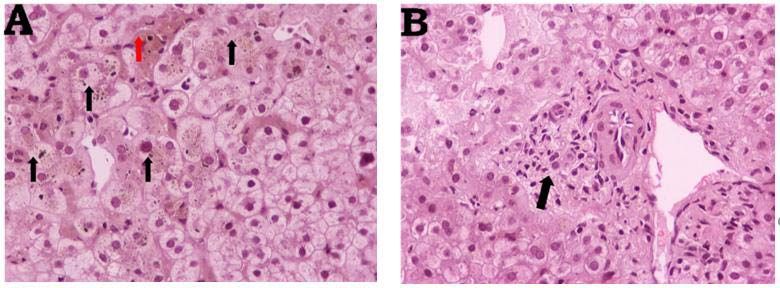
department with jaundice, pruritus and cervical lymphadenopathy of liver biopsy was performed (Figure 1). For that purpose, the levels
one month. Her past medical history is significant for heterozygous of bilirubin needed to be reduced, so the patient was treated with
beta-thalassemia. Physical examination was repeated and confirmed dexamethasone (40mg/day for 4 days). The+ liver biopsy revealed
the existence of jaundice and cervical lymphadenopathy. Her medium to severe centrilobular cholestasis, no neoplastic cells and
abnormal lab tests were: bilirubin 21.5mg/dl [n.v.Papaioannou M Austin Publishing Group
Figure 1: Liver biopsy. A) Significant centrilobular cholestasis, hepatocytes with feathery degeneration and bile pigments in their cytoplasm (black arrows). Red
arrow, histiocyte accumulation with bile pigments and a bile cylinder. B) Portal area with mild lymphocytic infiltration and degenerative alterations of the bile duct
epithelium. Hematoxylin-Eosin stain; 1x200.
Table 1: Causes of Vanishing Bile Duct Syndrome.
Congenital Alagille Sydrome, Cystic Fibrosis, Alpha-1 Antitrypsin Deficiency, Progressive Familial Intrahepatic Cholestasis.
Acquired
Non-FDA approved weight loss supplements, Sertraline, Oxcarbazepine, Chlorpromazine, Temozolomide, Carbamazepine, Interferon
Medications Levofloxacin, Ibuprofen, Sulfamethoxazole-Trimethoprim, Meropenom, Lamotrigine, Valproic Acid, Azithromycin, Moxifloxacin, Mycophenolate
Mofetil, Anabolic Steroids, Allopurinol, Ciprofloxacin, Amoxicillin.
Infections CMV, HCV, HBV, EBV, Cryptosporidium, Reovirus type 3.
Malignancy Lymphoma (Hodgkin's, non- Hodgkin's), Histiocytosis X.
Immunologic Primary Biliary Cholangitis, Primary Sclerosing Cholangitis, Sarcoidosis, GvHD, Hepatic Allograft Rejection.
Not a comprehensive list.
After 6 cycles of treatment, her clinical and biochemical, profile Table 2: Summary of clinical features of patients with VBDS related to Hodgkin’s
lymphoma.
was dramatically improved, while blood and liver function tests were
within normal levels. A PET-CT scan was conducted, confirming the Jaundice 38/38 (100%)
complete metabolic remission of HL. Two months later, a follow-up Pruritus 18/18 (100%)
laboratory test revealed elevated levels of hepatic enzymes (SGOT Weight loss 21/22 (95.45%)
85U/L, SGPT 201U/L, ALP 250U/L, γ-GT 317U/L). For this reason,
Night Sweats 12/16 (71.43%)
ursodeoxycholic acid was administered for 6 months, and liver
biochemistry returned to normal. Although the regeneration of Fever 15/19 (78.95%)
bile ducts was never confirmed via, liver biopsy, due to the patient’s Lymphadenopathy 33/34 (97.06%)
disapproval, recent laboratory tests showed normal levels of hepatic Hepatomegaly 11/25 (44%)
enzymes and consequently normal liver function to this day.
Splenomegaly 10/27 (37.04%)
Discussion
of HL-related VBDS, until recently, including those, which were not
A wide variety of conditions may lead to jaundice in HL since it is confirmed as VBDS via liver biopsy even though there is significant
considered to be a relatively common manifestation among patients evidence that, can prove so.
with HL. These conditions include biliary obstruction secondary to
lymph node enlargement, biliary and hepatic infiltration, hemolysis, We collected the data from 38 confirmed VBDS related to HL
viral illnesses (most commonly CMV and hepatitis), drug toxicity, cases. The mean age of patients is 32 years (range 3.5-75 years) with
Idiopathic Cholestasis (IC) and VBDS [1]. a male predominance (2:1). All patients presented with jaundice,
while other common manifestations were pruritus and weight loss.
VBDS is an uncommon condition characterized by ductopenia, Night sweats and fever appeared in many cases. In nearly half of the
which results in cholestasis and eventually in secondary biliary patients, clinical examination revealed hepatomegaly and in 37%
cirrhosis and liver failure. Apart from HL, VBDS has been associated splenomegaly (Table 2). It is interesting that while almost all patients
with numerous other etiologies, congenital or acquired (Table 1). had lymphadenopathy due to HL, in four cases the diagnosis of VBDS
Bouroncle had noticed the phenomenon of HL-related IC in two preceded the diagnosis of HL, because the pathological lymph nodes
patients with known HL, but without lymphomatous involvement of were not noted initially [4-7]. As a result, VBDS was attributed to
the liver or any other cause for liver dysfunction [2]. Until 1993, when other causes, until HL could be identified. There is one case in which
Hubscher first used the term “vanishing bile duct syndrome” and lymphadenopathy was absent since VBDS appeared six months after
described the association between VBDS and HL [3], all cases were the full remission of HL [8].
attributed to IC and other similar diagnoses. A literature review was The stage of HL was reported in 29 cases. The majority of
conducted, and Supplementary Table 1 presents all published cases them were distributed in stages III and II, 14 and 12 respectively.
Submit your Manuscript | www.austinpublishinggroup.com Ann Hematol Oncol 8(9): id1365 (2021) - Page - 02Papaioannou M Austin Publishing Group
Approximately 25% of all patients had previous or active infections corticosteroids. UDCA is a secondary bile acid. Experimental evidence
from viruses that affect the liver (EBV, CMV, hepatotropic viruses). suggests that UDCA not only stimulates hepatobiliary secretion but
Both total and direct bilirubin levels (as measured on admission in also protects cholangiocytes and hepatocytes against apoptosis and
all patients) were elevated with a mean value of 17.35 mg/dL (n=35) toxicity [15]. Thus, UDCA was administered to 9 out of 39 patients,
and 10.86 mg/dL (n=19). It was observed that in cases, in which full including our own patient (Supplementary Table 1), in order to
remission was achieved, the mean value of total bilirubin was 15.1 improve liver function and allow a liver biopsy or chemotherapy to
mg/dL, while the respective value in deceased patients was 20.01 mg/ be conducted. Corticosteroids have also been used successfully for the
dL. The vast majority of all cases (27/39) were treated with various aforementioned reasons, due to their proven anti-inflammatory effect
chemotherapy regimens (with or without corticosteroids and/or [16,17].
UDCA). In this group, the remission rate was up to 48%. Ten patients
received chemotherapy combined with radiotherapy, with a similar Conventional drug combinations and effective chemotherapeutic
remission rate (5/10). Overall remission rate was calculated as 43.6% doses are often difficult to be administered to patients with HL-related
(17/39), while two patients were awaiting further treatment, at the VBDS, since their liver function is severely affected. Treatment for
time of publication [9,10]. The predominant cause of death was those patients must balance between chemoradiation and the need
hepatic failure (52.6%), followed by sepsis (26.3%). to preserve liver function. The current treatment strategy revolves
around chemotherapy regimens, which are safe for the liver [7]. Once
The etiologic relationship between VBDS and HL is poorly liver function improves, standard regimens can be administered
understood, although it seems that two pathogenic mechanisms [18]. Nevertheless, the available literature includes cases in which full
exist. The first mechanism (direct) involves hepatic infiltration by doses of chemotherapy were used, leading to a plethora of outcomes,
lymphoma cells, leading to bile duct destruction. Hubscher [3] including remission [1,11]. Rota Scalabrini et al. [1] proposed that
continued by suggesting a different, indirect mechanism, which in order to achieve complete remission, an aggressive approach is
can be described as a paraneoplastic phenomenon. According to necessary, consisting of full dose high-intensity chemotherapy. It is
the latter, toxic cytokines released from HL cells, induce a cell- essential that patients, whose liver function is severely affected, are
mediated destruction. There is evidence that biliary epithelial cells
taken into consideration for liver transplantation. However, some
express Major Histocompatibility Complex (MHC), antigens (both
authors suggest that all cases of HL-related VBDS should be treated
class I and II) and adhesion molecules, which respond to cytokines
as potential candidates for transplantation [2,3,11,12,19].
produced by HL. T-lymphocytes adhere to this complex [1,11] and
attack the bile ducts, resulting in their disappearance [12]. Noticeably, Given the presumed immunological reaction causing VBDS,
most of the reported cases specify that a liver biopsy was performed treatments targeting this response have also been attempted. Keeping
prior to treatment, which leads to the conclusion that the second in mind that a humoral immune reaction may be the cause of VBDS,
mechanism may prevail [13]. Hubscher was unable to exclude the Hallen proposed the use of Rituximab, a chimeric monoclonal anti-
lymphomatous hepatic infiltration as the leading cause of VBDS, CD20-antibody used against CD20-positive B-cell lymphomas.
since he had administered chemotherapy before the liver biopsies The subtype of HL that can be treated with Rituximab is the non-
[3]. According to recent investigations, immunoglobulins are found classical nodular lymphocyte predominant, in which the tumor cells
in interlobular bile ducts in some cases, suggesting that a humoral are CD20- positive. Although Rituximab is traditionally used for the
immune reaction may be happening as well [11,10]. treatment of this unusual subtype, it has been reported that it may be
effective against other types of HL as well [11,20]. Rituximab has also
The diagnosis of VBDS can be challenging. Laboratory findings
include nonspecific elevations of alkaline phosphatase, γ-glutamyl been used in two other known cases [9,7].
transpeptidase, alanine aminotransferase and bilirubin levels, all Conclusion
of which compose a clinical profile, characterized by jaundice and
pruritus [13]. Ultrasound, CT-scanning and MRI do not contribute Vanishing bile duct syndrome is considered a rare condition;
significantly to the diagnostic procedure. In order to confirm thus, it can easily elude the attention of health care professionals.
the diagnosis, it is necessary that subsequent liver biopsies reveal Laboratory findings are not pathognomonic and the imaging
intrahepatic cholestasis and severe bile duct destruction. In particular, examinations do not contribute to the diagnostic procedure.
it is defined by the loss of more than 50% of interlobular bile ducts Therefore, raising the index of suspicion is the key to diagnosis. It is
in a pathologic specimen that includes at least 10 portal triads [14]. crucial that practitioners are aware of the association between HL and
Along with the histological evidence, it is important that all other VBDS and the fact that the manifestations of VBDS may precede the
causes for cholestasis are excluded, such as large duct disease, EBV diagnosis of HL.
and CMV infections, viral hepatitis, mechanical obstruction, primary References
biliary cirrhosis and primary sclerosing cholangitis [1]. 1. Rota Scalabrini D, Caravelli D, Carnevale Schianca F, D’Ambrosio L, Tolomeo
F, Boccone P, et al. Complete remission of paraneoplastic vanishing bile duct
Treating the underlying cause is considered the main objective. It syndrome after the successful treatment of Hodgkin’s lymphoma: a case
is crucial that therapy is initiated prior to the histological confirmation report and review of the literature. BMC Res Notes. 2014; 7: 529.
of VBDS, due to the fact that a delayed start may be proven 2. Bouroncle BA, Old JW Jr, Vazques AG. Pathogenesis of jaundice in Hodgkin’s
detrimental to the outcome. Patients with HL are traditionally treated disease. Arch Intern Med. 1962; 110: 872-883.
with the ABVD (Adriamycin, Bleomycin, Vinblastine, Dacarbazine)
3. Hubscher SG, Lumley MA, Elias E. Vanishing bile duct syndrome: a possible
regimen and targeted radiotherapy [14]. Supportive care includes mechanism for intrahepatic cholestasis in Hodgkin’s lymphoma. Hepatology.
ursodeoxycholic acid (UDCA) and immunosuppressants, such as 1993; 17: 70-77.
Submit your Manuscript | www.austinpublishinggroup.com Ann Hematol Oncol 8(9): id1365 (2021) - Page - 03Papaioannou M Austin Publishing Group
4. Ballonoff A, Kavanagh B, Nash R, Drabkin H, Trotter J, Costa L, et al. Hodgkin immunodeficiency preceding the diagnosis of Hodgkin lymphoma. Intern Med
lymphoma-related vanishing bile duct syndrome and idiopathic cholestasis: J. 2014; 44: 1240-1244.
statistical analysis of all published cases and literature review. Acta Oncol.
2008; 47: 962-970. 25. Nader K, Mok S, Kalra A, Harb A, Schwarting R, Ferber A. Vanishing bile
duct syndrome as a manifestation of Hodgkin’s lymphoma: a case report and
5. DeBenedet AT, Berg CL, Enfield KB, Woodford RL, Bennett AK, Northup review of the literature. Tumori. 2013; 99: e164-168.
PG. A case of vanishing bile duct syndrome and IBD secondary to Hodgkin’s
lymphoma. Nat Clin Pract Gastroenterol Hepatol. 2008; 5: 49-53. 26. Aleem A, Al-Katari M, Alsaleh K, AlSwat K, Al-Sheikh A. Vanishing bile
duct syndrome in a Hodgkin’s lymphoma patient with fatal outcome despite
6. Das A, Mitra S, Ghosh D, Modi SK, Roy P, Das J, et al. Vanishing Bile lymphoma remission. Saudi J Gastroenterol. 2013; 19: 286-289.
Duct Syndrome following cytomegalovirus infection in a child with Hodgkin
lymphoma. J Pediatr Hematol Oncol. 2018; 40: 83-84. 27. Umit H, Unsal G, Tezel A, Soylu AR, Pamuk GE, Turgut B, et al. Vanishing
bile duct syndrome in a patient with Hodgkin’s lymphoma and asymptomatic
7. Anugwom C, Goetz G, Hassan M. Vanishing Bile Duct Syndrome preceding hepatitis B virus infection. Acta Gastroenterol Belg. 2009; 72: 277-278.
the diagnosis of Hodgkin lymphoma. ACG Case Rep J. 2020; 7: e00336.
28. Leeuwenburgh I, Lugtenburg EP, van Buuren HR, Zondervan PE, de Man
8. Amer S, Muqeetadnan M, Rahman A, Nusrat S, Hassan S. Vanishing bile RA. Severe jaundice, due to vanishing bile duct syndrome, as presenting
duct syndrome: a rare cause of jaundice in Hodgkin’s lymphoma. Turk J symptom of Hodgkin’s lymphoma, fully reversible after chemotherapy. Eur J
Gastroenterol. 2013; 24: 444-446. Gastroenterol Hepatol. 2008; 20: 145-147.
9. Pass AK, McLin VA, Rushton JR, Kearney DL, Hastings CA, Margolin JF. 29. Schmitt A, Gilden DJ, Saint S, Moseley RH. Clinical problem solving.
Vanishing bile duct syndrome and Hodgkin disease: a case series and review Empirically incorrect. N Engl J Med. 2006; 354: 509-514.
of the literature. J Pediatr Hematol Oncol. 2008; 30: 976-980.
30. Han WS, Jung ES, Kim YH, Kim CH, Park SC, Lee JY, et al. [Spontaneous
10. Greca RD, Cunha-Silva M, Costa LBE, Costa JGF, Mazo DFC, Sevá-Pereira resolution of vanishing bile duct syndrome in Hodgkin’s lymphoma]. Korean J
T, et al. Vanishing bile duct syndrome related to DILI and Hodgkin lymphoma Hepatol. 2005; 11: 164-168.
overlap: A rare and severe case. Ann Hepatol. 2020; 19: 107-112.
31. Córdoba Iturriagagoitia A, Iñarrairaegui Bastarrica M, Pérez de Equiza E,
11. Bakhit M, McCarty TR, Park S, Njei B, Cho M, Karagozian R, et al. Vanishing Zozaya Urmeneta JM, Martínez-Peñuela JM, Beloqui Pérez R. Recuperación
bile duct syndrome in Hodgkin’s lymphoma: A case report and literature ductular en el síndrome de los conductillos biliares evanescent en paciente
review. World J Gastroenterol. 2017; 23: 366-372. con linfoma de Hodgkin [Ductal regeneration in vanishing bile duct syndrome
in Hodgkin’s lymphoma]. Gastroenterol Hepatol. 2005; 28: 275-278.
12. Rossini MS, Lorand-Metze I, Oliveira GB, Souza CA. Vanishing bile duct
syndrome in Hodgkin’s disease: case report. Sao Paulo Med J. 2000; 118: 32. Guliter S, Erdem O, Isik M, Yamac K, Uluoglu O. Cholestatic liver disease
154-157. with ductopenia (vanishing bile duct syndrome) in Hodgkin’s disease: report
of a case. Tumori. 2004; 90: 517-520.
13. Wong KM, Chang CS, Wu CC, Yin HL. Hodgkin’s lymphoma-related
vanishing bile duct syndrome: a case report and literature review. Kaohsiung 33. Kömürcü S, Ozet A, Altundag MK, Arpaci F, Oztürk B, Celasun B, et al.
J Med Sci. 2013; 29: 636-641. Vanishing bile duct syndrome occurring after high-dose chemotherapy and
autologous peripheral stem cell transplantation in a patient with Hodgkin’s
14. LiverTox: Clinical and Research Information on Drug-Induced Liver Injury disease. Ann Hematol. 2002; 81: 57-58.
[Internet]. Bethesda (MD): National Institute of Diabetes and Digestive and
Kidney Diseases. 2012. 34. Ripoll C, Carretero L, Sabin P, Alvarez E, Marrupe D, Bañares R. Colestasis
idiopática asociada a ductopenia progresiva en dos pacientes con linfoma
15. Pusl T, Beuers U. Ursodeoxycholic acid treatment of vanishing bile duct de Hodgkin [Idiopathic cholestasis associated with progressive ductopenia in
syndromes. World J Gastroenterol. 2006; 12: 3487-3495. two patients with hodgkin’s disease]. Gastroenterol Hepatol. 2002; 25: 313-
16. Paumgartner G, Beuers U. Ursodeoxycholic acid in cholestatic liver disease: 315.
mechanisms of action and therapeutic use revisited. Hepatology. 2002; 36: 35. Ozkan A, Yoruk A, Celkan T, Apak H, Yildiz I, Ozbay G. The vanishing bile
525-531. duct syndrome in a child with Hodgkin disease. Med Pediatr Oncol. 2001;
17. Piawah S, Hyland C, Umetsu SE, Esserman LJ, Rugo HS, Chien AJ. A 36: 398-399.
case report of vanishing bile duct syndrome after exposure to pexidartinib 36. Allory Y, Métreau J, Zafrani E. Raréfaction paranéoplasique des canaux
(PLX3397) and paclitaxel. NPJ Breast Cancer. 2019; 5: 17. biliaires interlobulaires au cours d’un cas de maladie de Hodgkin
18. Sathyanarayanan V, Foo WC, Fanale M, Westin J. Deeper insights into [Paraneoplastic vanishing bile duct syndrome in a case of Hodgkin’s disease].
Vanishing Bile Duct Syndrome in Lymphoma: A perplexing entity. Clin Ann Pathol. 2000; 20: 52-55.
Lymphoma Myeloma Leuk. 2016; 16: e65-70. 37. Yusuf MA, Elias E, Hübscher SG. Jaundice caused by the vanishing bile duct
19. Gottrand F, Cullu F, Mazingue F, Nelken B, Lecomte-Houcke M, Farriaux JP. syndrome in a child with Hodgkin lymphoma. J Pediatr Hematol Oncol. 2000;
Intrahepatic cholestasis related to vanishing bile duct syndrome in Hodgkin’s 22: 154-157.
disease. J Pediatr Gastroenterol Nutr. 1997; 24: 430-433. 38. de Medeiros BC, Lacerda MA, Telles JE, da Silva JA, de Medeiros CR.
20. Hallén K, Sangfelt P, Nilsson T, Nordgren H, Wanders A, Molin D. Vanishing Cholestasis secondary to Hodgkin’s disease: report of 2 cases of vanishing
bile duct-like syndrome in a patient with Hodgkin lymphoma - pathological bile duct syndrome. Haematologica. 1998; 83: 1038-1040.
development and restitution. Acta Oncol. 2014; 53: 1271-1275. 39. Crosbie OM, Crown JP, Nolan NP, Murray R, Hegarty JE. Resolution of
21. Fong M, Boyle S, Gutta N. Brentuximab vedotin in combination with sequential paraneoplastic bile duct paucity following successful treatment of Hodgkin’s
procarbazine, cyclophosphamide and prednisolone for the management of disease. Hepatology. 1997; 26: 5-8.
Hodgkin’s lymphoma-associated vanishing bile duct syndrome (VBDS) with 40. Barta SK, Yahalom J, Shia J, Hamlin PA. Idiopathic cholestasis as a
severe obstructive liver failure. BMJ Case Rep. 2019; 12: e227676. paraneoplastic phenomenon in Hodgkin’s lymphoma. Clin Lymphoma
22. Das A, Mitra S, Ghosh D, Modi SK, Roy P, Das J, et al. Vanishing Bile Myeloma. 2006; 7: 77-82.
Duct Syndrome following cytomegalovirus infection in a child with Hodgkin 41. Liangpunsakul S, Kwo P, Koukoulis GK. Hodgkin’s disease presenting as
lymphoma. J Pediatr Hematol Oncol. 2018; 40: 83-84. cholestatic hepatitis with prominent ductal injury. Eur J Gastroenterol Hepatol.
23. Bakhit M, McCarty TR, Park S, Njei B, Cho M, Karagozian R, et al. Vanishing 2002; 14: 323-327.
Bile Duct Syndrome in Hodgkin’s lymphoma: A single center experience and 42. Dourakis SP, Tzemanakis E, Deutsch M, Kafiri G, Hadziyannis SJ. Fulminant
clinical pearls. J Clin Gastroenterol. 2016; 50: 688. hepatic failure as a presenting paraneoplastic manifestation of Hodgkin’s
24. Yeh P, Lokan J, Anantharajah A, Grigg A. Vanishing bile duct syndrome and disease. Eur J Gastroenterol Hepatol. 1999; 11: 1055-1058.
Submit your Manuscript | www.austinpublishinggroup.com Ann Hematol Oncol 8(9): id1365 (2021) - Page - 04Papaioannou M Austin Publishing Group
43. Yalçin S, Kars A, Sökmensüer C, Atahan L. Extrahepatic Hodgkin’s disease 49. Piken EP, Abraham GE, Hepner GW. Investigation of a patient with Hodgkin’s
with intrahepatic cholestasis: report of two cases. Oncology. 1999; 57: 83-85. disease and cholestasis. Gastroenterology. 1979; 77: 145-147.
44. Warner AS, Whitcomb FF. Extrahepatic Hodgkin’s disease and cholestasis. 50. Perera DR, Greene ML, Fenster LF. Cholestasis associated with extrabiliary
Am J Gastroenterol. 1994; 89: 940-941. Hodgkin’s disease. Report of three cases and review of four others.
Gastroenterology. 1974; 67: 680-685.
45. Jansen PL, van der Lelie H. Intrahepatic cholestasis and biliary cirrhosis
associated with extrahepatic Hodgkin’s disease. Neth J Med. 1994; 44: 99- 51. Groth CG, Hellström K, Hofvendahl S, Nordenstam H, Wengle B. Diagnosis
102. of malignant lymphoma at laparotomy disclosing intrahepatic cholestasis.
Acta Chir Scand. 1972; 138: 186-189.
46. Birrer MJ, Young RC. Differential diagnosis of jaundice in lymphoma patients.
Semin Liver Dis. 1987; 7: 269-277. 52. Juniper K Jr. Prolonged severe obstructive jaundice in Hodgkin’s disease.
Gastroenterology. 1963; 44: 199-204.
47. Lieberman DA. Intrahepatic cholestasis due to Hodgkin’s disease. An elusive
diagnosis. J Clin Gastroenterol. 1986; 8: 304-307.
48. Trewby PN, Portmann B, Brinkley DM, Williams R. Liver disease as presenting
manifestation of Hodgkin’s disease. Q J Med. 1979; 48: 137-150.
Submit your Manuscript | www.austinpublishinggroup.com Ann Hematol Oncol 8(9): id1365 (2021) - Page - 05You can also read