Seasnakes and their bites - Anslem de Silva and Malik Fernando - The Sri Lanka Medical Association
←
→
Page content transcription
If your browser does not render page correctly, please read the page content below
1

                                   Seasnakes and their bites
                                    Anslem de Silva and Malik Fernando

Introduction

        Seasnakes have evolved to spend their entire lives in
the sea, including coastal estuaries and brackish lagoons.
Members of one genus (Laticauda, sea kraits) come ashore to
lay eggs, but the majority never leave the water, giving birth to
live young. They were previously grouped in their own family
Hydrophiidae (or in the subfamily Hydrophiinae of the family
Elapidae) but are now included in the family Elapidae together
with the cobras, coral snakes and kraits (Somaweera &                 Jayaindra Fernando - watercolour
Somaweera, 2009). They are all highly venomous, but mostly
non-aggressive, biting usually under provocation. Currently 15
species of true sea snakes (in two genera) are recognised in Sri Lankan
waters (de Silva & Ukuwela, 2017).

Biology
         Sea snakes reported from Sri Lanka                                   Identification
 Hydrophis bituberculatus          Peter’s sea snake
 Hydrophis curtus                  Shaw’s sea snake                            The head is often small in
 Hydrophis cyanocinctus            Annulated sea snake                comparison to the body, with a
 Hydrophis fasciatus               Striped sea snake                  slender neck and fore body. The
 Hydrophis jerdonii                Jerdon’s sea snake                 midbody is large, deep and laterally
 Hydrophis lapemoides              Persian Gulf sea snake             compressed; the belly V-shaped and
 Hydrophis mammilaris              Bombay Gulf sea snake              belly scales reduced except in
 Hydrophis ornatus                 Gray’s sea snake                   Hydrophis viperinus which has fairly
 Hydrophis schistosus              Hook-nosed sea snake               distinct ventral scales in the anterior
 Hydrophis spiralis                Narrow-banded sea snake            half of the body. Tail short, laterally
 Hydrophis stokesii                Stoke’s sea snake                  compressed, paddle-shaped, with a
 Hydrophis stricticollis           Guenther’s sea snake               rounded tip. Most sea snakes are
 Hydrophis platurus                Yellow-belly sea snake             silvery in colour with dark bands, the
 Hydrophis viperinus               Viperine sea snake                 back darker than the belly. The
 Microcephalophis gracilis         John’s sea snake                   notable exception is the yellow-belly
                                                                      sea snake (Hydrophis platurus) that is
                             - after de Silva and Ukuwela, 2017       chocolate brown or black on the back
                                                                      with a bright yellow belly; the tail is
patterned in the same colours [heading image].

        Sea snakes may be confused with fresh water-dwelling water snakes that are occasionally
washed out to sea from rivers. These snakes have rounded bellies, the belly scales being wide (except
in the file snake Acrochordus granulatus). The tail is cylindrical, tapering to a point and not paddle-
2
shaped. They are non-venomous and variously coloured, none silvery with black bands. The file snake or
cloth snake (A. granulatus) of the family Acrochordidae is sometimes found in shallow sea waters and
the dog-faced water snake (Cerberus rynchops) and the glossy marsh snake (Gerarda prevostiana) of the
family Homalopsidae live in the intertidal zones of Sri Lankan coastal waters (Somaweera & Somaweera,
2009).
Marine eels may also be mistaken for sea snakes,
some being silvery with black bands. They are types of fish,
with two pairs of nostrils, one pair being tubular, and a gill
opening on each side of the neck. Most have paired pectoral
fins. Some have a dorsal fin and an anal fin confluent with
the caudal (tail) fin, the tail itself ending in a point. They
have no scales. (Images of sea snakes and eels may be found
on the Guidelines2017 page:-More on identification of
venomous snakes / Pictures of medically important snakes /
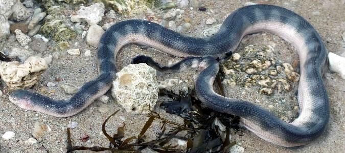
A sea snake washed up on the beach at family Elapidae subfamily Hydrophiinae.)
Mount Lavinia. The head is to the right.
Circa 2 m long.
Photo Naqueeb Hussain Behaviour
Sea snakes generally have a placid disposition, biting
only under provocation, a character first reported many years ago (Reid, 1956, Reid and Lim, 1957).
They are seen by scuba divers unconcernedly inspecting holes and crevices in shallow reefs, searching
for the fish and eels that they prey on. They take no notice of divers around them (personal
observations by MF). The hook-nosed seasnake however, (Hydrophis schistosus, previously Enhydrina
schistosa) is known to be a particularly dangerous animal (Kularatne et al, 2014). Sea snakes are to be
found all around Sri Lanka, but are particularly common in certain areas, such as the Gulf of Mannar and
around Jaffna in the north. During an island wide survey conducted by one of us (AdeS), we were able to
collect specimens of 12 species of sea snakes. Some were confined to specific localities like coastal
brackish lagoons (Hydrophis schistosus), while others were widely distributed (Hydrophis curtus and
Hydrophis spiralis). The yellow-belly seasnake Hydrophis platurus was collected from many localities
around the island; it is known to be the most widely distributed snake species in the world. The
aggressive, highly venomous viperine seasnake Hydrophis viperinus was observed only around the
coasts of
northern Sri
Lanka. As
mentioned
earlier, it is the
only seasnake
found in Sri
Lanka whose Hydrophis viperinus, the distinct anterior ventral scales arrowed
ventral scales are - photo Anslem de Silva
large and
distinct, species of the genus Aipysurus also sharing this character.
Many sea snakes are caught in fishing nets laid from boats, and when hauled aboard have to be
disentangled and thrown overboard (de Bruin, 1983 pers. comm. in Fernando and Gooneratne, 1983).
Fishermen are exposed to bites on their fingers and hands during this process (Karunaratne and
3

Panabokke, 1972) and may be bitten on their feet if accidently stepped on (Kularatne et al, 2014).
However, such bites are rare, as most species appear to be reluctant to bite, and dry bites are common.
The snakes are picked up by the tail and thrown overboard. They are unable to reach up when held by
the tail, unlike a terrestrial snake, because of their weak musculature developed for side-to-side
undulations in water only. At present fishermen around Kalpitiya use a heavy stick they carry in their
boats to kill sea snakes promptly if inadvertently hauled into the boat in the net (personal observations,
AdeS).

Toxinology

        Sea snake venoms are a mixture of various toxic polypeptides, proteins and other substances.
Common toxins in venoms are short-chain neurotoxins (60-62 amino acid residues), long-chain
neurotoxins (66-74 amino acid residues) and phospholipases. These are either neurotoxins or
myotoxins. The former block nerve conduction at the neuromuscular junction, either pre-synaptically or
post-synaptically, and may lead to paralysis and death through asphyxiation caused by failure of the
nerve supply to the diaphragm.
        The myotoxins cause breakdown of muscle tissue releasing myoglobin and creatine kinase in the
process. Myoglobin is excreted by the kidneys but if present in large quantities precipitates as plugs in
the tubules and causes extensive necrosis and kidney damage. Some toxins have multiple effects and act
both as neurotoxins and myotoxins (Tamiya et al, 1983). This study was based on the venoms of two sea
snakes viz. Hydrophis ornatus and Hydrophis lapemoides. A more recent publication is a systematic
review of references to the toxins of Hydrophis schistosus, Hydrophis cyanocinctus, Hydrophis
lapemoides, Hydrophis spiralis, and Lapemis curtus (Mohebi et al, 2016). The authors concluded thus:
“There is scant variability in the venom composition in the same and different species of sea snakes. Our
study revealed that there is a rather simple venom profile with an affinity towards a lethal mixture of
high abundance of neurotoxins and PLA2s, and lower amounts of toxins such as CRISP, SVMP and
LAAO1”.

Epidemiology
         Seasnake-human conflict occurs predominantly in estuaries and lagoons, particularly with the
dangerous hook-nosed sea snake. The victims are usually
fishermen but bathers and swimmers in estuaries and river
mouths may also be bitten (Reid & Lim, 1957). Sea snake bites               Seasnake bites are
are encountered infrequently in Sri Lanka, with non-                      painless with no local
envenoming ‘dry bites’ being frequent (Somaweera and                           inflammation.
Somaweera, 2009). Kularatne et al (2014) in their paper had            Puncture marks with rapid
this to say: “The first victim was bitten on a finger but despite a    onset      of       pain       and
bleeding injury he continued to work because ‘his fellow               inflammation would be due
fishermen reassured him that sea snakes of this sort are               to a fish or sea urchin sting
abundant in the area, are frequently trapped in nets and bite
                                                                                   [Cite your source here.]
people with no untoward effects’. Almost all of the bites were said to be asymptomatic     with few, if any,
needing hospital treatment.”

PLA = phospholipase A2, CRISP = cysteine-rich secretory protein, SVMP = snake venom Zn2+-
1

metalloproteinase , LAAO = L-amino acid oxidase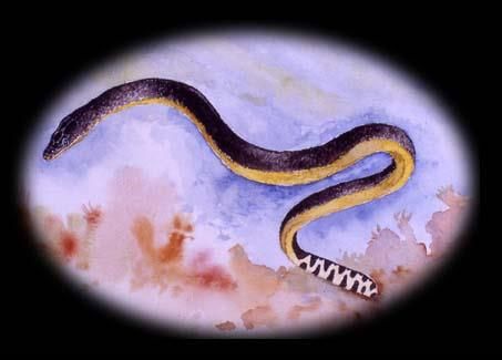
4

         Sea snake bites are characteristically painless with no inflammation unlike stings by venomous
fish that are very painful with inflammation. Sea urchin stings are also painful, the spine remnants being
often visible in the puncture holes. Although sea snake bites are reportedly painless, this may not always
be true as shown in the first reported case of sea snake bite reported from Sri Lanka (Amarasekera et al,
1994). This case was notable in that the authors commented as follows “… unusual features observed in
our patient were the occurrence of pain at the site of the bite, regional lymph node enlargement and
absence of muscle pain and tenderness.”

      Reports of sea snake bites in Sri Lanka are few. The literature has been summarised by
Somaweera & Somaweera, 2009 with an additional report by Kularatne et al in 2014 (see Annex I).

Clinical manifestations

        Early investigations of sea snake envenoming were by H. A. Reid and his co-workers in Malaysia
and the surrounding area in the nineteen-fifties. Their findings have been summarised in the 1983 CMJ
publication on snakebite (Fernando & Gooneratne, 1983). Kularatne et al, 2014 point out that recent
studies have shown that there are two distinct types of sea snake envenoming: myotoxic envenoming
and dominant flaccid paralytic envenoming, “the latter mediated by the neurotoxins that are found in
abundance in the venom of many species of sea snakes (White, 1995; Komori et al, 2009; Takasani,
1998; Tamiya et al, 1983)”.

        There are seven published reports of sea snake bite in Sri Lanka that describe signs and
symptoms and the outcome. (See ANNEX I for a summary of published reports.) The spectrum of clinical
effects seen have been varied, including both myotoxic and neurotoxic effects, as well as no signs of
envenoming.

        Many bites do not cause envenoming. Symptoms can be mild with spontaneous resolution in a
few days while others would result in systemic envenoming that needs aggressive management.
Symptoms are usually seen between 30 and 60 minutes. If no symptoms are seen 2 hours after the bite,
serious envenoming can be ruled out. Symptoms start as aching, stiffness, slight or moderate muscle
movement pains involving the neck, trunk and limbs. Patients will be reluctant to move because of pain
as a result of rhabdomyolysis. Hyaline necrosis involves the muscle fibres only, leaving the sarcolemmal
sheaths unaffected. With resolution the muscle fibres re-grow within the original sarcolemmal sheaths
with minimal scarring and therefore there are no long-term effects attributable to muscle involvement
(Marsden & Reid, 1961).

        True paresis can also occur initiated by the neurotoxins. This will be usually flaccid, with
diminished or absent tendon reflexes. Trismus, dysphagia and dysarthria may occur, as well as ptosis,
ophthalmoplegia and respiratory muscle paralysis. Hypertension and renal failure can be seen.

          The summary of symptoms and signs described above are taken from the publications of Reid
and his co-workers. Many of these features have been seen in the cases described by Sri Lankan
workers. They are shown in tabular form, together with laboratory findings in Box A. The sequence of
appearance of the symptoms and signs and the laboratory findings give an indication of the expected
clinical progress and the possible need for aggressive therapies.
5
BOX A
Summary of symptoms, signs and investigations in seasnake envenoming
Based on the work of H. A. Reid
Trivial envenoming: About 10% of bite victims; No treatment needed, will resolve within 3 days
without specific seasnake antivenom.
Aching, stiffness, slight or moderate muscle No leucocytosis, raised AST, proteinuria.
movement pains involving neck, trunk, limbs.
Serious envenoming: About 20% of bite victims; Treatment needed, preferably with seasnake
antivenom.
Aggravated and rapidly increasing aching, Leucocytosis, raised ALT, AST, hyperkalaemia,
stiffness. Severe muscle movement pains. proteinuria, microscopic haematuria,
Paresis - usually flaccid with diminished or myoglobinuria, ECG changes.
absent tendon reflexes. Trismus, dysphagia,
dysarthria. Ptosis, ophthalmoplegia, respiratory
muscle paralysis. Hypertension. Renal failure.
After Fernando & Gooneratne, 1983, CMJ 28/3 p. 137
Management
Management of a sea snake bite victim is conservative as there is no effective antivenom
available in Sri Lanka. The locally available polyvalent antivenom should not be administered. Specific
sea snake antivenom is manufactured in Australia by the Commonwealth Serum Laboratories (CSL) that
is reported to be effective against the venom of a number of sea snake species found in Sri Lankan
waters. (Details in the on-line CSL Antivenom Handbook on their website; see Bibliography for URL.)
Observe the victim for the development of any of the following, in the meantime ensuring good
hydration with adequate urine flow.
Myoglobinuria — Myoglobinuria is associated with necrosis of striated muscle that presents
as stiffness and pain on attempted movement, particularly involving the jaw and neck muscles. It turns
the urine red-brown to black in colour, the depth of colour being proportionate to the amount of
myoglobin being excreted. This is confirmed spectroscopically. Resolution of the condition will be
indicated by a progressive lightening of the colour in serial samples of urine. Prolonged and high levels
of myoglobinuria lead to myoglobin casts in the tubules with distal tubular necrosis and acute renal
failure (Marsden & Reid, 1961; Sitprija et al, 1971). More recently it has been postulated that the6
mechanisms that lead to kidney injury are direct venom effects as well as indirect effects such as
myoglobinuria (Pickwell, 1994; Kularatne et al, 2014). Kularatne et al (2014) point out that Sitprija et al
(1971) had reported two cases of severe myonecrosis and acute renal failure successfully managed with
early use of haemodialysis. This is an option that should be borne in mind. Hyperkalaemia and
cardiovascular collapse secondary to severe venom-induced rhabdomyolysis is well known and
anticipation of such cardiovascular collapse is important (Kularatne et al, 2014). The blood chemistry
and the ECG should be closely monitored. Hyperkalaemia can be treated with calcium gluconate,
dextrose with insulin, salbutamol or sodium bicarbonate. Haemodialysis is also effective but may not
always be practical.
Paresis— Paresis should be distinguished from the reluctance to use voluntary muscles due to
the pain of myonecrosis, described by Reid as “muscle movement pain”. Paresis is usually flaccid with
diminished or absent tendon reflexes. If involving respiratory muscles mechanical ventilation is
indicated.
Laboratory investigations— Laboratory investigations that are useful in a case of sea snake
envenoming are set out in the table in Box A, which is based on Reid’s publications. One that is not in
that list is creatine kinase (CK). Raised CK levels will be seen when there is rhabdomyolysis. Congestion
and centrilobular necrosis of the liver was a common finding reported by Marsden & Reid, 1961 and
Karunaratne & Panabokke, 1972 (Kularatne et al, 2014), accounting for raised liver enzymes ALT and
AST. A rise in AST alone indicates muscle necrosis and will be seen in the milder form of envenoming.
ECG changes—Reid has reported two possible changes in the ECG: where there is
hyperkalaemia the characteristic T-wave changes will be seen (such as prolongation of the PR interval
and development of peaked T-waves) and in others, changes indicative of right ventricular dominance (a
dominant R wave in chest lead V4R 2). The serum potassium level at which ECG changes occur is said to
be variable and interpretation of the recording can be difficult, so these changes should not be relied on
to detect hyperkalaemia.
Anslem de Silva MSc, DSc (Honoris Causa) Malik Fernando MBChB (Bristol), MIOB (SL)
University of Peradeniya Retired Physician
Herpetologist Past President
Regional Chairman IUCN Crocodile Sri Lanka Medical Association
Specialist Group for South Asia and Iran,
Co-Chair IUCN Amphibian Specialist Group
for Sri Lanka
April 2018
Acknowledgements: We wish to thank Dr. Kanishka Ukuwela and Prof. S. A. M. Kularatne for
reviewing the text and making helpful comments.
2
Chest lead V4R is a lead placed on the right side of the chest in the same anatomical location as the left-sided lead
V4 in the standard 12-lead ECG.7 Bibliography Amarasekera N, Jayawardena A, Ariaratnam A, Hewage UC, de Silva A (1994). Bite of a sea snake (Hydrophis spiralis): a case report from Sri Lanka. J Trop Med & Hyg, 97(4):195-198] CSL Sea Snake Antivenom: http://www.toxinology.com/generic_static_files/cslavh_antivenom_seasnake.html Das I, de Silva Anslem (2011). A Photographic Guide to Snakes and other Reptiles of Sri Lanka. New Holland, UK, 144 pp. de Silva A, Kanishka Ukuwela, (2017). Reptiles of Sri Lanka, Sri Lanka Edition, 176 pp, Vijitha Yapa, Colombo. de Silva Anslem (1994). An account of the sea snakes (Serpentes: Hydrophiidae) of Sri Lanka. IN: Sea Snake Toxinology, (Editor) P. Gopalakrishnakone, National University of Singapore, pp. 234-249. de Silva Anslem (2011). Sea snake survey of Sri Lanka. Sea Snake Specialist Group Newsletter, 1: 1-2. de Silva Anslem, Ukuwela Kanishka, Sivaruban Abyerami, Sanders Kate L (2011). Preliminary observations on the reproductive biology of six species of Sri Lankan sea snakes (Elapidae: Hydrophiinae). Salamandra,47(4) 193–198. de Silva Anslem, Sivaruban A, Ukuwela Kanishka, Rasmussen Arne R, Sanders Kate L (2011). First record of a sea snake (Lapemis curtus) feeding on a Gastropod. Herpetology Notes, volume 4: 373-375 (published online on 2 November 2011). Fernando Malik, Gooneratne Walter (1983). Sea-snake Envenoming, CMJ, 28, 131-143. Jahubar M, Subramanium A, James RF (1984). An analysis of snake bite in Base Hospital Mannar. 2ndAnnual Sessions of the Jaffna Medical Association: 9. Karunaratne KEdeS, Panabokke RG (1972). Sea-snake poisoning - Case Report, J Trop Med & Hyg, 75 (5), 91. Marsden ATH, Reid HA (1961). Pathology of Sea-snake Poisoning.Br Med J1, 1290-1293. Komori Y, Nagamizu M, Uchiya K, Nikai T, Tu Anthony (2009). Comparison of sea snake (Hydrophiidae) neurotoxin to cobra (Naja) neurotoxin. Toxins 1, 151–161. Kularatne SAM, R. Hettiarachchi, J. Dalpathadu, ASV Mendis, PDSAN Appuhamy, HDJ Zoysa, K Maduwage, VS Weerasinghe, A de Silva (2014). Enhydrina schistosa (Elapidae: Hydrophiinae) the most dangerous sea snake in Sri Lanka: Three case studies of severe envenoming, Toxicon77, 78–86. Mohebbi G, Seyedian R, Nabipour I (2016). The toxinology of sea snakes: A systematic review. Iran South Med J, 19(4), 662-703.
8

Pickwell VG (1994). A review of contemporary sea snake toxinology: chemistry, pharmacology,
immunology and clinico-pathological aspects. In: Gopalakrishnakone, P. (Ed.), Sea Snake Toxinology.
Singapore University Press, Singapore Read, pp. 93–166.

Reid HA (1956). Sea-snake bite research, Trans R Soc Trop Med & Hyg, 50, 517.

Reid HA, Lim KJ (1957). Sea-snake bite, a survey of fishing hazards in N. W. Malaya, Br Med J, 2, 1266.

Senanayake M P, Ariaratnam CA, Abeywickrema S, Belligaswatte A (2005). Two Sri Lankan cases of
identified sea snake bites, without envenoming. Toxicon, 45: 861-863.

Sitprija V, Sribhibhadh R, Benyajati C (1971). Haemodialysis in poisoning by sea snake venom. Br Med J 3,
218–219.

Somaweera Ruchira, Somaweera Nilusha (2009). An overview of Sri Lankan sea snakes with an
annotated checklist and a field key, Taprobanica, 1/1, 43-54, 3 pls.

Takasani C (1998). The Toxinology of sea snake venom. J. Toxicol -Toxin Rev 17, 361–372.

Tamiya N, Maeda N, Cogger HG (1983). Neurotoxins from the venoms of the sea snakes Hydrophis
ornatus and Hydrophis lapemoides. Biochem J 312, 31–38.

Thanabalasundrum RS, Vidyasagara NW (1969). Snake bites and its treatment. CMJ 14 (4): 188-191.

Ukuwela, Kanishka, de Silva Anslem, Mumpuni, Fry BG, Lee MSY, Sanders Kate L (2013). Molecular
evidence that the deadliest sea snake Enhydrina schistosa (Elapidae: Hydrophiinae) consists of two
convergent species. Molecular Phylogenetics and Evolution 66: 262–269.

Ukuwela, KDB, De Silva A, Mumpuni, Fry BG, Sanders KL (2014). Multi-locus phylogeography of the sea
snake Hydrophis curtus reveals historical vicariance and cryptic speciation. Zoologica Scripta. Royal
Swedish Academy of Sciences, 43, 5, pp 472–484.

Ukuwela, KDB, de Silva A, Lee MSY, Sanders KL (2016). Diversity, systematics and conservation of the
marine snakes of Sri Lanka. Proceedings of the annual Session – Wildlanka Symposium. (Abstracts). 23 &
24 August, 2016: p 44-45.

Vithanage KK, Thirumavalavan K (2012). A case of a sea snake bite resulting in fatal envenoming, CMJ 57:
174-175.

White J (1995). Clinical toxicology of sea snakebites. In: Meier J, White J (Eds.), Handbook of Clinical
Toxicology of Animal Venoms and Poisons. CRC Press, Boca Raton (FL), pp. 159–170.
9

                  ANNEX I - Summary of reported seasnake bites in Sri Lanka

Jahubar et al, 1984 & Subramaniam & James, 1985; Reports of three bites in fishermen at Mannar
during a period of five months.
Amarasekera et al, 1994; Hydrophis spiralis; Pain at the bite site, regional lymph node enlargement
and absence of muscle pain and tenderness.
Karunaratne & Panabokke, 1972; Un-identified, Pelamis platurus ?; Adult fisherman; Ptosis, difficulty
in talking, swallowing but no heart or respiratory difficulties. Later developed severe pains, renal
failure, hyperkalaemia which lasted for 24 days, and the patient died.
Senanayake et al, 2005; Pelamis platurus; 7-year-old boy; No local or systemic effects recorded other
than a 2.5cm linear scratch mark. Hospitalised for one and a half days.
Senanayake et al, 2005; Enhydrina schistosa; 39-year-old male; Mild redness around two bite marks
but no pain or local/ systemic effects. Only prescribed tetanus toxoid.
                                                                After Somaweera & Somaweera, 2009
Kularatne et al, 2014; Enhydrina schistosa; Three lagoon fishermen: a 26-year-old fisherman, severe
myalgia with very high creatine kinase (CK) levels lasting longer than 7 days, a 32-year-old fisherman,
gross myoglobinuria, high CK levels and hyperkalaemia, both recovering; a 41-year-old man who trod
on a sea snake in a river mouth, severe myalgia seven hours later, severe rhabdomyolysis, died three
days later due to cardiovascular collapse.
                                                                                 Kularatne et al, 2014
You can also read