Heterogeneity of Brain Structural Variation and the Structural Imaging Endophenotypes in Schizophrenia
←
→
Page content transcription
If your browser does not render page correctly, please read the page content below
Neuropsychobiology 2012;66:44–49                                 Received: July 5, 2011
                                                                                                                    Accepted after revision: March 29, 2012
                                                   DOI: 10.1159/000338547
                                                                                                                    Published online: July 13, 2012

Heterogeneity of Brain Structural
Variation and the Structural Imaging
Endophenotypes in Schizophrenia
Igor Nenadic Christian Gaser Heinrich Sauer
Department of Psychiatry and Psychotherapy, Jena University Hospital, Jena, Germany

Key Words                                                              tal changes, but show divergence in structural deficits in
Endophenotype ⴢ Hippocampus ⴢ Intermediate                             other areas such as the thalamus, hippocampus, or cerebel-
phenotype ⴢ Magnetic resonance imaging ⴢ Prefrontal                    lum. Altogether, these studies demonstrate that brain struc-
cortex ⴢ Schizophrenia                                                 ture per se is not a uniform endophenotype, but rather a
                                                                       combination of regional deficits highly heterogeneous in
                                                                       both meeting endophenotype criteria as well as in their dis-
Abstract                                                               tribution within the disease category.
Schizophrenia is often assumed to comprise a group of bio-                                                             Copyright © 2012 S. Karger AG, Basel
logically distinct disorders, yet it has been difficult to dissect
subgroups using biological markers. We review recent brain
imaging morphometry studies addressing the issue of het-                   Clinical Heterogeneity and Subtypes of
erogeneity within the diagnostic category of schizophrenia.                Schizophrenia
Studies of subgroups of schizophrenia patients have mostly
used either symptom structure or clinical course for the de-               Kraepelin’s original concept of dementia praecox orig-
lineation of potentially meaningful subgroups. Studies de-             inates in a synthesis of the previously separately classified
fining subgroups according to outcome, i.e. good versus                disorders of hebephrenia, catatonia, and dementia para-
poor outcome (or ‘non-Kraepelinian’ vs. ‘Kraepelinian’, re-            noides. Hence, although the ‘Kraepelinian dichotomy’
spectively) have shown that while these two subgroups                  separating schizophrenia from manic-depressive illness
might overlap in the extent (and possibly also strength) of            provides a delineation of psychotic disorders in two large
prefrontal deficits, they differ in temporal and occipital ar-         groups, it also relies on grouping together entities previ-
eas, where poor-outcome patients show stronger deficits.               ously conceived of as distinct. Similarly, Eugen Bleuler’s
More recent studies have investigated subgroups of schizo-             conception of the ‘group of schizophrenias’ acknowledg-
phrenia based on factor analysis of psychopathology. They              es the clinical (and putatively etiological) heterogeneity of
have demonstrated a complex pattern of regional changes,               schizophrenia. However, there has been only limited suc-
where the typical three subgroups might overlap in prefron-            cess in delineating different types of schizophrenia on

                          © 2012 S. Karger AG, Basel                   Dr. Igor Nenadic
                          0302–282X/12/0661–0044$38.00/0               Department of Psychiatry and Psychotherapy
Fax +41 61 306 12 34                                                   Jena University Hospital, Philosophenweg 3
E-Mail karger@karger.ch   Accessible online at:                        DE–07743 Jena (Germany)
www.karger.com            www.karger.com/nps                           Tel. +49 3641 939 0127, E-Mail igor.nenadic @ uni-jena.de
clinical grounds, either cross-sectional psychopathology tial resolution, for example by making use of anatomical
or course of disease. Even though the current classifica- likelihood estimation techniques [2]. The pattern of brain
tion in DSM-IV (and similarly in ICD-10) defines sub- structural changes in these studies typically involves the
types of paranoid, disorganized, catatonic, undifferenti- medial temporal lobe (the hippocampus, amygdala, and
ated, and residual schizophrenia, it is far from being clear partially also the parahippocampal cortex), the superior
whether these prototypes correspond to distinct biologi- temporal cortex (including both the superior temporal
cal entities or disease mechanisms. gyrus and the transverse temporal gyrus/gyri or Heschl
Biological research on schizophrenia today is mostly gyrus), as well as the thalamus, medial and lateral parts
directed at unveiling potential biological markers or en- of the prefrontal cortex, and the insula.
dophenotypes, or identifying common pathways leading Following the endophenotype strategy laid out by
to the disease. As both the phenotype and genotype ap- Gottesman and Gould [1], one of the major goals of iden-
pear to be complex in nature, the heterogeneity of schizo- tifying an endophenotype is the ability to delineate a sub-
phrenia is one of the main problems in understanding the group of patients, which might then be linked to a par-
relation between putative genetic markers and their ex- ticular genotype (more) specific to this subgroup than the
pression into a clinically identifiable phenotype (e.g. a whole group of patients. The above set of anatomical re-
combination of symptoms, disease course, etc.). Hence, gions, therefore, might reliably differ between schizo-
the heterogeneity of biological abnormalities is a core phrenia and healthy control subjects, but we do not know
problem in identifying reliable biological markers. It is, whether it might actually be a subset of patients that con-
therefore, surprising that there are relatively few studies tributes more to this association than others. If brain im-
investigating potential subgroups of schizophrenia ac- aging markers were useful to ‘deconstruct’ the biological
cording to the distribution of a particular biological basis of the disorders, we would not only need a better
marker or endophenotype. understanding of the association with different sub-
In this paper, we aim to provide a selective review on groups, but also consider that this association might vary
studies using magnetic resonance imaging (MRI) to as- across implicated brain regions [3].
sess brain structural differences in subgroups of schizo- While the delineation of subgroups might be possible
phrenia. For the discussion of subgroups, our review fo- using either phenotype or genotype, we will limit our-
cuses selectively on recent morphometry studies that selves to an overview of studies using the former ap-
have studied subgroups or ‘subsyndromes’ using mor- proach: firstly, those studies linking single symptoms to
phometric techniques. For this purpose, we conducted a brain structure; secondly, those using dichotomies such
Medline-based literature search using the phrases schizo- as clinical course/outcome to define subgroups, and
phreni* and (subtype OR subgroup OR subsyndrome) as thirdly, a few more recent studies using psychopathology
well as the phrase ‘Kraepelinian’; from the identified pa- ratings to define subgroups of schizophrenia.
pers, we selected those that compared different schizo-
phrenia samples with different phenotypic/clinical char-
acteristics, hence not including those that compared only Correlations of Symptoms and Brain Structure
one putative schizophrenia subtype (e.g. deficit schizo-
phrenia) to controls. A rather large number of morphometry studies have
aimed to link specific symptoms of schizophrenia to
brain structure, either using correlations (e.g. severity of
Brain Imaging as a Schizophrenia Endophenotype a symptom correlated with volume) or a dichotomous
variable (e.g. hallucinating vs. nonhallucinating patients).
Brain structural changes as detected with MRI have In earlier studies, such associations were often performed
been put forward as a putative endophenotype for schizo- in an exploratory fashion, and many findings were not
phrenia. In fact, they appear to meet most of the stringent replicated in subsequent studies. A few symptoms such as
criteria for endophenotypes [1], e.g. being associated with auditory hallucinations and formal thought disorder
the disorder, heritable (as shown in twin studies), and rel- have more consistently been linked to the superior tem-
atively stable over the course of disease. Also, there are poral cortices in several volumetric MRI studies [4].
now several meta-analyses, especially on studies employ- While a subsequent parcellation study linked anterior
ing voxel-based morphometry (VBM). These provide ev- parts of the left superior temporal cortex to hallucina-
idence for the regional distribution with increasing spa- tions [5], our own studies using deformation-based mor-
Heterogeneity in Schizophrenia Neuropsychobiology 2012;66:44–49 45phometry and VBM have provided evidence for the re- show stronger progressive volume loss in poor-outcome
gional specificity of a part of Heschl gyrus showing a sig- patients [15]. In a cross-sectional study using atlas-based
nificant correlation with the current severity of auditory parcellation, approximating the volume of cortical Brod-
hallucinations in schizophrenia patients [6, 7]. While rep- mann regions, the authors found both good- and poor-
licated in a smaller sample [8], a recent VBM study assess- outcome patients to overlap in volume reductions of lat-
ing this correlation in a sample of hallucinating patients eral prefrontal areas (BA 45, 44, 46, 47), while differences
(the previous studies had included patients irrespective of between these patient subgroups appeared more in the
the presence or history of hallucinations) did not find an occipital cortices, the superior parietal, and certain me-
association with the left superior temporal cortex [9]. dial temporal/parahippocampal regions [16]. While this
Similarly, studies comparing subgroups of patients based finding is somewhat surprising, as some areas discrimi-
on a history of hallucinations have shown rather diver- nating the subgroups do not show strong effects of diag-
gent findings regarding the superior temporal and pre- nosis per se, it is partially replicated in a recent VBM
frontal cortices [10, 11]. study where the ‘Kraepelinian’ subgroup of patients
While these studies provide an interesting heuristic showed stronger grey matter deficits in the basal ganglia
approach to certain common pathways in the expression and occipital cortices [17].
of a phenotype, they also illustrate the limitations of us- Taken together, these findings provide evidence that
ing single symptom correlations to resolve heterogeneity poor outcome is associated with stronger deficits (and
across the schizophrenia population. One challenging possibly also accelerated volume loss over time). Howev-
problem is that many symptoms are in fact highly corre- er, for the purpose of delineating endophenotypes, there
lated to others, especially within the group of positive and are several limitations. As this dichotomy makes use of
negative symptoms, respectively. For example, auditory clinical variables related to outcome, it might instead be
hallucinations are often highly correlated with the pres- a categorization of a dimensional marker, reflecting over-
ence or severity of delusional symptoms. Although statis- all disease severity. This might bear little relation to bio-
tical approaches have included the removal of variance logically valid discriminator markers.
related to other positive symptoms [7], findings will in- A second approach has been to divide schizophrenia
evitably vary according to the degree of intercorrelation samples into subgroups based on factor analysis of psy-
in the sample studied. Also, no brain region appears to chopathology ratings. This goes back to some classical
show an exclusive correlation to a particular symptom. It studies by Liddle et al. [18], who identified three syn-
appears, therefore, that single symptoms might not be a dromes of schizophrenia, termed psychomotor poverty,
reliable phenotype marker in providing an additional ex- disorganization, and reality distortion, which were relat-
planation for the heterogeneity of brain structure in ed to different patterns of cerebral blood flow. Recently,
schizophrenia. two large VBM studies have used factor analysis to divide
their schizophrenia patient samples into three subgroups
with predominantly negative, disorganization, and para-
Imaging Findings in Subgroups/Subsyndromes of noid symptom profiles [19, 20]. The three-factor solution
Schizophrenia is an often-replicated delineation of subgroups based on
psychopathology, which is also replicated in chronic and
Two alternative approaches have been used in a series old-age populations [21]. Both VBM studies, although
of recent studies to address the issue of heterogeneity of differing in several details, showed that there is consider-
brain structure in schizophrenia. The first approach is able heterogeneity of spatial distribution and extent of
based on using clinical parameters of outcome to classify structural deficits across the three schizophrenia sub-
patients into poor-outcome (also termed ‘Kraepelinian’ groups. Our own study showed that the areas of overall
type) or good-outcome (‘non-Kraepelinian’) groups. This disease activity of all subgroups were mostly in the pre-
dichotomy takes into account a number of clinical as- frontal areas, while the thalamus, superior temporal cor-
pects, including longitudinal aspects, and has been used tices, and cerebellum were only affected in one or two
in a series of imaging studies by Mitelman and Buchs- subgroups [20] (fig. 1). In an extension of that study, we
baum [12]. Cross-sectional MRI studies indicate that sev- also assessed whether the three subgroups showed differ-
eral brain areas show stronger reductions in poor-out- ent age-related progression (i.e. an interaction of group by
come versus good-outcome patients, including the thala- age in a cross-sectional design), demonstrating that the
mus [13] and cingulate [14], while areas like the putamen paranoid and to some extent also the negative subgroup
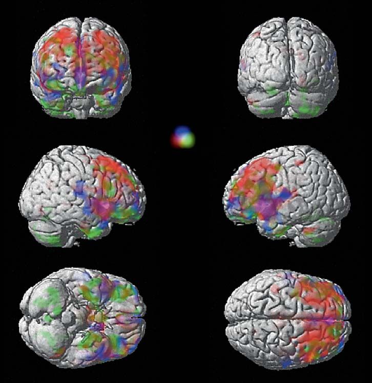
46 Neuropsychobiology 2012;66:44–49 Nenadic/Gaser/SauerColor version available online
Paranoid
Negative Disorganized
Fig. 1. VBM comparison of schizo-
phrenia subgroups versus healthy
controls (p ! 0.01, FDR-corrected for
multiple comparisons; extent thresh-
old p = 0.05 [for details and original
analyses, see 20]): composite image of
group-wise comparisons with nega-
tive subgroup (red), disorganized
subgroups (green), and hallucinatory
subgroup (blue); intermediate colors
reflect overlap (superimposed on cor-
tical surface of a single subject T1
scan). Colors refer to the online ver-
sion only.
showed a stronger decline in the superior temporal and increasing number of imaging studies in prodromal
some smaller lateral prefrontal areas compared to healthy schizophrenia and the early detection of the disease,
control subjects [22]. which would depend on a genetically mediated marker
The latter studies clearly demonstrate that our under- (or pattern) that is relatively stable to changes of clinical
standing of brain structure as a putative endophenotype state and/or medication. An example of the model is giv-
will have to consider a significant variability of effects en in figure 2.
both within single areas as well as across the different
brain regions.
Based on the reviewed findings, we put forward the Conclusions
hypothesis that regional brain structural changes could
be considered as separate putative endophenotypes, each Brain structural alterations are among the most prom-
linked to a specific form of schizophrenia. Pursuing such ising endophenotypes for schizophrenia. However, an
a strategy could ultimately allow for the delineating of overview of recent studies exploring the heterogeneity of
subtypes of schizophrenia based on a biological marker brain structural patterns in subgroups of schizophrenia
with stable endophenotype properties. For use as a bio- clearly emphasizes the fact that different subgroups of pa-
logical marker, additional studies would be useful to in- tients show different degrees and extent of structural def-
dicate a biological subtype of the disease based on a de- icits, and that some regional effects might only be evident
tected brain structural pattern (i.e. a combination of re- in a subset of patients. The anatomical set of regions de-
gional deficits). This would also have implications for the rived from meta-analyses is, therefore, a suitable indica-
Heterogeneity in Schizophrenia Neuropsychobiology 2012;66:44–49 47Genotype
Endophenotype MRI ... PFC thal STG ...
Phenotype Sz Sz1 Sz2 Sz3 Sz4
Fig. 2. Brain-imaging endophenotypes and biologically distinct sibly specific) relation to a particular imaging endophenotype,
subgroups in schizophrenia. The diagram illustrates the problem e.g. a particular brain structure such as the prefrontal cortex
of identifying endophenotypes in brain imaging. While a simple (PFC), the thalamus (thal), or the superior temporal gyrus (STG).
genotype-phenotype relation (left side model) has been refuted Each of these imaging endophenotypes, however, might also dif-
for schizophrenia, many approaches rely on the identification of ferentially be influenced by the genotype, e.g. specific risk genes.
multiple endophenotypes for schizophrenia (middle model). The dotted lines (right side model) indicate potential different
However, taking into account that there might be different sub- boundaries of diagnostic classification systems, which might in-
groups of schizophrenia (right side model; subgroups delineated clude different biologically related subgroups into disease entities
as Sz1–4), each of these subgroups might have a different (and pos- like ‘schizophrenia’.
tor of the general pattern of brain structural alteration in ‘deconstruction’ of biologically meaningful subtypes of
schizophrenia, but not a useful schizophrenia endophe- the disease. Finally, such viable intermediate phenotypes
notype per se. Rather, specific brain regions or a combi- might then be linked to a specific genotype, which might
nation thereof (i.e. anatomical patterns) might show suit- provide a clearer link between genetic variation and phe-
able endophenotype characteristics that would allow the notypes [23].
References
1 Gottesman II, Gould TD: The endopheno- 4 Sun J, Maller JJ, Guo L, Fitzgerald PB: Supe- 7 Nenadic I, Smesny S, Schlosser RG, Sauer H,
type concept in psychiatry: etymology and rior temporal gyrus volume change in schizo- Gaser C: Auditory hallucinations and brain
strategic intentions. Am J Psychiatry 2003; phrenia: a review on region of interest volu- structure in schizophrenia: voxel-based
160:636–645. metric studies. Brain Res Rev 2009;61:14–32. morphometric study. Br J Psychiatry 2010;
2 Fornito A, Yucel M, Patti J, Wood SJ, Pantelis 5 Takahashi T, Suzuki M, Zhou SY, Tanino R, 196:412–413.
C: Mapping grey matter reductions in Hagino H, Kawasaki Y, Matsui M, Seto H, 8 Neckelmann G, Specht K, Lund A, Ersland L,
schizophrenia: an anatomical likelihood es- Kurachi M: Morphologic alterations of the Smievoll AI, Neckelmann D, Hugdahl K: MR
timation analysis of voxel-based morphom- parcellated superior temporal gyrus in morphometry analysis of grey matter vol-
etry studies. Schizophr Res 2009; 108: 104– schizophrenia spectrum. Schizophr Res ume reduction in schizophrenia: association
113. 2006;83:131–143. with hallucinations. Int J Neurosci 2006;116:
3 Braff DL, Freedman R, Schork NJ, Gottes- 6 Gaser C, Nenadic I, Volz HP, Buchel C, Sauer 9–23.
man II: Deconstructing schizophrenia: an H: Neuroanatomy of ‘hearing voices’: a fron-
overview of the use of endophenotypes in or- totemporal brain structural abnormality as-
der to understand a complex disorder. sociated with auditory hallucinations in
Schizophr Bull 2007;33:21–32. schizophrenia. Cereb Cortex 2004;14:91–96.
48 Neuropsychobiology 2012;66:44–49 Nenadic/Gaser/Sauer9 Garcia-Marti G, Aguilar EJ, Lull JJ, Marti- 15 Mitelman SA, Canfield EL, Chu KW, Brick- 20 Nenadic I, Sauer H, Gaser C: Distinct pattern
Bonmati L, Escarti MJ, Manjon JV, Moratal man AM, Shihabuddin L, Hazlett EA, of brain structural deficits in subsyndromes
D, Robles M, Sanjuan J: Schizophrenia with Buchsbaum MS: Poor outcome in chronic of schizophrenia delineated by psychopa-
auditory hallucinations: a voxel-based mor- schizophrenia is associated with progressive thology. Neuroimage 2010;49:1153–1160.
phometry study. Prog Neuropsychopharma- loss of volume of the putamen. Schizophr Res 21 Sauer H, Hornstein C, Richter P, Mortimer
col Biol Psychiatry 2008; 32:72–80. 2009;113:241–245. A, Hirsch SR: Symptom dimensions in old-
10 Hubl D, Dougoud-Chauvin V, Zeller M, Fe- 16 Mitelman SA, Shihabuddin L, Brickman age schizophrenics. Relationship to neuro-
derspiel A, Boesch C, Strik W, Dierks T, Koe- AM, Hazlett EA, Buchsbaum MS: MRI as- psychological and motor abnormalities.
nig T: Structural analysis of Heschl’s gyrus sessment of gray and white matter distribu- Schizophr Res 1999;39:31–38.
in schizophrenia patients with auditory hal- tion in Brodmann’s areas of the cortex in pa- 22 Nenadic I, Sauer H, Smesny S, Gaser C: Ag-
lucinations. Neuropsychobiology 2010; 61: tients with schizophrenia with good and ing effects on regional brain structural
1–9. poor outcomes. Am J Psychiatry 2003; 160: changes in schizophrenia. Schizophr Bull
11 Shapleske J, Rossell SL, Chitnis XA, Suckling 2154–2168. 2011, E-pub ahead of print.
J, Simmons A, Bullmore ET, Woodruff PW, 17 Molina V, Hernandez JA, Sanz J, Paniagua 23 Greenwood TA, Lazzeroni LC, Murray SS,
David AS: A computational morphometric JC, Hernandez AI, Martin C, Matias J, Cala- Cadenhead KS, Calkins ME, Dobie DJ,
MRI study of schizophrenia: effects of hal- ma J, Bote B: Subcortical and cortical gray Green MF, Gur RE, Gur RC, Hardiman G,
lucinations. Cereb Cortex 2002; 12: 1331– matter differences between Kraepelinian Kelsoe JR, Leonard S, Light GA, Nuechter-
1341. and non-Kraepelinian schizophrenia pa- lein KH, Olincy A, Radant AD, Schork NJ,
12 Mitelman SA, Buchsbaum MS: Very poor tients identified using voxel-based mor- Seidman LJ, Siever LJ, Silverman JM, Stone
outcome schizophrenia: clinical and neuro- phometry. Psychiatry Res 2010;184:16–22. WS, Swerdlow NR, Tsuang DW, Tsuang MT,
imaging aspects. Int Rev Psychiatry 2007;19: 18 Liddle PF, Friston KJ, Frith CD, Hirsch SR, Turetsky BI, Freedman R, Braff DL: Analysis
345–357. Jones T, Frackowiak RS: Patterns of cerebral of 94 candidate genes and 12 endopheno-
13 Brickman AM, Buchsbaum MS, Shihabud- blood flow in schizophrenia. Br J Psychiatry types for schizophrenia from the Consor-
din L, Byne W, Newmark RE, Brand J, 1992;160:179–186. tium on the Genetics of Schizophrenia. Am
Ahmed S, Mitelman SA, Hazlett EA: Thala- 19 Koutsouleris N, Gaser C, Jager M, Bottlender J Psychiatry 2011;168:930–946.
mus size and outcome in schizophrenia. R, Frodl T, Holzinger S, Schmitt GJ, Zetzsche
Schizophr Res 2004;71:473–484. T, Burgermeister B, Scheuerecker J, Born C,
14 Mitelman SA, Shihabuddin L, Brickman Reiser M, Moller HJ, Meisenzahl EM: Struc-
AM, Hazlett EA, Buchsbaum MS: Volume of tural correlates of psychopathological symp-
the cingulate and outcome in schizophrenia. tom dimensions in schizophrenia: a voxel-
Schizophr Res 2005;72: 91–108. based morphometric study. Neuroimage
2008;39:1600–1612.
Heterogeneity in Schizophrenia Neuropsychobiology 2012;66:44–49 49You can also read