Objective Outcomes Analysis Following Microvascular Gracilis Transfer for Facial Reanimation A Review of 10 Years' Experience
←
→
Page content transcription
If your browser does not render page correctly, please read the page content below
Research

                 Original Investigation

                 Objective Outcomes Analysis Following Microvascular Gracilis
                 Transfer for Facial Reanimation
                 A Review of 10 Years’ Experience
                 Prabhat K. Bhama, MD; Julie S. Weinberg, BA; Robin W. Lindsay, MD; Marc H. Hohman, MD; Mack L. Cheney, MD;
                 Tessa A. Hadlock, MD

                                                                                                                                  Journal Club Slides at
                    IMPORTANCE Objective assessment of smile outcome after microvascular free gracilis                            jamafacialplasticsurgery.com
                    transfer is challenging, and quantification of smile outcomes in the literature is inconsistent.              CME Quiz at
                                                                                                                                  jamanetworkcme.com and
                                                                                                                                  CME Questions page 160
                    OBJECTIVE To report objective excursion and symmetry outcomes from a series of free
                    gracilis cases and investigate the predictive value of intraoperative measurements on final
                    outcomes.

                    DESIGN, SETTING, AND PARTICIPANTS A retrospective medical chart review was undertaken of
                    all patients who underwent microvascular free gracilis transfer for smile at our institution over
                    the past 10 years.

                    MAIN OUTCOMES AND MEASURES Outcome measures included the following: smile excursion,
                    angle of smile with respect to the vertical midline, and facial symmetry during repose and
                    with smile. Measurements were obtained using an automated tool for assessment of facial
                    landmarks (FACE-Gram). An exhaustive set of intraoperative parameters including degree of
                    recoil of the gracilis muscle following harvest, the degree to which the muscle foreshortened
                    during stimulation of the obturator nerve, final stretched length of the inset muscle, surgeon
                    assessment of neurorrhaphy and pulse pressure, ischemia time, number of sutures used
                    during neurorrhaphy, nerve used to innervate the flap, and surgeon assessment of oral
                    commissure overcorrection were recorded and placed into a linear regression model to
                    investigate correlations with smile.

                    RESULTS From March 2003 to March 2013, 154 microvascular free gracilis transfers were
                    performed for facial reanimation at our institution, 14 (9%) of which were deemed failures. Of
                    the remaining 140 flaps, 127 fulfilled inclusion criteria and constituted the study cohort. Smile
                    excursion, angle excursion, and symmetry of the oral commissure at repose and with smile all
                    improved following gracilis free flap (P < .05). Associations between selected outcomes
                    measures and intraoperative gracilis measurements were identified.

                    CONCLUSIONS AND RELEVANCE Facial reanimation using free gracilis transfer results in
                    quantifiable improvements in oral commissure excursion and facial symmetry both at rest                   Author Affiliations: Division of Facial
                    and with smiling. Associations between contractility and internal recoil of the flap and final            Plastic and Reconstructive Surgery,
                    outcome were identified.                                                                                  Department of Otolaryngology,
                                                                                                                              Harvard Medical School,
                                                                                                                              Massachusetts Eye and Ear Infirmary,
                    LEVEL OF EVIDENCE 4.                                                                                      Boston (Bhama, Weinberg, Lindsay,
                                                                                                                              Cheney, Hadlock); Harvard School of
                                                                                                                              Public Health, Boston, Massachusetts
                                                                                                                              (Bhama); Madigan Army Medical
                                                                                                                              Center, Tacoma, Washington
                                                                                                                              (Hohman).
                                                                                                                              Corresponding Author: Prabhat K.
                                                                                                                              Bhama, MD, Division of Facial Plastic
                                                                                                                              and Reconstructive Surgery,
                                                                                                                              Department of Otolaryngology,
                                                                                                                              Harvard Medical School,
                                                                                                                              Massachusetts Eye and Ear Infirmary,
                                                                                                                              Harvard School of Public Health, 243
                    JAMA Facial Plast Surg. 2014;16(2):85-92. doi:10.1001/jamafacial.2013.2463                                Charles St, Boston, MA 02114
                    Published online January 30, 2014.                                                                        (pbhama@gmail.com).

                                                                                                                                                                        85

                                                       Copyright 2014 American Medical Association. All rights reserved.

Downloaded From: https://jamanetwork.com/ on 03/23/2020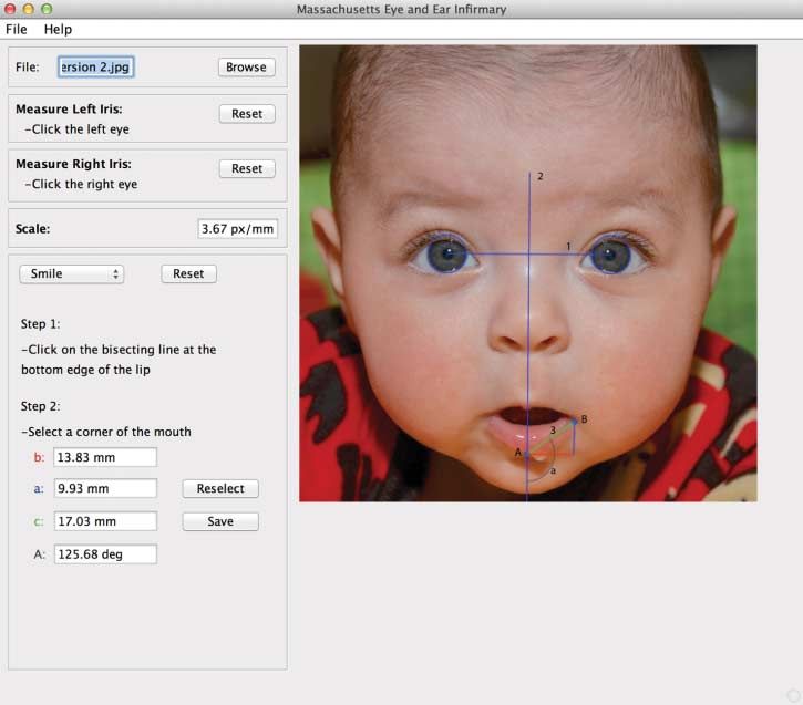
Research Original Investigation                                                                           Objective Assessment of Smile Outcomes

               S
                      ince its introduction in 1976 by Harii et al,1 gracilis free     went GFTT at the Massachusetts Eye and Ear Infirmary (MEEI)
                      tissue transfer (GFTT) has become the gold standard for          from March 2003 to March 2013 were initially included in this
                      dynamic reanimation of the oral commissure and is per-           study. Patients who underwent revision surgery or had necro-
               formed at many centers worldwide. Outcomes data following               sis of the flap because of infection or vascular insufficiency
               GFTT is sparse in the literature despite the frequency with             were excluded, as GFTT was counted as having failed in these
               which it is used. The most important barrier in advancing our           patients. Patients with missing data or in whom outcomes data
               understanding of outcomes following GFTT has been the lack              were not acquired following a sufficient recovery period were
               of an objective, comprehensive tool to measure facial func-             excluded.
               tion following surgical intervention.2
                    Because of the lack of available objective outcome mea-            Preoperative Data Collection
               sures for smile, predicting outcomes based on specific intra-           Demographic data including age and sex were recorded. Pre-
               operative events and parameters has been impractical. To meet           operative photographs and videos were used to measure base-
               these challenges, we recently developed an automated soft-              line oral commissure excursion and angle of the oral commis-
               ware tool (FACE-Gram) that can be used to analyze zonal fa-             sure with respect to the vertical midline of the face at repose
               cial movements with accuracy and precision3 and have also me-           and with smile (Figure 1) using FACE-Gram software, an au-
               ticulously recorded intraoperative parameters that may be               tomated tool for measuring facial landmarks.3
               predictive of flap outcome. In this study, we demonstrate the
               use of FACE-Gram to analyze smile outcomes in a large series            Operative Procedure and Intraoperative Data Collection
               of patients and investigate relationships between intraopera-           All operations were performed as previously described.4 Briefly,
               tive parameters and outcomes.                                           a segment of gracilis muscle was harvested from a medial in-
                                                                                       cision in the thigh along with the adductor artery, its venae
                                                                                       comitantes, and the obturator nerve. The muscle was trimmed
                                                                                       on the back table and inset into the face as described in the fol-
               Methods                                                                 lowing subsection (Figure 2).
               Selection Criteria
               Institutional review board approval was obtained from the Mas-          Gracilis Parameters
               sachusetts Eye and Ear Infirmary (MEEI) Human Studies Com-              Prior to harvest, the gracilis muscle length was measured and
               mittee prior to beginning this study. All patients who under-           recorded as the “marked length.” The muscle foreshortens to

               Figure 1. FACE-Gram Software

                                                                                                                            1, Horizontal interpupillary line; 2,
                                                                                                                            vertical line bisecting and
                                                                                                                            perpendicular to horizontal
                                                                                                                            interpupillary line; A, intersection of
                                                                                                                            line 2 and vermilliocutaneous border
                                                                                                                            of lower lip; B, oral commissure; 3,
                                                                                                                            line with distance spanning points A
                                                                                                                            and B; and a, angle between line 2
                                                                                                                            and line 3.

        86     JAMA Facial Plastic Surgery March/April 2014 Volume 16, Number 2                                                      jamafacialplasticsurgery.com

                                                     Copyright 2014 American Medical Association. All rights reserved.

Downloaded From: https://jamanetwork.com/ on 03/23/2020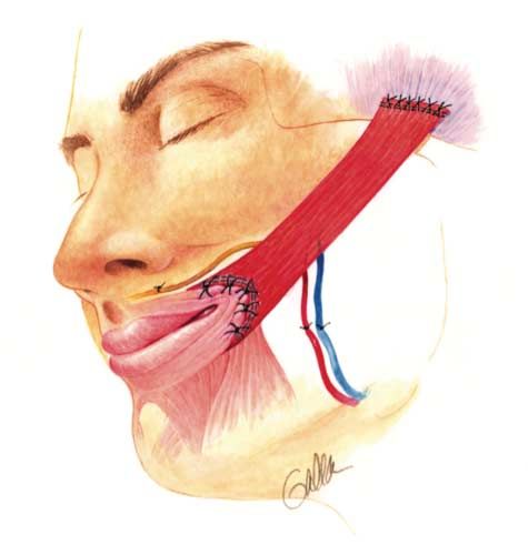
Objective Assessment of Smile Outcomes                                                                                Original Investigation Research

                 Figure 2. Gracilis Muscle Following Inset                                    Box. Intraoperative Measurements and Variables

                                                                                              Prior to Gracilis Harvest
                                                                                              In vivo marked length
                                                                                              Internal recoil length
                                                                                              Stimulated length

                                                                                              After Gracilis Harvest
                                                                                              Harvested length
                                                                                              Harvested width
                                                                                              Harvested thickness
                                                                                              Harvested weight
                                                                                              Trimmed weight

                                                                                              During Gracilis Inset
                                                                                              Inset length
                                                                                              Inset width
                                                                                              Inset thickness
                                                                                              Surgeon neurorrhaphy grade
                                                                                              No. of sutures placed for neurorrhaphy
                                                                                              Type of tissue sealant used for neurorrhaphy
                                                                                              Nerve(s) used to innervate free flap
                                                                                              Surgeon assessment of pulse pressure of donor artery
                 a variable degree after the muscle cuts are made, and this                   Surgeon assessment of oral commissure overcorrection
                 new length was recorded as the “recoil length.” The muscle                   Ischemia time
                 was then stimulated with a Montgomery Nerve Stimulator
                                                                                              Variables
                 (Boston Medical Products) at 2 V, and the length during
                 stimulation was recorded as the “stimulated length.” After                   Stretch = (harvest length/inset length) × 100.

                 the muscle was trimmed and prepared on the back table, the                   Internal recoil proportion = internal recoil length/in vivo marked
                 final length was recorded as the “harvest length.” During                    length.

                 inset, the muscle was stretched and sutured superiorly to the                Contractility proportion = (marked length−stimulated length/
                 deep temporal fascia to create a neoorigin, and inferiorly to                marked length) × 100.

                 the modiolus to create a neoinsertion. This new length was
                 recorded as the “inset length.” Several parameters were cal-
                 culated based on these measurements, as displayed in the
                 Box, including stretch, recoil proportion, and contractility              assessment included photographs and videographs, which
                 proportion. Thickness and width of the gracilis were also                 were used to obtain measurements with the FACE-Gram soft-
                 recorded before and after inset.                                          ware as was done preoperatively. In several cases, recovery had
                                                                                           stabilized prior to the expected recovery periods. These pa-
                 Operative Parameters                                                      tients were included in the final analysis.
                 The nerve used to innervate the free flap was noted. During
                 neurorrhaphy, a subjective surgeon neurorrhaphy grade was                 Excursion Analysis
                 assigned on an ordinal scale to the repair based on absence or            Objective comparisons of preoperative and postoperative pho-
                 presence of tension at the coaptation site, nerve diameter mis-           tographs were performed using FACE Gram software (Figure 1).
                 match, and extravasation of axonal contents from the suture               FACE-Gram software scaled each photograph based on the size
                 line. The number of sutures used and the type of tissue seal-             of the iris (approximately 11.8 mm).3 After the operator out-
                 ant used during the neurorrhaphy was also noted. A sub-                   lined each iris, a horizontal line (line 1) was created by the soft-
                 jective assessment of the pulse pressure of the donor artery              ware through the pupils, followed by a perpendicular line (line
                 and oral commissure overcorrection was also performed and                 2) bisecting the former. The user then marked the intersec-
                 recorded.                                                                 tion of the perpendicular line with the vermilliocutaneous bor-
                                                                                           der of the lower lip (point A). The user then defined the oral
                 Postoperative Data Collection                                             commissure (point B), and the software was able to calculate
                 Outcome assessment in the postoperative phase was per-                    the distance from point A to point B (line 3), which represents
                 formed after recovery stabilized—9 months for a flap inner-               the distance from the midline of the vermilliocutaneous bor-
                 vated by the ipsilateral masseteric branch of the trigeminal              der of the lower lip and the oral commissure. The change in
                 nerve and 18 months for a flap innervated by the contralat-               this distance between repose and smile is referred to as the
                 eral facial nerve via cross-face nerve graft. The postoperative           “smile excursion.” The change in angle “a” between repose and

                 jamafacialplasticsurgery.com                                                    JAMA Facial Plastic Surgery March/April 2014 Volume 16, Number 2        87

                                                      Copyright 2014 American Medical Association. All rights reserved.

Downloaded From: https://jamanetwork.com/ on 03/23/2020
Research Original Investigation Objective Assessment of Smile Outcomes
Table 1. Demographics and Operative Characteristics
Study Cohort Failure Cohort
Characteristic (Flaps, n = 127; Patients, n = 124) (n = 14)
Demographics
Sex, No. (%)
Male 52 (42) 10 (71)
Female 72 (58) 4 (29)
Age, mean (SD) [range], y 35 (18) [6-80] 35 (12) [10-57]
Motor nerve, No. (%)
Ipsilateral CN V 66 (52) 4 (29)
Contralateral CN VII 52 (41) 10 (71)
Ipsilateral CN V + contralateral CN VII 6 (5) 0
Ipsilateral CN VII 1 (1) 0
No. of neurorrhaphy sutures, No. (%)
1 1 (1) 0
2 37 (29) 1 (7)
3 24 (19) 3 (21)
4 38 (30) 5 (36)
≥5 22 (17) 5 (36)
Not recorded 5 (4) 0
Measurements, mean (SD) [range]
Preharvest
In vivo length, cm 14.4 (1.11) [12-19] 14 (0) [14-14]
Cut recoil length, cm 9.2 (1.2) [6-12] 9.67 (0.6) [9-10]
Stimulated length, cm 5.4 (1.0) [3.5-8.5] 5.57 (1.0) [4.5-6.5]
Harvest
Length, cm 7.99 (1.0) [6.0-13.0] 8.1 (0.6) [7.5-9.0]
Width, cm 3.99 (0.78) [2.4-6.0] 4.2 (0.6) [3.3-5.0]
Thickness, cm 0.95 (0.23) [0.2-1.5] 1 (0.1) [0.8-1.1]
Inset
Length, cm 10.6 (1.16) [8.5-13.0] 11.2 (1.4) [10.0-13.0]
Width, cm 3.6 (0.62) [2.5-5.0] 3.8 (0.4) [3.5-4.6]
Thickness, cm 0.71 (0.17) [0.2-1.2] 0.69 (0.13) [0.45-0.80]
Weight, g 28.5 (10.1) [10.5-51.2] 33.7 (13.0) [2.0-53.3]
Operative variables, mean (SD) [range]
Stretch 75 (8.0) [60-100] 71 (62) [62-78]
Contractility 59 (9.25) [44-85] 58 (7.6) [50-65]
Contractility proportion 0.448 (0.064) [0.29-0.63] 0.45 (0.02) [0.40-0.50]
Ischemia time, mean (SD) [range], min 132 (30) [60-262] 119 (22) [95-154]
Time from onset to operation, mean (SD) [range], d 3841 (5483) [328-38 166] 5825 (7071) [624-19 309]
Abbreviation: CN, cranial nerve.
smile is referred to as “angle excursion.” The smile and angle Hypothesis Testing and Regression Analysis
excursions on both the affected and healthy sides were then Differences between continuous variables were assessed using
calculated. a 2-tailed paired t test for preoperative-postoperative compari-
sons. A Wilcoxon signed-rank test was used to compare con-
Symmetry Analysis tinuous variables with a nonparametric distribution.
Symmetry was measured by calculating the difference in length Linear regression models were used to assess the associa-
of line 3 between the normal and affected sides, and the dif- tion of demographic and operative factors with postoperative
ference in angle “a” between the normal and affected sides. This angle and length excursion on the affected side and postop-
calculation was performed at repose and with smile, both in erative symmetry with smile. A robust empirical variance struc-
the preoperative and postoperative setting. A perfectly sym- ture was used to overcome slight deviations from normality.
metric smile yields a symmetry score of zero. Surgeon neurorrhaphy grade, number of sutures used for the
88 JAMA Facial Plastic Surgery March/April 2014 Volume 16, Number 2 jamafacialplasticsurgery.com
Copyright 2014 American Medical Association. All rights reserved.
Downloaded From: https://jamanetwork.com/ on 03/23/2020Objective Assessment of Smile Outcomes Original Investigation Research
neurorrhaphy, type of tissue sealant used for the neurorrha- Outcome Assessment
phy, assessment of pulse pressure of donor artery, and assess- Excursion on the healthy side decreased from 8.4 mm pre-
ment of oral commissure were analyzed continuously. Age, is- operatively to 7.2 mm postoperatively, while excursion on
chemia time, stretch, internal recoil proportion, and the affected side increased from −0.86 mm preoperatively to
contractility proportion were individually divided into quar- 7.8 mm postoperatively. Angle excursion decreased as well
tiles. All statistical analysis was performed using STATA/SE 12.1 on the healthy side, and increased on the affected side fol-
(StataCorp). lowing GFTT. Symmetry at repose with respect to angle
improved following surgery. Symmetry during smile with
respect to angle and length improved after GFTT (Table 3).
Flaps innervated by the trigeminal nerve had a mean of 2.2
Results mm greater excursion than those innervated by the contra-
Patient and Operative Characteristics lateral facial nerve. Those innervated by the contralateral
A total of 154 free flaps were performed on 148 patients dur- facial nerve had better postoperative symmetry during smile
ing the study period, of which 14 (9%) were failures. Flaps still with regard to length (Table 4). There was no statistically sig-
within the predetermined recovery period that had not pla- nificant difference between the nerve used to innervate the
teaued and those with lack of adequate data were excluded flap and failure rate (Figure 3).
from the analysis, leaving 127 flaps performed on 124 patients Regression analysis revealed an association between con-
in the study cohort. tractility proportion and excursion on the affected side. Spe-
More than half of the patients in the study cohort were fe- cifically, those patients in the third quartile (75th percentile) had
male, whereas most in the failure cohort were male. The mean a higher excursion compared with those in the first quartile (25th
age of the study population was 35 years. Half of the free flaps percentile) (95% CI, 0.002 to 5.6). An association between in-
were innervated by the masseteric branch of the trigeminal ternal recoil proportion and symmetry with smile using length
nerve, and just under half were innervated by the contralat- as a measurement index was also identified. Similarly, pa-
eral facial nerve. In 1 patient, a functioning branch of the ip- tients in the third quartile had better symmetry compared with
silateral facial nerve was used to drive the gracilis, and in 6 pa- their first quartile cohorts (95% CI, −7.2 to −1.2). We found no
tients, both the masseteric nerve and contralateral facial nerve correlation between age, ischemia time, stretch, neurorrha-
were used. phy grade and number of sutures used, type of tissue sealant
Typically, fewer than 4 sutures were used during neuror-
rhaphy. The marked length of gracilis for harvest was typi-
cally 14 cm, with a recoil length of approximately 9 cm. After Table 2. Cause of Facial Paralysis
stimulation of the obturator nerve, the cut gracilis contracted Cause No. (%)
to a mean of 5 cm. Harvest and inset measurements are all also Intracranial neoplasm 33 (22)
listed in the Table 1. Ischemia time averaged approximately 2 Vestibular schwannoma 33 (22)
hours, but examination of the raw data revealed an obvious Congenital 20 (13)
trend toward decreased ischemia time with increased sur-
Temporal bone fracture 10 (6)
geon experience (Table 1).
Iatrogenic 10 (6)
In 22% of cases, intracranial neoplasm was the cause of the
Malignant parotid neoplasm 10 (6)
facial paralysis. Vestibular schwannoma was the specific cause
Bell palsy 9 (6)
in another 22%. The remainder were congenital, temporal bone
Benign facial nerve neoplasm 8 (5)
fracture, iatrogenic injury, malignant parotid neoplasia, and
Other 21 (14)
others (Table 2).
Table 3. Outcome Assessment in 74 Flaps a
The P values are based on a 2-tailed
Mean (SD) paired t test.
b
Excursion refers to oral commissure,
Outcome Preoperative Postoperative P Valuea
b
measured in millimeters.
Excursion c
Δ Angle refers to change in angle
Healthy side 8.4 (4.5) 7.2 (4.3) .03 with smile, measured in degrees.
Affected side −0.86 (3.6) 7.8 (3.3)Research Original Investigation Objective Assessment of Smile Outcomes
Table 4. Postoperative Outcome Assessment With Respect to the Nerve Used
Mean (SD)
Trigeminal Facial
(n = 43)a (n = 35)b P Valuec
Affected side
Excursiond 8.7 (3.5) 6.5 (2.9) .006 a
Trigeminal refers to innervation via
Δ Angle d
5.2 (6.3) 7.1 (5.5) .39 the ipsilateral masseteric branch of
the trigeminal nerve.
Symmetry (angle)d b
Facial refers to innervation from the
Repose 4.7 (3.5) 4.5 (2.3) .91 contralateral facial nerve via a
Smile 4.3 (4.3) 4.8 (3.1) .21 cross-face nerve graft.
c
Symmetry (length)d The P values are based on a
Repose 4.7 (3.9) 4.2 (3.5) .47 Wilcoxon rank-sum test.
d
See Table 3 footnotes for
Smile 5.9 (3.9) 4.1 (3.4) .03
description.
reinnervation of the muscle and eventually translation of
Figure 3. Gracilis Flap Failures by Nerve
muscle contraction to the modiolus and oral commissure. Al-
though many of the outcome parameters improved signifi-
18
cantly following GFTT, symmetry at repose with regard to
16
length did not, though lack of statistical power may have
14 masked a potential true difference in a larger population
Flap Failure Rate, %
12 (Table 3).
10 The decrease in excursion with smile on the healthy
8
side following GFTT is not surprising. In the flaccid face, the
normal side of the face may contract unopposed by contra-
6
lateral mimetic muscle contraction. However, following
4
gracilis inset, the normal side of the face experiences the
2 force of an opposing vector from the gracilis muscle, which
0 results in decreased smile excursion on the normal side. A
Cranial Nerve V Cranial Nerve VII
similar rationale explains the preoperative negative excur-
Nerve Used for Innervation
sion with smile on the affected side. During smile, the normal
side contracts, thereby displacing the contralateral oral com-
The y-axis shows the percentage of total flaps innervated by the nerve denoted
on the x-axis that failed over the study period. P = .09 by Fisher exact test. missure to the normal side. This displacement is considered
negative because the vector of movement is toward the con-
tralateral side of the face.
used, assessment of pulse pressure, oral commissure overcor- Patient-reported outcome measures such as the Facial
rection, and outcome in the study or failure cohorts (Table 5). Clinimetric Evaluation (FaCE) Scale may provide more clini-
cally relevant indices5 from which to measure success. In the
future, we plan to analyze FaCE scores in patients following
GFTT to determine if associations between the outcomes
Discussion assessed in Table 3 and FaCE scores exists. Moreover, observer-
Our centers’ goal was to perform an analysis of smile excur- based studies that show less ocular scrutiny of facial abnor-
sion, assess improvements in facial symmetry, and track dif- malities following GFTT could also provide a sensitive mea-
ferent quantitative properties of the gracilis muscle and intra- sure of success.
operative clinical variables to determine if these factors are Smile excursion may not be the most relevant outcome
associated with clinical outcome. Herein, we present data dem- measure of smile reanimation. Facial anatomic differences and
onstrating that in the experience at a single institution, smile discrepancies in zygomaticus major muscle size may account
excursion in the frontal plane reaches a mean of 8 mm follow- for substantial differences in smile excursion between indi-
ing GFTT. This value does not represent total excursion be- viduals. Cultural differences in smile appearance also exist and
cause it fails to address Z-plane excursion; nonetheless, it is can account for measurable differences in smile excursion be-
similar to smile excursion reported in the normal population.3 tween individuals.6 Nonetheless, studies have found an asso-
The measure simply provides an objective 2-dimensional mea- ciation between smile intensity and lifespan,7 indicating that
sure of outcomes and a gauge from which to assess success in greater smile excursion may be confer a health benefit.
the future. From a strictly anatomic and physiologic stand- In the present study, we observed that patient age, sur-
point, excursion with smile presents a useful tool for measur- geon perception of quality of the neurorrhaphy, number of su-
ing surgical success because gracilis contraction following tures used during the neurorrhaphy, flap ischemia time, and
transfer is dependent on the surgeon successfully perform- pulse pressure were not associated with excursion or symme-
ing a number of surgical steps, permitting revascularization and try outcomes. However, this study was limited by its sample
90 JAMA Facial Plastic Surgery March/April 2014 Volume 16, Number 2 jamafacialplasticsurgery.com
Copyright 2014 American Medical Association. All rights reserved.
Downloaded From: https://jamanetwork.com/ on 03/23/2020Objective Assessment of Smile Outcomes Original Investigation Research
Table 5. Regression Analysis
95% Confidence Intervala a
95% Confidence intervals were
Independent Excursion, Δ Angle, Symmetry With obtained using robust regression
Variable Affected Side Affected Side Smile (Length) and represent the change in the
Contractility 0.002 to 5.6b −3.3 to 5.6 −1.7 to 4.5 dependent variable associated with
proportion a change from quartile 1 to quartile 3
Internal recoil −2.7 to 3.3 −8.3 to 3.7 −7.2 to −1.2b in the independent variable.
proportion b
P < .05.
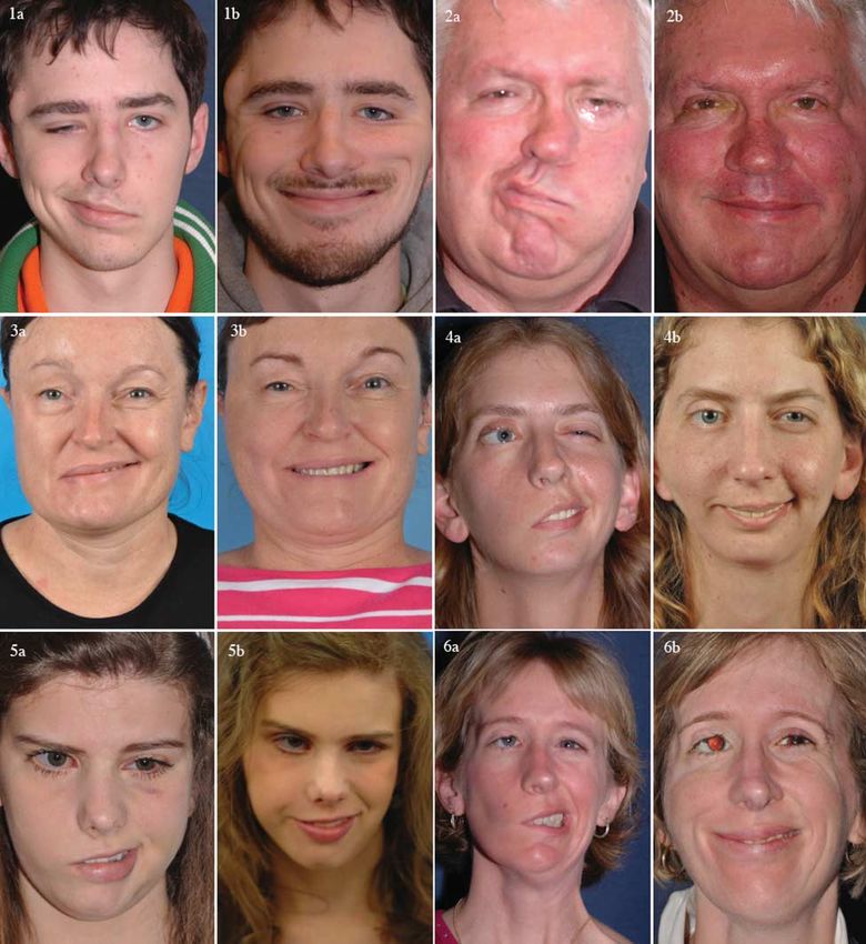
Figure 4. Preoperative and Postoperative Images Following Gracilis Free Tissue Transfer
A B C D
E F G H
I J K L
Preoperative (A, C, E, G, I, and K) and postoperative (B, D, F, H, J, and L) cross-face nerve graft coapted to the contralateral facial nerve. Patient 4 (G and
photographs. Patients 1, 2, and 3 (A-F) had innervation of the gracilis from the H) had a gracilis innervated by both the masseteric branch of the trigeminal
masseteric branch of the trigeminal nerve, and patients 5 and 6 (I-L) via a nerve and the contralateral facial nerve via a cross-face nerve graft.
jamafacialplasticsurgery.com JAMA Facial Plastic Surgery March/April 2014 Volume 16, Number 2 91
Copyright 2014 American Medical Association. All rights reserved.
Downloaded From: https://jamanetwork.com/ on 03/23/2020Research Original Investigation Objective Assessment of Smile Outcomes
size, therefore lacking power to detect small associations that (Figure 3), although the tendency toward higher failure rates
may be statistically significant. in cross-face innervated flaps is consistent with current ob-
We identified 2 statistically significant associations be- servations regarding flap failure rates. Figure 4 demonstrates
tween intraoperative measurements and outcome. Specifi- several typical examples of outcomes following GFTT inner-
cally, patients in the third quartile of contractility proportion vated by the trigeminal and facial nerves.
had greater excursion with smile compared with those in the The number of GFTT performed on an annual basis has in-
first quartile; flaps with more contraction during stimulation creased since the inception of the Facial Nerve Center at our
of the obturator nerve had increased postoperative smile ex- institution. It appears that failure rates were higher on aver-
cursion. Similarly, patients in the third quartile of internal re- age during the first several years of our practice; the decrease
coil proportion exhibited better postoperative symmetry com- in failure rate may be associated with the technological ad-
pared with those in the first quartile, indicating that less vances that we have used (venous couplers, color Doppler ul-
foreshortening of the gracilis following the muscle cuts (prior trasonography to monitor blood flow through the pedicle, il-
to nerve stimulation) is optimal. luminated retractors, and split operating beds for harvest of
The finding that smile excursion was greater in trigemi- the flap). It would be interesting to investigate whether case
nally innervated flaps is consistent with other reports in the volume and these technological advances are associated with
literature.8 The masseteric branch of the trigeminal nerve has improved outcomes. Multi-institutional collaboration and a
a robust axon count, and increased throughput of these axons larger sample size would be required for such studies.
into the obturator nerve compared with those from a cross-
face nerve graft likely accounts increased smile excursion. In
addition, 2 neural coaptation sites are required when innervat-
ing the gracilis from a cross-face nerve graft, introducing more
Conclusions
opportunity for axonal loss at the neurorrhaphy sites. The find- Gracilis free tissue transfer is a reliable procedure for dy-
ing that smile symmetry was better in flaps innervated by the namic reanimation of the paralyzed oral commissure. Suc-
facial nerve is not surprising because the neural stimulus to both cessful muscle transfers result in measureable improve-
the zygomaticus major on the normal side and the gracilis on ments in smile excursion and oral commissure symmetry, and
the paralyzed side arise from a common neural origin. How- both muscle recoil and contractility parameters relate to final
ever, this does not account for the differences in muscle char- outcome. We were unable to discern a difference between fail-
acteristics between the zygomaticus major and the trans- ure rates based on the nerve used, but such a difference may
planted gracilis. exist. Larger, prospective studies are indicated to better de-
In this study, we were not able to identify a difference in fine associations between intraoperative gracilis muscle para-
flap failure rate based on the nerve used for flap innervation meters and outcomes.
ARTICLE INFORMATION Funding/Support: This research was supported by reanimation. Arch Facial Plast Surg.
Accepted for Publication: September 16, 2013. grant R01NS071067 from the National Institutes of 2012;14(4):277-282.
Health (NIH). 4. Hohman MH, Hadlock TA. Microneurovascular
Published Online: January 30, 2014.
doi:10.1001/jamafacial.2013.2463. Role of the Sponsor: The NIH had no role in the free gracilis transfer for smile reanimation.
design and conduct of the study; collection, Operative Techniques Otolaryngol.
Author Contributions: Drs Bhama and Hadlock management, analysis, and interpretation of the 2012;23(4):262-267.
had full access to all of the data in the study and data; preparation, review, or approval of the
takes responsibility for the integrity of the data and 5. Kahn JB, Gliklich RE, Boyev KP, Stewart MG,
manuscript; and decision to submit the manuscript Metson RB, McKenna MJ. Validation of a
the accuracy of the data analysis. for publication.
Study concept and design: Bhama, Weinberg, patient-graded instrument for facial nerve paralysis:
Lindsay, Cheney, Hadlock. Previous Presentation: This research was the FaCE scale. Laryngoscope. 2001;111(3):387-398.
Acquisition of data: Bhama, Weinberg, Lindsay, presented at the American Academy of Facial 6. Ozono H, Watabe M, Yoshikawa S, et al. What’s
Hohman, Cheney, Hadlock. Plastic and Reconstructive Surgery section of the in a smile? cultural differences in the effects of
Analysis and interpretation of data: Bhama, Combined Otolaryngological Spring Meetings; April smiling on judgments of trustworthiness. Letters
Weinberg, Lindsay, Cheney, Hadlock. 14, 2013; Orlando, Florida. Evolutionary Behav Sci. 2010;1(1):15-18.
Drafting of the manuscript: Bhama, Cheney, Additional Contributions: Chris Sudfeld, ScD, 7. Abel EL, Kruger ML. Smile intensity in
Hadlock. provided guidance with statistical analysis. photographs predicts longevity. Psychol Sci.
Critical revision of the manuscript for important 2010;21(4):542-544.
intellectual content: Bhama, Weinberg, Lindsay, REFERENCES
Hohman, Hadlock. 8. Hontanilla B, Marre D, Cabello A. Facial
1. Harii K, Ohmori K, Torii S. Free gracilis muscle reanimation with gracilis muscle transfer
Statistical analysis: Bhama, Weinberg, Lindsay. transplantation, with microneurovascular
Obtained funding: Hadlock. neurotized to cross-facial nerve graft versus
anastomoses for the treatment of facial paralysis: masseteric nerve: a comparative study using the
Administrative, technical, or material support: a preliminary report. Plast Reconstr Surg.
Hadlock. FACIAL CLIMA evaluating system. Plast Reconstr
1976;57(2):133-143.
Study supervision: Bhama, Lindsay, Cheney, Surg. 2013;131(6):1241-1252.
Hadlock. 2. Hadlock T. Facial paralysis: research and future
directions. Facial Plast Surg. 2008;24(2):260-267.
Conflict of Interest Disclosures: None reported.
3. Hadlock TA, Urban LS. Toward a universal,
automated facial measurement tool in facial
92 JAMA Facial Plastic Surgery March/April 2014 Volume 16, Number 2 jamafacialplasticsurgery.com
Copyright 2014 American Medical Association. All rights reserved.
Downloaded From: https://jamanetwork.com/ on 03/23/2020You can also read