FATAL EXACERBATION OF CHADOX1-NCOV-19-INDUCED THROMBOTIC THROMBOCYTOPENIA SYNDROME AFTER INITIAL SUCCESSFUL THERAPY WITH INTRAVENOUS ...
←
→
Page content transcription
If your browser does not render page correctly, please read the page content below
Fatal exacerbation of ChadOx1-nCoV-19-induced
thrombotic thrombocytopenia syndrome after initial
successful therapy with intravenous immunoglobulins - a
rational for monitoring immunoglobulin G levels

by Jonathan Douxfils, Caroline Vayne, Claire Pouplard, Thomas Lecompte, Julien Favresse,
Florence Potier, Emy Gasser, Valérie Mathieux, Jean-Michel Dogné, Yves Gruel, Jérôme Rollin,
and François Mullier

Received: June 28, 2021.
Accepted: August 3, 2021.

Citation: Jonathan Douxfils, Caroline Vayne, Claire Pouplard, Thomas Lecompte, Julien Favresse,
Florence Potier, Emy Gasser, Valérie Mathieux, Jean-Michel Dogné, Yves Gruel, Jérôme Rollin
and François Mullier. Fatal exacerbation of ChadOx1-nCoV-19-induced thrombotic thrombocytopenia
syndrome after initial successful therapy with intravenous immunoglobulins - a rational for monitoring
immunoglobulin G levels.
Haematologica. 2021 Aug 12. doi: 10.3324/haematol.2021.279509. [Epub ahead of print]

Publisher's Disclaimer.
E-publishing ahead of print is increasingly important for the rapid dissemination of science.
Haematologica is, therefore, E-publishing PDF files of an early version of manuscripts that
have completed a regular peer review and have been accepted for publication. E-publishing
of this PDF file has been approved by the authors. After having E-published Ahead of Print,
manuscripts will then undergo technical and English editing, typesetting, proof correction and
be presented for the authors' final approval; the final version of the manuscript will then
appear in a regular issue of the journal. All legal disclaimers that apply to the
journal also pertain to this production process.
Fatal exacerbation of ChadOx1-nCoV-19-induced thrombotic thrombocytopenia syndrome after

         initial successful therapy with intravenous immunoglobulins - a rational for monitoring

                                         immunoglobulin G levels

Journal:
Haematologica – Case reports & case series
Authors:
Jonathan Douxfils*1,2, Caroline Vayne*3, Claire Pouplard3, Thomas Lecompte4, Julien Favresse1,5,
Florence Potier6, Emy Gasser7, Valérie Mathieux8, Jean-Michel Dogné1, Yves Gruel3, Jérôme Rollin*3,
François Mullier*9
1
 University of Namur, Department of Pharmacy, Namur Research for Life Sciences, Namur Thrombosis and
Hemostasis Center, Namur, Belgium
2
    QUALIblood s.a., Namur, Belgium
3
    University of Tours, EA7501 GICC, CHRU de Tours, Department of Haemostasis, Tours, France
4
 Département de Médecine, Hôpitaux Universitaires de Genève, service d’angiologie et d’hémostase et
Faculté de Médecine, Geneva Platelet Group (GpG), Université de Genève, Geneva, Switzerland
5
    Clinique Saint-Luc Bouge, Department of Laboratory Medicine, Bouge, Belgium
6
    Service de gériatrie, CHU UCL Namur site Sainte-Elisabeth, Namur, Belgium
7
 Université Catholique de Louvain, service de gériatrie, CHU UCL Namur site Sainte-Elisabeth, Namur,
Belgium
8
 CHU UCL Namur | site Sainte-Elizabeth, Université catholique de Louvain, Department of Hematology,
Namur Research Institute for Life Sciences, Namur Thrombosis and Hemostasis Center, Yvoir, Belgium.
9
  Université catholique de Louvain, CHU UCL Namur, Namur Thrombosis and Hemostasis Center,
Hematology Laboratory, Namur Research Institute for Life Sciences, Yvoir, Belgium
*Contributed equally
Acknowledgements:

The authors would like to thank the technical staff of the CHU UCL Namur, Mrs Justine Baudar, Mrs

Maïté Guldenpfennig and the technical staff of the CHRU Tours, Mrs Séverine Augereau.

Contribution:

J.D. and C.V., analyzed the results, wrote the first draft of the manuscript and designed the figures. J.R,

Y.G, C.P, C.V. provided and analyzed the results and revised the manuscript. T.L. analyzed the results

and thoroughly revised the manuscript. J.F. provided and analyzed the results and revised the manuscript.

F.P., E.G., and V.M. managed the patient and thoroughly revised the manuscript. J-M Dogné analyzed the

                                                    1/10
results. F.M. designed and supervised the experiments, provided and analyzed the data, and interpreted
the results.
Conflict of interest disclosure:
Among the authors, J.D. is the CEO and founder of QUALIblood s.a., a contract research organization
manufacturing the DP-Filter, is a coinventor of the DP-Filter (patent application number: PCT/ET2019/
052903) and reports personal fees from Daiichi-Sankyo, DOASense Gmbh, Gedeon Richter, Mithra
Pharmaceuticals, Norgine, Portola, Stago, Roche and Roche Diagnostics outside the submitted work; T.L.
reports non-personal fees from IRIS and Stago. F.M. reports institutional fees from Stago, Werfen, Nodia,
Roche Sysmex and Bayer. He also reports speaker fees from Boehringer Ingelheim, Bayer Healthcare,
Bristol- Myers Squibb-Pfizer, Stago, Sysmex and Aspen all outside the submitted work. The other authors
have no conflict of interest to disclose.
Corresponding author:
Prof. Jonathan Douxfils,
University of Namur, Department of Pharmacy, Namur Research for Life Sciences, Namur Thrombosis
and Hemostasis Center, Namur, Belgium
E-mail: jonathan.douxfils@unamur.be
Mail : Rue de Bruxelles, 61 – 5000 Namur, Belgium
Phone: +32 496 22 31 53
Fax: +32 81 72 42 91
Number of words: 1,484
Number of figures/tables: 2
Number of references: 14
Keywords: Thrombocytopenia, Thrombosis, PF-4 antibodies, VITT, ChadOx1-nCoV-19
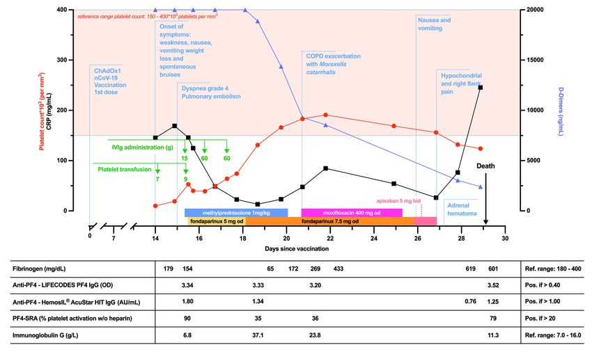
2/10The present report describes a vaccine-induced thrombotic thrombocytopenia (VITT) case with
fatal exacerbation after initial improvement following initial IVIg administration and
anticoagulation. An 83-year-old woman presented at the emergency room with an alteration of
her general condition. She presented with symptoms of weakness, nausea, vomiting, weight loss
and spontaneous bruises without any obvious reason, 14 days after having received her first dose
of ChadOx1 nCov-19. According to our medical records, she did not receive heparin or
derivative during the last four months. Clinical examination unraveled bruising on the upper
limbs. Computer Tomography (CT) of thorax and abdomen was normal. Oxygen saturation was
98% at admission and the patient was tested negative for SARS-CoV-2 infection as assessed by
reverse-transcriptase polymerase chain reaction (RT-PCR). The initial laboratory investigations
on the day of admission revealed that the patient was suffering from marked thrombocytopenia
(i.e. platelet count of 10,000 per mm3), dramatically increased D-dimers plasma levels (i.e. >
20,000 ng/mL) and slightly low plasma fibrinogen (i.e. 179 mg/dL) (Figure 1). She was
transfused with a platelet concentrate (roughly 3.5x10¹¹ platelets) on the day of admission.
During the night, she suffered from dyspnea grade NYHA 4. Pulmonary ventilation and
perfusion (V/Q) scan was performed and disclosed bilateral pulmonary embolism. Anti-PF4 IgG
antibodies (i.e. 1.80 AU/mL, Figure 1) were detected on day 1 post-admission using a
PF4/polyvinylsulfonate rapid assay (HemosIL® AcuStar HIT IgG assay, Instrumentation
Laboratory Belgium NV, Zaventem, Belgium). The diagnosis of VITT was confirmed using a
heparin-induced multi-electrode aggregometry method.(1) In face of the clinical picture, i.e.
thrombocytopenia and thrombosis, with the presence of anti-PF4 antibodies and positive platelet
activation tests within 30 days after vaccination with ChadOx1 nCov-19, VITT was
diagnosed.(2) The patient therefore promptly received 15 grams of IVIg (Privigen®, CSL
3/10Behring Gmbh, Marburg, Germany) and methylprednisolone 1 mg/kg. A second platelet
concentrate (roughly 4.5x10¹¹ platelets) was administered for allowing initiation of
anticoagulation as platelet count was still below 30,000 per mm3.(3) Platelet count rapidly
improved, i.e. 53,000 per mm3, and anticoagulation was started with fondaparinux 5 mg od
subcutaneously from day 1 to day 3 (taking into account renal failure, i.e. Cockcroft-Gault
creatinine clearance < 50 mL/min). She received additional IVIg on day 2 and 3, at the dose of
60 grams per day for a total IVIg dose of 135 grams corresponding to 1.7 grams of IVIg per kg
administered over a period of 48 hours. Fondaparinux dose was increased to 7.5 mg od from day
4 to day 11 since renal function improved. On day 6, the patient was stabilized, and her global
health status was improved as witnessed by normalized platelet count, decrease in D-dimers (i.e.
from > 20,000 ng/mL at admission to 14,380 ng/mL) and CRP (from 145 mg/dL at admission to
23 mg/dL). Later that day, however, oxygen saturation dropped below 80%. Cough with sputum
production was noted and exacerbation of COPD with Moraxella catarrhalis infection was
diagnosed. Oxygen supplementation was then started (2 liters per minute) combined with oral
moxifloxacin 400 mg od for 5 days. On day 12 post-admission, anticoagulation was switched
from fondaparinux to apixaban 5 mg bid.
Unfortunately, the clinical status worsened on day 12 post-admission with a de novo reduction of
platelet count. Abdominal CT scan showed right adrenal hematoma with left adrenal infiltrate,
which is a usual presentation of adrenal infarction, as described in auto-immune heparin-induced
thrombocytopenia and also recently in VITT.(4, 5) Four units of 500 IU/mL of prothrombin
complex concentrate were administered on day 14 as an attempt to control the adrenal
hematoma, but she died later that day from hypovolemic shock probably secondary to adrenal
hemorrhage. A causal adrenal infarction may have existed but could not be confirmed as neither
4/10an injected CT scan nor an autopsy was performed. Nevertheless, adrenal insufficiency was not
documented and cortisol levels on day 14 was still in the upper range (i.e. 21 µg/dL, normal
range: 6.2 to 18 µg/dL). Moreover, it must be noted that although apixaban was last administered
on day 13 in the morning and never reintroduced, its plasma level on day 14 was 353 ng/mL
(usual Ctrough range: 22 to 177 ng/mL) and was still 132 ng/mL on day 15. The accumulation of
apixaban may thus have contributed to, or even triggered, this bleeding event. The initial
infusion of platelet concentrates may also have contributed to disease progression, as well as to
the pulmonary embolism observed at diagnosis. An immediate treatment with IVIg, as now
recommended by the American Society of Hematology,(6) could have been beneficial but the
presence of active bruising, the absence of documented thrombosis and the marked
thrombocytopenia guided our therapeutic choice at that time. This case further highlights how
VITT is a dynamical condition, which should not be discounted in a recently vaccinated patient
with only thrombocytopenia and increased D-dimer levels, even in the absence of documented
thrombosis, and that IVIG should be considered promptly, along with anti-PF4 testing and
thrombosis screening.
The rapid fatal outcome, occurring two weeks after VITT diagnosis while an improvement was
noticed, raised the question of an early relapse or an exacerbation of the initial event. In an
attempt to understand the possible cause(s), additional laboratory investigations were performed,
as reported in Figure 1. Blood samples collected post-admission on day 1 (diagnosis), day 4
(after IVIg administration), day 7 (marked clinical improvement with platelet count
normalization) and day 15 (deterioration of patient’s status, leading to death) were selected to
assess time-related changes of anti-PF4 antibodies, platelet activation with PF4-SRA (7) and
total IgG levels.
5/10Result of the enzyme-linked immunosorbent assay (ELISA) with immobilized PF4/PVS
complexes (LIFECODES PF4 IgG, Immucor Lifecodes, Jette, Belgium) remained strongly
positive during the whole hospital stay with OD > 3.00 measured with all samples collected from
day 1 to day 15. Positive results were also obtained using a modified in-house ELISA in which
the wells are only coated by PF4 (8), demonstrating the presence of IgG antibodies that bound
PF4 alone (data not shown). Such characteristics of VITT antibodies, shared with those of highly
pathogenic auto-immune HIT antibodies (9), indicate a different specificity and affinity towards
PF4 compared to classical HIT antibodies, and explain why their detection by HIT-dedicated
immunoassays may be inadequate.(7) The fact that anti-PF4 IgG antibodies were detected using
a PF4/polyvinylsulfonate rapid assay (HemosIL® AcuStar HIT IgG assay) is particular since this
assay failed to detect anti-PF4 antibodies in most of reported VITT cases. Nevertheless, the
HemosIL® Acustar HIT IgG assay may rarely give weak positive results in patients with likely
VITT diagnosis, as stated by Platton et al., who reported 2 positive results out of 31 patients, all
of whom had anti-PF4 IgG detected by ELISA.(10)
PF4-SRA was performed with the same samples, as previously described.(7) Platelet activation,
measured through maximal serotonin release, was 90% in absence of heparin on day 1, decreased
by more than 50% after IVIg administration (day 4, 35%) and remained low when the patient
was getting better (day 7, 36%). In contrast, platelet activation returned to a high level at the time
of clinical deterioration, day 15 (79%). Time related changes of total IgG plasma levels mirrored
those of PF4-SRA (Figure 1). While total IgG level was rather low at diagnosis, it rose above 30
g/L after IVIg administration and then rapidly decreased, as observed on day 15, with a return to
normal values for a healthy adult. These data support the hypothesis that the rebound of platelet
activation observed in PF4-SRA at the time of clinical deterioration could be due to a rapid
6/10elimination of the infused immunoglobulins and the loss of their competing effect with platelet
activating anti-PF4 antibodies on platelet FcRIIa.
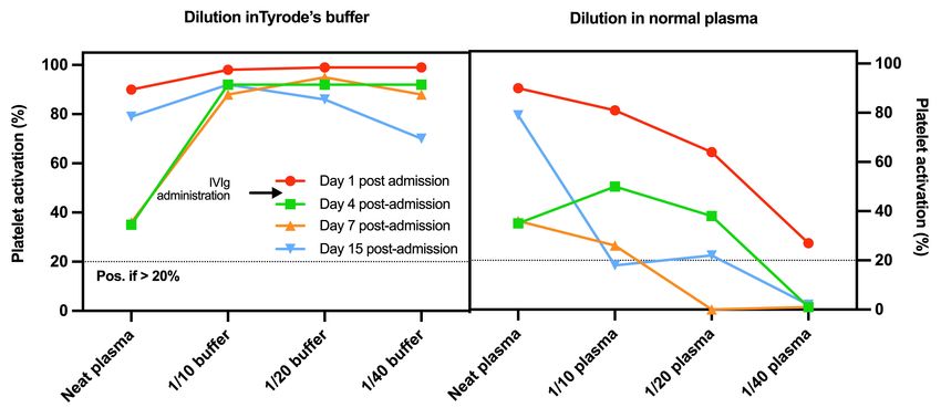
Additional experiments were performed with PF4-SRA to consolidate our hypothesis that the
levels of IgG can explain the exacerbation The same samples were diluted either in normal
plasma (containing normal IgG level) or in modified Tyrode’s buffer at 1/10, 1/20, 1/40 dilution
ratios. When samples were diluted in normal plasma, platelet activation decreased proportionally
with the dilution, and was completely abolished at 1/40, except for the day 1 sample, which still
slightly activate platelets under these conditions (Figure 2). In contrast, when the samples were
diluted in Tyrode’s buffer, platelet activation remained high (near 100%) for the samples
collected on day 1 and day 15 but increased for those obtained at days 5 and 7, approaching
100% (Figure 2). These results strongly support that platelet activation by VITT antibodies was
inhibited by normal IgG, and that lowering the concentrations of normal IgG led to the
reappearance of platelet activation by loss of competition between the IVIg and anti-PF4 IgG.
This case supports the concept that a proper monitoring using an appropriate functional assay
could help in clinical decision making since PF4-SRA mirrored with the clinical evolution of the
patient. Such observation has also been made in a recent study, which demonstrated that platelet
activation by VITT antibodies was inhibited in patients treated with IVIg.(11) Nevertheless, this
needs to be confirmed. Interestingly, the inhibitory effect of normal polyclonal IgG on the
platelet activation induced by PF4-specific antibodies could also vary from one patient to
another, as previously demonstrated in HIT patients.(12) It is also important to note that
administration of IVIg reduces the activation of platelets as assessed by the PF4-SRA. This has
major consequences when collecting samples for confirmation of VITT diagnosis and dilution in
appropriate buffer, as we did in our experiments, could be recommended to assess the
7/10competitive interaction between anti-PF4 IgG and IVIg. However, this test lacks worldwide
availability, and cannot easily be used for emergency patients monitoring. Therefore, as total IgG
concentration measured in the patient inversely correlated with platelet activation in PF4-SRA,
quantitatively assaying anti-PF4 IgG antibodies levels and total IgG concentration in the
patient’s plasma could help in identifying situations where the competition between normal
polyclonal IgG and anti-PF4 IgG on FcRIIa may switch in favor of the platelet activating
antibodies.(13)
Despite data on IVIg clearance parameters and target concentrations are lacking for such a very
peculiar condition, a rapid decrease in total IgG concentrations within the normal range (i.e. 7-16
g/l) (14) could alert to possible therapeutic escape, and the need for re-administration of IVIg,
especially in situation where anti-PF4 IgG remains high. In the patient, total IgG concentration
was divided by two within eight days, which is pretty faster than the median half-life of 30 days
generally reported in the literature for IVIg.(15) Although further studies are needed to
understand that accelerated clearance and assess clinical relevance of total IgG measurement to
monitor the efficacy of IVIg, it appears a very affordable tool in practice in combination with
anti-PF4 IgG antibodies testing.
Informed consent and ethical committee approval:
As the patient died, an independent review board of the Clinique Sainte-Elisabeth CHU UCL Namur
(OM070) has been consulted to decide if the reporting of this case is ethically acceptable. This ethical
committee (decision number: 27-21) was favorable to the publication of this case and did not see any
ethical issue in publishing this case-report. All the data has been anonymized.
8/10References:
1. Morel-Kopp MC, Mullier F, Gkalea V, et al. Heparin-induced multi-electrode aggregometry method
for heparin-induced thrombocytopenia testing: communication from the SSC of the ISTH. J Thromb
Haemost. 2016;14(12):2548-2552.
2. Robert T. Proposed Brighton Collaboration process for developing a standard case definition for study
of new clinical syndrome X, as applied to Thrombosis with Thrombocytopenia Syndrome (TTS) -
V10.16.3. Accessed on May 30, 2021. Available from: https://brightoncollaboration.us/wp-
content/uploads/2021/04/TTS-Case-Finding-and-Definition-Process.v9.0-April-16-202115853.pdf.
3. Guidance produced from the Expert Haematology Panel (EHP) focussed on Covid-19 Vaccine induced
Thrombosis and Thrombocytopenia (VITT) Updated Guidance on Management. Version 1.3. Accessed
on Apr 7, 2021. Available from: https://b-s-h.org.uk/media/19530/guidance-version-13-on-mngmt-of-
thrombosis-with-thrombocytopenia-occurring-after-c-19-vaccine_20210407.pdf.
4. Rosenberger LH, Smith PW, Sawyer RG, et al. Bilateral adrenal hemorrhage: the unrecognized cause
of hemodynamic collapse associated with heparin-induced thrombocytopenia. Crit Care Med.
2011;39(4):833-838.
5. Scully M, Singh D, Lown R, et al. Pathologic Antibodies to Platelet Factor 4 after ChAdOx1 nCoV-19
Vaccination. N Engl J Med. 2021;384(23):2202-2211.
6. Bussel JB, Connors J, Cines DB, et al. Thrombosis with Thrombocytopenia Syndrome (also termed
Vaccine-induced Thrombotic Thrombocytopenia). version 1.5. Accessed on Jul 26, 2021. Available from:
https://www.hematology.org/covid-19/vaccine-induced-immune-thrombotic-thrombocytopenia.
7. Vayne C, Rollin J, Gruel Y, et al. PF4 Immunoassays in Vaccine-Induced Thrombotic
Thrombocytopenia. N Engl J Med. 2021;385(4):376-378.
8. Pouplard C, Leroux D, Rollin J, et al. Incidence of antibodies to protamine sulfate/heparin complexes
incardiac surgery patients and impact on platelet activation and clinical outcome. Thromb Haemost.
2013;109(6):1141-1147.
9. Nguyen T-H, Medvedev N, Delcea M, Greinacher A. Anti-platelet factor 4/polyanion antibodies
mediate a new mechanism of autoimmunity. Nat Commun. 2017;8:14945.
10. Platton S, Bartlett A, MacCallum P, et al. Evaluation of laboratory assays for anti-platelet factor 4
antibodies after ChAdOx1 nCOV-19 vaccination. J Thromb Haemost. 2021;19(8):2007-2013.
11. Bourguignon A, Arnold DM, Warkentin TE, et al. Adjunct Immune Globulin for Vaccine-Induced
Thrombotic Thrombocytopenia. N Engl J Med. 2021 Jun 09. [Epub ahead of print].
9/1012. Rollin J, Pouplard C, Sung HC, et al. Increased risk of thrombosis in FcgammaRIIA 131RR patients
with HIT due to defective control of platelet activation by plasma IgG2. Blood. 2015;125(15):2397-2404.
13. Warkentin TE. High-dose intravenous immunoglobulin for the treatment and prevention of heparin-
induced thrombocytopenia: a review. Expert Rev Hematol. 2019;12(8):685-698.
14. Svendsen PJ, Ward AM, Dati F, et al. New international reference preparation for proteins in human
serum (RPPHS). Clin Chem. 1994;40(6):934-938.
15. Koleba T, Ensom MH. Pharmacokinetics of intravenous immunoglobulin: a systematic review.
Pharmacotherapy. 2006;26(6):813-827.
Figures
Figure 1: Clinical and laboratory data of the case.
Figure 2: Dilution experiments on samples collected at day 1 post-admission (diagnosis), days 4
post-admission (after IVIg administration), day 7 post-admission (marked clinical improvement
with platelet count normalization) and day 15 post-admission (deterioration of patient’s status and
death). Dilutions were made in normal heated plasma (containing normal IgG level) or in modified
Tyrode’s buffer at 1/10, 1/20, 1/40 dilution ratios. Platelet activation was assessed without heparin in
PF4-SRA and results are expressed in percentage of serotonin release.
10/10You can also read