Use of Bead-Based Serologic Assay to Evaluate Chikungunya Virus Epidemic, Haiti - CDC
←
→
Page content transcription
If your browser does not render page correctly, please read the page content below

Use of Bead-Based Serologic
        Assay to Evaluate Chikungunya
             Virus Epidemic, Haiti
          Eric W. Rogier, Delynn M. Moss, Kimberly E. Mace, Michelle Chang, Samuel E. Jean,
       Stevan M. Bullard, Patrick J. Lammie, Jean Frantz Lemoine, Venkatachalam Udhayakumar

The index case of chikungunya virus (CHIKV) in Haiti was                Confirmation of active CHIKV infection is accom-
reported during early 2014; the vector, the pervasive Aedes        plished through reverse transcription PCR or detection of
aegypti mosquito, promoted rapid spread throughout the             CHIKV IgM (3,4). Although confirming infection aids in
country. During December 2014–February 2015, we col-               determining the causative agent of symptoms, only sup-
lected blood samples from 4,438 persons at 154 sites (62           portive care is currently available for chikungunya, be-
urban, 92 rural) throughout Haiti and measured CHIKV IgG
                                                                   cause CHIKV-specific antiviral drugs have not been iden-
by using a multiplex bead assay. Overall CHIKV seropreva-
lence was 57.9%; differences between rural (mean 44.9%)
                                                                   tified (5). Furthermore, using these assays would require
and urban (mean 78.4%) areas were pronounced. Logistic             persons to have been sampled during active or recent vire-
modeling identified the urban environment as a strong pre-         mia, whereas CHIKV IgG could persist for longer periods
dictor of CHIKV exposure (adjusted odds ratio 3.34, 95%            of time (4,6). We present data from a nationwide survey
CI 2.38–4.69), and geographic elevation provided a strong          in Haiti in which we used a bead-based serologic assay to
negative correlation. We observed no correlation between           determine the overall presence of CHIKV IgG, which pro-
age and antibody positivity or titer. Our findings demonstrat-     vides evidence of past and current exposure.
ed through serologic testing the recent and rapid dissemina-
tion of the arbovirus throughout the country. These results        Materials and Methods
show the utility of serologic data to conduct epidemiologic
studies of quickly spreading mosquitoborne arboviruses.
                                                                   Study Population
                                                                   Our group, which includes Population Services Internation-

C    hikungunya virus (CHIKV) is an arbovirus, transmitted
     by Aedes aegypti and Ae. albopictus mosquitoes, that
can cause transient but debilitating disease in humans. The
                                                                   al (PSI), conducted a survey to evaluate malaria prevalence
                                                                   in Haiti during December 2014–February 2015 as part of
                                                                   the malaria control activities supported by the Global Fund
World Health Organization reported the first cases of CHIKV        (https://www.theglobalfund.org/en/malaria/). In addition to
on the island nation of Haiti in April 2014; by June 2014, a       planned multiplex serologic testing for malaria, we chose
total of 6,318 cases had been reported there and in 16 other       antigens for nonmalarial febrile diseases before starting
countries or territories in the Caribbean and South America;       data collection; these tests were approved by the Haitian
103,018 suspected cases were reported (1). Additional evi-         Ministry of Health. The Institut Haïtien de Statistiques et
dence that CHIKV was introduced into Haiti in 2014 came            d’Informatique (http://www.ihsi.ht/) had previously subdi-
from evaluation of a longitudinal cohort of children in the        vided the nation into 12,000 enumeration areas (sections
coastal town of Leogane during 2011–2014. Before 2014,             d’énumération, SDEs) on the basis of population density;
these children tested negative for CHIKV antibodies, but           we chose 154 of these SDEs for this survey through propor-
samples collected in 2014 showed CHIKV IgG; 78.9% of all           tional sampling of predicted malaria risk strata within the
children seroconverted within the span of 1 year (2).              country, as had been determined by predictive modeling
                                                                   (7). The SDEs were classified as urban if they were within
Author affiliations: Centers for Disease Control and Prevention,
                                                                   the administrative boundary of any of the 140 municipal
Atlanta, Georgia, USA (E.W. Rogier, D.M. Moss, K.E. Mace,
                                                                   cities in Haiti and were otherwise considered rural. Field
M. Chang, S.M. Bullard, P.J. Lammie, V. Udhayakumar);
                                                                   teams randomly selected 20 households within each SDE;
Population Services International/Organisation Haïtienne de
                                                                   all members of selected households were offered participa-
Marketing Social pour la Santé, Port-au-Prince, Haiti
                                                                   tion. Following verbal consent by the participant (or par-
(S.E. Jean); Programme National de Contrôle de la Malaria/
                                                                   ents or guardian if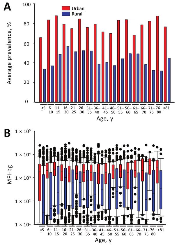
RESEARCH

individually stored in plastic bags with desiccant. We as-        minus background (OD-bg). We tested serum samples (n =
signed samples unique identification numbers that were not        50) from CHIKV endemic and nonendemic regions by us-
traceable to the individual persons. The study protocol was       ing ELISA and the bead assay; test comparisons for continu-
approved by the Haitian Ministry of Health and approved           ous data are shown in online Technical Appendix Figure 1,
as a nonresearch activity by the US Centers for Disease           panel A (https://wwwnc.cdc.gov/EID/article/24/6/17-1447-
Control and Prevention (CDC; Center for Global Health             Techapp1.pdf) and binary IgG positive/IgG negative data in
determination #2015-04).                                          online Technical Appendix Figure 1, panel B. When we used
     A total of 4,438 participants, 1–99 years of age, were       ELISA as the standard assay, the sensitivity of the bead as-
included in the survey; the median number of persons sam-         say was estimated to be 90% and specificity was 85%. The
pled per site was 30. During sample collection, we logged         bead assay signal for anti-CHIKV IgG in Haitians was basi-
global positioning system coordinates for each SDE; we            cally nonexistent before CHIKV introduction (online Tech-
later obtained elevation above sea level in meters by using a     nical Appendix Figure 2).
digital elevation map of Haiti that was accurate within 1 m.
                                                                  Dried Blood Spot Elution and Data Acquisition
Antigen Coupling to Microbeads and Direct                         Elution of whole blood from dried blood spots (DBSs), de-
Comparison with Anti-CHIKV IgG ELISA                              tection of the IgG bound to the CHIKV antigen-coupled mi-
The CHIKV IgG bead assay was designed by CDC labo-                crobeads, and data acquisition by the multiplex bead assay
ratories (2), and the assay was conducted at CDC labora-          have been described (8). In brief, we took a 6-mm circular
tories (Atlanta, GA, USA). Carboxyl groups on the surface         punch corresponding to 14 µL whole blood from the center
of specifically classified spectral polystyrene microspheres      of each DBS for elution. Samples were shaken overnight at
(BioPlex microbeads; Bio-Rad, Hercules, CA, USA) were             room temperature in 140 µL protein elution buffer containing
converted to reactive esters by using the 1-ethyl-3-(3-di-        PBS (pH 7.2), 0.05% Tween-20, and 0.05% sodium azide.
methylaminopropyl) carbodiimide method (Calbiochem,               Samples were then stored at 4°C until analysis. Elution from
Woburn, MA, USA). The recombinant CHIKV wild-type                 blood spots provided an initial 1:10 dilution of whole blood,
and mutant (A226V) envelope 1 (E1) antigens (CTK Bio-             and samples were further diluted 1:40 in sample diluent for
tech, San Diego, CA, USA), 7.5 µg each, were covalently           a final whole blood dilution of 1:400, corresponding to a se-
linked to 1 mL (12.5 × 10⁶ microbeads) of activated micro-        rum dilution of ≈1:800 on the basis of the assumption of 50%
spheres by amide bonds by using phosphate-buffered saline         hematocrit in whole blood. We diluted samples in a blocking
(pH 7.2). To confirm the coupling reaction, the serum we          buffer (sample diluent) containing 0.5% polyvinyl alcohol
tested was previously found to be highly reactive to the          (Sigma, St. Louis, MO, USA), 0.8% polyvinylpyrrolidine
antigens; these showed high median fluorescence intensity         (Sigma), 0.1% casein (ThermoFisher Scientific, Waltham,
(MFI) minus background (MFI-bg) values, indicating ap-            MA, USA), 0.5% bovine serum albumin (Millipore, Bur-
propriate antigen coupling to the microbeads. A seropositiv-      lington, MA, USA), 0.3% Tween-20, 0.1% sodium azide,
ity MFI-bg cutoff value for the antigen-coupled microbeads        and 0.01% E. coli extract to prevent nonspecific binding.
was determined by using 86 serum specimens from adults            Assay reagent diluent (Buffer C) consisted of PBS-Tween
from the United States who had not traveled internationally.      (ThermoFisher Scientific, Waltham, MA, USA) plus 0.5%
Of the 86 specimens, 2 outliers had MFI-bg readings >2 SDs        bovine serum albumin and 0.02% sodium azide. We prewet-
above the mean and were eliminated from the analysis as in-       ted filter bottom plates (Multiscreen 1.2 µmol/L, Millipore)
fluential outliers. We defined the lower limit for seropositiv-   with PBS-Tween, added 1,500 microbeads/classification
ity to IgG as 594 MFI-bg, which was the fluorescence inten-       each well, and incubated with sample in duplicate for 1.5
sity 3 SD above the mean MFI for the remaining 84 samples.        h under gentle shaking. We then added secondary antibod-
      We evaluated the sensitivity and specificity of the bead    ies tagged with biotin (1:500 anti-human IgG1–3; Southern
assay in comparison to the anti-CHIKV IgG ELISA proto-            Biotech, Birmingham, AL; 1:2,500 anti-human IgG4; Sig-
col used by CDC’s Division of Vector-Borne Diseases, Na-          ma) and incubated for 45 min. Next, we added streptavidin-
tional Center for Emerging and Zoonotic Infectious Diseases       phycoerythrin (1:200; Invitrogen, Carlsbad, CA, USA) and
(2–4). For the ELISA, the viral antigen came from the brain       incubated for 30 min. Plates had a final wash incubation with
of a suckling mouse and was captured with a monoclonal            Buffer C for 30 min and were read on a Bio-Plex 200 instru-
antibody. Any IgG from a test serum that reacted with this        ment by generating the median fluorescence signal for 50
antigen was probed by using goat anti-human IgG linked to         microbeads/analyte. We calculated the mean from duplicate
alkaline phosphatase. We developed color by using disodi-         wells, each with an MFI (1 – 32,766 channels) by using Bio-
um p-nitrophenyl phosphate and read at 405 nm. We sub-            Plex Manager 6.1 software (Bio-Rad). We subtracted back-
tracted optical density (OD) of a blank well containing sam-      ground from a DBS blank from all sample MFI values to
ple diluent from the test serum to report a final value of OD     give a final MFI-bg value that we used for analysis.

996                         Emerging Infectious Diseases • www.cdc.gov/eid • Vol. 24, No. 6, June 2018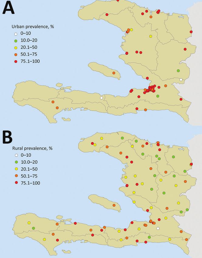
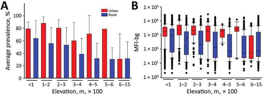
Bead Serologic Assay to Evaluate CHIKV Epidemic

Statistical Methods                                                 category, only the difference within the 100–200 m category
We used the Mann-Whitney rank sum test to determine dif-            was found to be significant (p = 0.024), although urban/ru-
ferences between groups for continuous variables and the            ral differences for other categories at elevations
RESEARCH

Relationship between Seroprevalence and IgG Titer                      population would have evidence of exposure to this arbo-
and Age by Urban and Rural Setting                                     virus within 9 months of introduction. The fast spread of
Seroprevalence of CHIKV IgG and a plot of median MFI-                  CHIKV has been observed before: in the French island of
bg (magnitude of IgG response) by age categories for per-              La Reunion in the 2005–2006 outbreak, more than one third
sons living in urban versus rural areas are shown in Figure            of the population was believed to have been exposed, and
3. These graphs show similar patterns; seroprevalence and              63% of the persons living on the island of the Union of the
MFI-bg responses were consistently higher from persons                 Comoros were exposed to CHIKV during the initial out-
living in urban versus rural areas, regardless of age. Median          break there (11,12).
MFI-bg was higher in urban areas when compared with ru-                     One of our most striking findings was that, even after
ral areas for all age groups (p80 y, p = 0.03) (Figure 3, panel B). By using regres-             had a >3-fold increase in odds (adjusted odds ratio 3.34)
sion models, we did not find age to be a significant predictor         of CHIKV exposure. This result, in combination with the
when considering either seroprevalence of CHIKV IgG (p                 increased likelihood of CHIKV exposure at lower eleva-
= 0.34) or magnitude of the IgG response (p = 0.19), in-               tion, is directly in line with the consistent observation that
dicating equivalent probability of lifetime exposure for all           the Aedes vector house index (HI) is typically higher in
Haiti residents, regardless of age. Children 0–10 years of             these settings. Multiple studies have observed an increase
age in rural areas showed exceptionally low median MFI-                in Ae. aegypti vector HI and breeding sites in urban areas
bg compared with urban children of the same age group.                 when compared with rural locations (13–17). Previous find-
                                                                       ings showing increased prevalence of Ae. aegypti mosquito
Discussion                                                             colonization in areas of high-density housing and higher
From this serosurvey, we found 57.9% of the 4,438 resi-                water temperature allude to the importance of the urban
dents of Haiti that were tested had IgG responses to CHIKV             environment for this vector (18). These similar behavioral
by early 2015. Although our sample was not nationally                  patterns have recently been observed in Haiti, where Ae.
representative, we may have underestimated true expo-                  aegypti mosquitoes are readily observed in urban and peri-
sure rates, because 38.8% of persons sampled resided in                urban settings and have a propensity for manmade habitats
urban areas, and population estimates for Haiti calculated             (19,20). Although Aedes sp. mosquito populations have
the percentage of the population in cities at 48.8% (9). We            not been mapped by elevation in recent surveys in Haiti,
found a seroprevalence of 78.4% in urban areas, similar to             past studies showed substantial decreases in prevalence of
the 78.9% seroprevalence previously found in the urban                 hemagglutination inhibition antibodies to dengue virus (as
area of Leogane in August 2014 (10). Considering that the              well as to the vector) in moving from low-lying coastal to
first confirmed cases of CHIKV in Haiti were reported in               mountainous inland areas (21). We observed a gradual and
April 2014 (the same month confirmed cases were seen in                consistent decrease in seroprevalence to CHIKV IgG for
the Dominican Republic [1]), it is notable that >50% of the            rural areas of increasing elevation >600 m, but this same

Figure 2. Seroprevalence and chikungunya IgG levels among persons living in urban and rural areas at different elevations, Haiti,
December 2014–February 2015. A) Seroprevalence mean of persons sampled in urban or rural areas at different elevations; error bars
indicate SEM. B) Chikungunya IgG median fluorescence intensity minus background signal by urban and rural sampling sites at different
elevations. Boxes indicate interquartile ranges; horizontal lines within boxes indicate medians; black dots indicate values 90th
percentiles; error bars indicate 10th and 90th percentiles of data. M, meters; MFI-bg, median fluorescence intensity minus background.

998                            Emerging Infectious Diseases • www.cdc.gov/eid • Vol. 24, No. 6, June 2018
Bead Serologic Assay to Evaluate CHIKV Epidemic
Figure 3. Seroprevalence
and chikungunya IgG levels
among persons living in
urban and rural areas, by
age group, Haiti, December
2014–February 2015. A)
Mean seroprevalence by
urban or rural setting and age
category. B) Range of median
fluorescence intensity minus
background (IgG responses)
to chikungunya antigens for
the same age categories. Bars
indicate interquartile ranges;
horizontal lines within bars
indicate medians; black dots
indicate values >10th or 600 m elevation showed no differ- persons acquiring CHIKV infection at the geographic loca-
ence in seroprevalence or CHIKV IgG titers regardless of tion of residence, as this was the location in which persons
whether they lived in an urban or rural setting. This find- were sampled. Travel history was not gathered for partici-
ing shows that urban environments in Haiti become less pants in this study.
Emerging Infectious Diseases • www.cdc.gov/eid • Vol. 24, No. 6, June 2018 999RESEARCH
In our study, all age groups living in urban areas References
showed substantially higher MFI-bg signal intensity (IgG 1. Fischer M, Staples JE; Arboviral Diseases Branch, National Center
for Emerging and Zoonotic Infectious Diseases, CDC. Notes from
responses) to the CHIKV antigens compared with the same the field: chikungunya virus spreads in the Americas—Caribbean
age groups living in rural areas. Differences were not seen and South America, 2013–2014. MMWR Morb Mortal Wkly Rep.
between IgG prevalence or titer between younger and older 2014;63:500–1.
age categories, which is atypical for serologic studies of 2. Poirier MJ, Moss DM, Feeser KR, Streit TG, Chang GJ,
Whitney M, et al. Measuring Haitian children’s exposure to
infectious diseases (2,9,24,25), because an older age indi- chikungunya, dengue and malaria. Bull World Health
cates more life-years of potential exposure to an endemic Organ. 2016;94:817–825A. http://dx.doi.org/10.2471/
infectious disease and, thus, acquisition of IgG. These sero- BLT.16.173252
logic findings provide strong evidence for the rapid dissem- 3. Johnson AJ, Martin DA, Karabatsos N, Roehrig JT. Detection of
anti-arboviral immunoglobulin G by using a monoclonal
ination of CHIKV by the Aedes vector. It appears all age antibody-based capture enzyme-linked immunosorbent assay.
groups in the population were uniformly susceptible to a J Clin Microbiol. 2000;38:1827–31.
high rate of transmission, with a preference for urban popu- 4. Johnson BW, Russell BJ, Goodman CH. Laboratory diagnosis
lations. Of note was the finding that childrenBead Serologic Assay to Evaluate CHIKV Epidemic
15. Codeço CT, Lima AW, Araújo SC, Lima JB, Maciel-de-Freitas R, urbanization. J Vector Ecol. 2015;40:46–58. http://dx.doi.org/
Honório NA, et al. Surveillance of Aedes aegypti: comparison of 10.1111/jvec.12131
house index with four alternative traps. PLoS Negl 21. Ventura AK, Ehrenkranz NJ. Endemic dengue virus infection
Trop Dis. 2015;9:e0003475. http://dx.doi.org/10.1371/ in Hispaniola. I. Haiti. J Infect Dis. 1976;134:436–41.
journal.pntd.0003475 http://dx.doi.org/10.1093/infdis/134.5.436
16. Tsuda Y, Suwonkerd W, Chawprom S, Prajakwong S, 22. Christophers SR. Aëdes aegypti, the yellow fever mosquito;
Takagi M. Different spatial distribution of Aedes aegypti and its life history, bionomics, and structure. New York: Cambridge
Aedes albopictus along an urban-rural gradient and the relating University Press; 1960. p. 38–40.
environmental factors examined in three villages in northern 23. Equihua M, Ibáñez-Bernal S, Benítez G, Estrada-Contreras I,
Thailand. J Am Mosq Control Assoc. 2006;22:222–8. Sandoval-Ruiz CA, Mendoza-Palmero FS. Establishment of
http://dx.doi.org/10.2987/8756-971X(2006)22[222:DSDOAA] Aedes aegypti (L.) in mountainous regions in Mexico:
2.0.CO;2 increasing number of population at risk of mosquito-borne
17. Braks MA, Honório NA, Lourenço-De-Oliveira R, Juliano SA, disease and future climate conditions. Acta Trop. 2017;166:316–27.
Lounibos LP. Convergent habitat segregation of Aedes aegypti http://dx.doi.org/10.1016/j.actatropica.2016.11.014
and Aedes albopictus (Diptera: Culicidae) in southeastern Brazil 24. Moss DM, Priest JW, Hamlin K, Derado G, Herbein J,
and Florida. J Med Entomol. 2003;40:785–94. http://dx.doi.org/ Petri WA Jr, et al. Longitudinal evaluation of enteric protozoa
10.1603/0022-2585-40.6.785 in Haitian children by stool exam and multiplex serologic assay.
18. Cox J, Grillet ME, Ramos OM, Amador M, Barrera R. Habitat Am J Trop Med Hyg. 2014;90:653–60. http://dx.doi.org/10.4269/
segregation of dengue vectors along an urban environmental ajtmh.13-0545
gradient. Am J Trop Med Hyg. 2007;76:820–6. 25. Neff JM, Morris L, Gonzalez-Alcover R, Coleman PH, Lyss SB,
19. Lenhart A, Orelus N, Maskill R, Alexander N, Streit T, Negron H. Dengue fever in a Puerto Rican community.
McCall PJ. Insecticide-treated bednets to control dengue vectors: Am J Epidemiol. 1967;86:162–84. http://dx.doi.org/10.1093/
preliminary evidence from a controlled trial in Haiti. Trop Med oxfordjournals.aje.a120722
Int Health. 2008;13:56–67. http://dx.doi.org/10.1111/
j.1365-3156.2007.01966.x Address for correspondence: Eric W. Rogier, Centers for Disease Control
20. Samson DM, Archer RS, Alimi TO, Arheart KL, Impoinvil DE,
and Prevention, 1600 Clifton Road NE, Mailstop D67, Atlanta, GA
Oscar R, et al. New baseline environmental assessment of
mosquito ecology in northern Haiti during increased 30329-4027, USA; email: erogier@cdc.gov
Emerging Infectious Diseases • www.cdc.gov/eid • Vol. 24, No. 6, June 2018 1001You can also read