Annals of Clinical Anatomy
←
→
Page content transcription
If your browser does not render page correctly, please read the page content below
Annals of Clinical Anatomy                                                                                                                       Research Article
                                                                                                                                          Published: 25 Apr, 2018

   Calcification Scarcely Occurs in Human Atrioventricular
                   Nodal Arteries in Old Age
                                             Yoshiyuki Tohno1*, Setsuko Tohno1, Takeshi Minami2, Pasuk Mahakkanukrauh1,3,4 and
                                             Noppadol Phasukdee1
                                             1
                                                 Department of Anatomy, Faculty of Medicine, Chiang Mai University, Thailand
                                             2
                                               Laboratory of Environmental Biology, Department of Life Science, Faculty of Science and Technology
                                             Kinki University, Japan
                                             3
                                               Forensic Osteology Center, Faculty of Medicine, Chiang Mai University, Thailand
                                             4
                                                 Excellence in Osteology Research and Training Center (ORTC), Chiang Mai University, Thailand

                                             Abstract
                                             To elucidate age-related changes of the Atrioventricular Nodal (AVN) artery, the authors investigated
                                             age-related changes of elements in the AVN artery by direct chemical analysis. In addition, the effects
                                             of different arterial origins, arterial sizes, and genders on element accumulation were investigated in
                                             the AVN arteries. Sixty-two formalin-fixed adult Thai hearts were dissected, and the following two
                                             types of the AVN artery were found: The first type was a single AVN artery arising from the Right
                                             Coronary Artery (RCA). The second type was a single AVN artery arising from the terminal part of
                                             the Left Circumflex Artery (LCX). For element analysis, both 55 and 7 AVN arteries arising from the
                                             RCA and the LCX, respectively, were used. After the arteries were incinerated with nitric acid and
                                             perchloric acid, element contents were determined by inductively coupled plasma-atomic emission
                                             spectrometry. It was found that the Zn content decreased significantly in the AVN arteries with
                                             aging, but six element contents such as Ca, P, S, Mg, Fe, and Na did not change significantly with
                                             aging. Regarding the relationships among seven elements in the AVN arteries, extremely significant
                                             direct correlations were found both between Ca and Mg contents and between P and S contents,
                                             and a significant direct correlation was found between S and Mg contents. However, no significant
                                             correlation was found between Ca and P contents in the AVN arteries. To examine an effect of
                    OPEN ACCESS              the different arterial origins on element accumulation, the AVN arteries were separated into the
                                             RCA and the LCX groups by the arterial origin and age-related changes of element contents were
                     *Correspondence:        compared between two groups. It was found that there were no significant differences between the
        Yoshiyuki Tohno, Department of       RCA and LCX groups in age-related changes of Ca and P contents. No gender differences and effect
  Anatomy, Faculty of Medicine, Chiang       of arterial size were found in age-related changes of Ca and P contents in the AVN arteries. To
    Mai University, Chiang Mai, 50200,       elucidate whether calcification occurred in the AVN arteries in old age, both the mass ratios of Ca/P
  Thailand, Tel: 66-53-935312; Fax: 66-      and Mg/Ca were estimated in the AVN arteries. The mass ratio of Ca/P increased progressively in
                            53-935304;       the AVN arteries with Ca increase, being not constant. The mass ratio of Mg/Ca decreased gradually
         E-mail: yoshiyuki.t@cmu.ac.th       in the AVN arteries with Ca increase, but the average mass ratio of Mg/Ca was moderate, being 8.9%
          Received Date: 27 Mar 2018         ± 0.9%. These results indicated that calcification scarcely occurred in the AVN arteries in old age,
          Accepted Date: 18 Apr 2018         independently of the arterial origin, arterial size and gender.
          Published Date: 25 Apr 2018
                                             Keywords: Atrioventricular nodal artery; Coronary artery; Calcium; Phosphorus; Magnesium;
                               Citation:     Aging
            Tohno Y, Tohno S, Minami T,
     Mahakkanukrauh P, Phasukdee N.          Introduction
Calcification Scarcely Occurs in Human
                                                 The conduction system of the heart is supplied by the Sinoatrial Nodal (SAN) artery, the
  Atrioventricular Nodal Arteries in Old
                                             Atrioventricular Nodal (AVN) artery, the first septal branch of the Left Anterior Descending artery
 Age. Ann Clin Anat. 2018; 1(1): 1002.       (LAD), and the posterior descending branch of the Right Coronary Artery (RCA). These arteries
    Copyright © 2018 Yoshiyuki Tohno.        ensure adequate blood supply to maintain the electrical properties in the heart. It is well known
          This is an open access article     that a high accumulation of Ca and P occurs in the proximal sites of the LAD, Left Coronary Artery
          distributed under the Creative     (LCA), RCA, and Left Circumflex Artery (LCX) with aging [1-3]. For example, Montenegro and
   Commons Attribution License, which        Eggen [1] studied the distribution of atherosclerotic lesions along the axis of the coronary artery
   permits unrestricted use, distribution,   (topography) and the relationship of topography to concepts of pathogenesis and reported that a
       and reproduction in any medium,       higher prevalence of atherosclerotic lesions in isolated coronary arteries occurred from the first to
   provided the original work is properly    second centimeters of both the LAD and the RCA, with a decrease in prevalence from 3 cm onward
                                    cited.   in the LAD and 5 cm onward in the RCA.

Remedy Publications LLC.                                                       1                                         2018 | Volume 1 | Issue 1 | Article 1002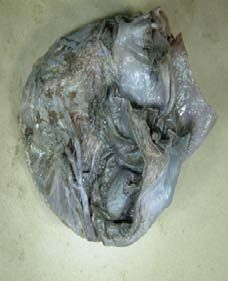
Yoshiyuki Tohno, et al., Annals of Clinical Anatomy
Table 1: Subjects used in the present study.
81 M Senility
Age (Years) Sex Cause of death
81 W Senility
36 M Epilepsy
83 M Septicemia
36 W Lymphoma
83 W Cerebrovascular disease
38 M Strangulation
83 W Infected emphysema
40 M Hepatic cancer
83 M Septicemia
47 M Pneumonia
84 W Senility
50 W Hepatic cancer
85 M Septicemia
51 M Leukemia
86 W Pneumonia
52 M Gunshot wound
87 W Heart failure
53 W Chronic renal failure
88 M Respiratory tract infection
55 M Intracerebral hemorrhage
88 W Myocardial infarction
57 M Alcoholism
89 M Pneumonia
57 M Hypertension
92 M Heart failure
57 W Cerebrovascular disease
93 W COPD and pneumonia
57 W Lung cancer
94 W Pneumonia
60 M Urinary tract infection
Note: M and W indicate man and woman.
63 M Bile duct cancer
Table 2: Incidence of the AVN artery with the Ca content more than 5.0 mg/g.
63 W Intracerebral hemorrhage
Age group (years) Incidence (%)
64 M Myocardial infarction
30s (n=3) 0 (0/3)
65 M Acute natural illness
40s (n=2) 50.0 (1/2)
66 M Intracerebral hemorrhage
50s (n=9) 44.4 (4/9)
66 M Liver cirrhosis
60s (n=11) 18.2 (2/11)
67 M Senility
70s (n=19) 31.6 (6/19)
67 M Rectal cancer
80s (n=15) 26.7 (4/15)
67 W Heart failure
90s (n=3) 33.3 (1/3)
69 W Intracerebral hemorrhage Note: The numbers of cases are indicated in parentheses.
70 M Traumatic head injury
There are a few reports on age-related changes of the AVN artery
72 M Acute myocardial infarction
[4-6]. Velican et al. [4,5] investigated histological changes of the
72 W Aspiration pneumonia vessels supplying the conduction system of the heart including the
73 M Cardiac hypertrophy AVN artery, with aging. They reported that in the healthy subjects,
73 M Pneumonia intimal thickening of the AVN artery started to occur in the 30s of
the subjects and then increased in the 40s and the 50s of the subjects.
73 M Septicemia
However, changes of the AVN artery in old age had not yet been
75 M Senility
studied. Therefore, the authors investigated age-related changes
75 W Senility of the AVN artery using adult Thai hearts (of the subjects between
76 W Pneumonia 36 and 94 years of age) from a viewpoint of elements. It was found
76 W Cerebrovascular disease
that calcification scarcely occurred in the AVN arteries in old age,
independently of the arterial origin, arterial size, and gender.
78 M Hepatic cancer
78 M Intracerebral hemorrhage
Materials and Methods
78 M Hyperglycemic crisis Dissection of the AVN arteries
78 M Tracheal obstruction The research was carried out on 62 adult Thai hearts (of subjects
78 W Senility
between 36 and 94 years of age: average age, 70.7 ± 14.5 years) received
from the Department of Anatomy, Faculty of Medicine, Chiang Mai
78 W Septicemia
University. The perivascular fatty tissue was carefully removed, where
79 M Pneumonia necessary, to visualize the epicardial course of the coronary arteries.
79 W Intestinal cancer The AVN arteries were carefully dissected in the hearts. The AVN
79 M Senility artery arising from the distal RCA penetrated into the posterior
interatrial septum base at the level of heart crux, and then coursed
80 W Senility
forwards and upwards, to be directly related to the upper angle of
80 W Cardiac arrest the Posterior Septal Space (PSS) until its apex. The AVN artery also
Remedy Publications LLC. 2 2018 | Volume 1 | Issue 1 | Article 1002Yoshiyuki Tohno, et al., Annals of Clinical Anatomy
a b
LCX AVNA
AVNA
RCA
Figure 1: Posterior views of the hearts with type 1 (a) and type 2 (b) of the
AVN artery. AVNA atrioventricular nodal artery; LCX left circumflex artery;
RCA right coronary artery.
Figure 3: Age-related changes of Ca (a) and P (b) contents in the RCA and
LCX groups of the AVN arteries which arise from the RCA and the LCX,
respectively. The open and solid circles indicate the RCA and LCX groups of
the AVN arteries, respectively. The straight and dotted lines of trend with age
indicate the RCA and LCX groups of the AVN arteries, respectively.
[11] are related to atherosclerosis; and Na is an important cation.
The resulting filtrates were analyzed by inductively coupled plasma-
atomic emission spectrometry (iCAP 7400 ICP-OES Duo; Thermo
Fisher Scientific Japan Inc., Kanagawa, Japan). The conditions
were as follows: 1.15 kW from the radiofrequency forward power,
an auxiliary gas flow rate of 0.5 l/min, a nebulizer gas flow rate of
0.55 l/min, a coolant gas flow rate of 12 l/min, a purge gas flow
rate of 3.2 l/min, and an exposure time of 10 s. Especially prepared
standard solutions of Ca, Mg, Zn, Fe, and Na for atomic absorption
spectrometry and phosphate and sulfate ions for ion chromatography
were purchased from Wako Pure Chemical Industries (Osaka, Japan)
and were used as standard solutions. The measurement of elements
was performed at a fixed wave length of 588.995 nm for Na, 393.366
nm for Ca, 279.553 nm for Mg, 259.940 nm for Fe, 213.856 nm for Zn,
Figure 2: Age-related changes of Ca (a), P (b), S (c), Mg (d), Zn (e), Fe (f), 180.731 nm for S and 177.495 nm for P. The amount of elements was
and Na (g) contents in the AVN arteries. The AVN arteries consisted of 55
and 7 AVN arteries arising from the RCA and the LCX, respectively.
expressed on a dry weight basis.
Scanning electron microscopy
arose from the terminal part of the LCX. The artery ran through the The arterial samples were carefully dissected longitudinally. The
fat-filled space located at the inferior wall of the right atrium. The samples were post-fixed in 2% osmium tetroxide, 0.1 M phosphate
diameter of the AVN artery was measured by the precise electronic buffer (pH 7.4) for 1h to 2 h and were rinsed in the phosphate buffer.
caliper. For element analysis, the 62 AVN arteries arising from the The samples were then dehydrated in a series of graded ethanol
RCA and the LCX were used in the present study. (50% to 100%) for 15 min. After critical point drying, the samples
Determination of elements were rendered conductive by sputtering them with gold. The coated
samples were examined by scanning electron microscopy (JSM-
The arterial samples were washed thoroughly with distilled water
6610LV; JEOL, Tokyo) operated at 15 kV.
and were dried at 95°C for 16 h. After 1 ml concentrated nitric acid
was added to the dry samples to incinerate, the mixtures were heated Statistical analysis
at 100°C for 2 h. After the addition of 0.5 ml concentrated perchloric Statistical analyses were performed using the GraphPad Prism
acid, they were heated at 100°C for an additional 2 h [7]. The samples version 7.0 (GraphPad Software, San Diego, CA, USA). Pearson’s
were adjusted to a volume of 10 ml by adding ultrapure water and correlation was used to investigate the association between parameters.
were filtered through filter paper (no. 7; Toyo Roshi, Osaka, Japan). It was analyzed whether significant differences were found between
Seven elements of Ca, P, S, Mg, Zn, Fe, and Na were selected for two slopes and between two intercepts of the regression lines. The
measurement because of the following reasons: Both Ca and P are two-tailed unpaired student t test was used to analyze differences
directly correlated with Mg on calcification [8]; smooth muscles between groups. A p value of less than 0.05 was considered to be
containing S decrease on atherosclerosis [9]; both Zn [10] and Fe significant. Data were expressed as the mean ± standard deviation.
Remedy Publications LLC. 3 2018 | Volume 1 | Issue 1 | Article 1002Yoshiyuki Tohno, et al., Annals of Clinical Anatomy
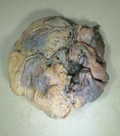
Figure 5: Age-related changes of Ca (a) and P (b) contents in the smaller
group less than 1.0 mm of the diameter and the larger group more than 1.0
Figure 4: Age-related changes of Ca (a) and P (b) contents in men and
mm of the diameter in the AVN arteries. The open and solid circles indicate
women’s AVN arteries arising from the RCA and the LCX. The open and solid
smaller and larger groups. The straight and dotted lines of trend with age
circles indicate men and women’s AVN arteries, respectively. The straight
indicate smaller and larger groups.
and dotted lines of trend with age indicate men and women’s AVN arteries,
respectively.
Results
Table 1 indicates ages, sexes, and causes of deaths of the 62
subjects used in the present study.
AVN arteries
The following two types of the AVN artery were found in 62 adult
hearts (Figure 1): The first type was a single AVN artery arising from
the RCA (Figure 1a). It arose from the distal RCA, penetrated into the
posterior interatrial septum base at the level of heart crux, and then
coursed forwards and upwards, to be directly related to the upper
angle of the PSS until its apex. The second type was a single AVN
artery arising from the LCX (Figure 1b). It arose from the terminal
part of the LCX and ran through the fat-filled space located at the
inferior wall of the right atrium. The incidences of types 1 and 2 were
88.7% (55/62) and 11.3% (7/62), respectively.
Measurements on the AVN arteries
Figure 6: Changes of mass ratios of Ca/P (a) and Mg/Ca (b) in the AVN
The mean diameters of the AVN arteries arising from the RCA arteries. The mass ratios of Ca/P and of Mg/Ca are plotted against the Ca
and the LCX were 1.01 mm ± 0.30 mm (n=55) and 0.76 mm ± 0.27 content.
mm (n=7), respectively. The mean diameter of the AVN arteries
arising from the RCA was significantly larger than that from the LCX mg/g for Ca, 0.658 mg/g ± 0.149 mg/g for P, 3.700 mg/g ± 0.446 mg/g
(p=0.046). for S, 423.3 μg/g ± 65.74 μg/g for Mg, 687.7 μg/g ± 579.2 μg/g for Zn,
Age-related changes of elements in the AVN arteries 767.9 μg/g ± 399.3 μg/g for Fe, and 80.45 μg/g ± 146.0 μg/g for Na. The
average content of Ca was highest, and it decreased in the order of S,
The 62 AVN arteries arising from both the RCA and the LCX Fe, Zn, P, Mg, and Na.
were used for the present study. Figure 2 shows age-related changes
of seven element contents in the AVN arteries. The correlation Table 2 indicates the incidence of the AVN artery with the Ca
coefficients between age and element contents were estimated to be content more than 5.0 mg/g, which is not contained in a normal artery
0.057 (p=0.665) for Ca, 0.013 (p=0.919) for P, 0.111 (p=0.395) for S, [12]. The incidence of the AVN artery with the Ca content more than
-0.079 (p=0.540) for Mg, -0.346 (p=0.006) for Zn, 0.059 (p=0.654) for 5.0 mg/g was 50% and 44% in the 40s and the 50s, respectively and did
Fe, and 0.024 (p=0.855) for Na. A very significant inverse correlation not increase in the age groups older than the 50s. The mean incidence
was found between age and Zn content in the AVN arteries, but was 29.6% between the 50s and the 80s of the subjects.
no significant correlations were found between age and the other Relationships among seven element contents in the AVN
element contents such as Ca, P, S, Mg, Fe, and Na. arteries
The average contents of seven elements were 4.746 mg/g ± 0.724 The relationships among seven element contents were examined
Remedy Publications LLC. 4 2018 | Volume 1 | Issue 1 | Article 1002Yoshiyuki Tohno, et al., Annals of Clinical Anatomy
in the AVN arteries described above. Table 3 lists the relationships
among seven element contents in the AVN arteries. Extremely
significant direct correlations were found both between Ca and Mg
contents and between P and S contents, and a significant direct
correlation was found between S and Mg contents in the AVN
arteries. However, no significant correlation was found between Ca
and P contents in the AVN arteries. As the Ca content increased in
the artery, the Mg content increased simultaneously in the artery, but
the P content did not increase.
Age-related changes of elements in the two different AVN
arteries
The AVN arteries were separated into the RCA (n=55) and the
LCX (n=7) groups by the arterial origin. The RCA group ranged in
age from 36 to 94 years (average age=70.8 ± 14.3 years), whereas the
Figure 7: Observation of the inner surface of the AVN artery from 82-year-old
LCX group ranged in age from 36 to 85 years (average age=69.9 ± 17.2
man by scanning electron microscopy. Scale bar: 50 µm.
years). The two groups were separately analyzed to examine whether
age-related changes of elements were different between two groups.
of the regression lines between age and P content in Figure 4b showed
Figure 3 shows age-related changes of Ca and P contents in both that no significant differences were found between the two slopes
the RCA and the LCX groups. The correlation coefficients between (p=0.757) and between the two intercepts (p=0.871) of the regression
age and Ca content were estimated to be 0.010 (p=0.945) in the RCA lines for men and women’s AVN arteries. Therefore, no gender
group and 0.563 (p=0.188) in the LCX group (Figure 3a). The Ca differences were found in the AVN arteries with regard to age-related
content did not change significantly in two groups with aging. The changes of Ca and P contents.
correlation coefficients between age and P content were estimated to
be 0.046 (p=0.744) in the RCA group and -0.176 (p=0.706) in the LCX Effect of arterial size on age-related changes of Ca and P
group (Figure 3b). No significant correlations were found between contents
age and either Ca or P content in two groups. To examine an effect of arterial size on age-related changes of
elements, the AVN arteries were separated into the smaller group
The analysis of the regression lines between age and Ca content in
(n=34) less than 1.0 mm of the diameter and the larger group (n=28)
Figure 3a showed that no significant differences were found between
more than 1.0 mm of the diameter. The smaller group ranged in age
the two slopes (p=0.403) and between the two intercepts (p=0.431) of
the regression lines for the RCA and the LCX groups. The analysis of from 36 to 94 years (average age=71.5 ± 16.5 years), whereas the
the regression lines between age and P content in Figure 3b showed larger group ranged in age from 38 to 89 years (average age=69.3
that no significant differences were found between the two slopes ± 12.3 years). The two groups were separately analyzed to examine
(p=0.558) and between the two intercepts (p=0.750) of the regression whether age-related changes of Ca and P contents were different
lines for the RCA and the LCX groups. Therefore, no significant between smaller and larger groups.
differences were found in age-related changes of Ca and P contents Figure 5 shows age-related changes of Ca and P contents in both
between the two groups. the smaller and larger groups. The correlation coefficients between
Gender differences in Ca and P contents of the AVN age and Ca content were estimated to be 0.057 (p=0.755) in the
arteries smaller group and 0.002 (p=0.992) in the larger group (Figure 5a).
No significant correlations were found between age and Ca content
Men’s samples consisted of 31 and 5 AVN arteries arising from
in two groups. The correlation coefficients between age and P content
the RCA and the LCX, respectively. The average age of men’s subjects
were estimated to be 0.072 (p=0.697) in the smaller group and -0.037
was 68.3 ± 14.5 years. Women’s samples consisted of 24 and 2 AVN
(p=0.855) in the larger group (Figure 5b). No significant correlations
arteries arising from the RCA and the LCX, respectively. The average
were found between age and P content in two groups.
age of women’s subjects was 74.0 ± 14.1 years.
Figure 4 shows age-related changes of Ca and P contents in men The analysis of the regression lines between age and Ca content in
and women’s AVN arteries. The correlation coefficients between age Figure 5a showed that no significant differences were found between
and Ca content were estimated to be 0.024 (p=0.893) in men’s AVN the two slopes (p=0.839) and between the two intercepts (p=0.170) of
arteries and 0.208 (p=0.308) in women’s AVN arteries (Figure 4a). the regression lines for the smaller and larger groups. The analysis of
The correlation coefficients between age and P content were estimated the regression lines between age and P content in Figure 5b showed
to be -0.014 (p=0.935) in men’s AVN arteries and 0.106 (p=0.613) in that no significant differences were found between the two slopes
women’s AVN arteries (Figure 4b). No significant correlations were (p=0.710) and between the two intercepts (p=0.226) of the regression
found between age and either Ca or P content in men and women’s lines for the smaller and larger groups. Therefore, no significant
ones. Both the Ca and P contents did not increase significantly in men differences were found in age-related changes of Ca and P contents
and women’s AVN arteries with aging. between the smaller and larger groups.
The analysis of the regression lines between age and Ca content in Mass ratios of Ca/P and Mg/Ca in the AVN arteries
Figure 4a showed that no significant differences were found between To elucidate whether calcification occurred in the AVN arteries
the two slopes (p=0.488) and between the two intercepts (p=0.111) of in old age, both the mass ratios of Ca/P and Mg/Ca were investigated
the regression lines for men and women’s AVN arteries. The analysis in 62 AVN arteries arising from both the RCA and the LCX.
Remedy Publications LLC. 5 2018 | Volume 1 | Issue 1 | Article 1002Yoshiyuki Tohno, et al., Annals of Clinical Anatomy
Table 3: Relationships among seven element contents in the AVN arteries.
Correlation coefficient and p value
Element
P S Mg Zn Fe Na
Ca -0.080 (0.546) 0.033 (0.802) 0.773 (Yoshiyuki Tohno, et al., Annals of Clinical Anatomy
in the SAN arteries of the subjects (age range, 36-94 years; average magnesium and decrease of sulfur in human arteries. Biol Trace Elem Res.
age, 70.6 ± 14.5 years), being 0.644 mg/g ± 0.259 mg/g [34]. All the 2001;82(1-3):9-19.
AVN, SAN, internal thoracic, and radial arteries are characterized 10. Little PJ, Bhattacharya R, Moreyra AE, Korichneva IL. Zinc and
by a low average content of P. In the absence of calcification, the P cardiovascular disease. Nutrition. 2010;26(11-12):1050-7.
content of tissue is mostly determined by the nucleic acid content
11. Kraml P. The role of iron in the pathogenesis of atherosclerosis. Physiol
(DNA and RNA) and the phospholipid content of tissue. Res. 2017;66(Suppl1):S55-67.
There are many studies on the relationship between Ca and cell 12. Tohno Y, Tohno S, Minami T, Utsumi M, Moriwake Y, Nishiwaki F, et al.
proliferation [35-39]. For example, Whitfield et al. [35] demonstrated Age-related changes of mineral contents in the human aorta and internal
that Ca positively controlled the proliferation of non-tumorigenic thoracic artery. Biol Trace Elem Res. 1998;61(2):219-26.
epithelial and mesenchymally derived bovine, human, and rodent 13. Thoma R. Uber die Abhangigkeit der Bindegewebsneubildung in der
cells in vitro. According to Velican et al. [4], the incidence of the Arterienintima von den mechanischen Bedingungen des Blutumlaufes:
AVN artery with intimal thickening was 28% in 51-55 years of age Erste Mitteilung. Virchows Arch Pathol Anat Physiol. 1883;93:443-505.
with regard to apparently healthy persons. According to Kozlowski et 14. Wolkoff K. Uber die histologische Struktur der Coronararterien des
al. [6], the incidence of the AVN artery with intimal thickening was menschlichen Herzens. Virchows Arch Pathol Anat Physiol. 1923;241:42-
about 50% in the normal subjects ranging in age from 17 to 86 years 58.
(average age, 56 ± 14 years). In the present study, the incidence of
15. Wolkoff K. Uber die Altersveranderungen der Arterien bei Tieren.
the AVN artery with the Ca content more than 5.0 mg/g was 44.4% Virchows Arch. 1924;252:208-28.
(4/9 cases) in the 50s of the subjects and 29.0% (18/62 cases) in all
the subjects of 36-94 years of age. The incidence of the AVN artery 16. Stary HC, Blankenhorn DH, Chandler AB, Glagov S, Insull W,
Richardson M, et al. A definition of the intima of human arteries and of its
with this Ca content appears to be similar to those by Velican et al.
atherosclerosis-prone regions. A report from the committee on vascular
[4] and Kozlowski et al. [6]. Therefore, there is a possibility that this lesions of the council on arteriosclerosis, American heart association.
Ca increase may be related to intimal thickening in the AVN artery. Circulation. 1992;85(1):391-405.
Conclusion 17. Nakashima Y, Chen YX, Kinukawa N, Sueishi K. Distributions of
diffuse intimal thickening in human arteries: preferential expression
Calcification scarcely occurred in the AVN arteries in old age, in atherosclerosis-prone arteries from an early age. Virchows Arch.
independently of the arterial origin, arterial size and gender. 2002;441(3):279-88.
Acknowledgement 18. Nakashima Y, Wight TN, Sueishi K. Early atherosclerosis in humans:
role of diffuse intimal thickening and extracellular matrix proteoglycans.
The authors wish to thank Ms. Thippawan Yasanga (Medical Cardiovasc Res. 2008;79(1):14-23.
Science Research Equipment Center, Faculty of Medicine, Chiang
19. Subbotin VM. Excessive intimal hyperplasia in human coronary arteries
Mai University) for observation by scanning electron microscopy.
before intimal lipid depositions is the initiation of coronary atherosclerosis
References and constitutes a therapeutic target. Drug Discov Today. 2016;21(10):1578-
95.
1. Montenegro MR, Eggen DA. Topography of atherosclerosis in the
coronary arteries. Lab Invest. 1968;18(5):586-93. 20. Adams CW. Multiple factors in the pathogenesis of atherosclerosis. Guys
Hosp Rep. 1963;112:222-53.
2. Azuma C, Tohno S, Mahakkanukrauh P, Tohno Y, Satoh H, Chomsung
R, et al. Different accumulation of elements in the rami of the coronary 21. Duggal K, Tandon HD, Karmarkar MG, Ramalingaswami V. Diffuse
arteries of Thai. Biol Trace Elem Res. 2003;95(3):211-8. intimal thickening of the aorta in India and its relation to atherosclerosis. J
Pathol Bacteriol. 1966;92(1):49-56.
3. Tohno Y, Tohno S, Mahakkanukrauh P, Minami T, Sinthubua A,
Suwannahoy P, et al. Accumulation of calcium and phosphorus in the 22. Nakahara T, Dweck MR, Narula N, Pisapia D, Narula J, Strauss HW.
coronary arteries of Thai subjects. Biol Trace Elem Res. 2012;145(3):275- Coronary artery calcification: From mechanism to molecular imaging.
82. JACC Cardiovasc Imaging. 2017;10(5):582-93.
4. Velican D, Serban-Piriu G, Petrescu C, Velican C. Prevalence of thick 23. Ohnishi Y, Tohno S, Mahakkanukrauh P, Tohno Y, Vaidhayakarn P,
intimas and of obstructive lesions in the vessels supplying the conduction Azuma C, et al. Accumulation of elements in the arteries and cardiac
system of the heart. Med Interne. 1989;27(3):197-208. valves of Thai with aging. Biol Trace Elem Res. 2003;96(1-3):71-92.
5. Velican C, Velican D. Study of coronary intimal thickening. Atherosclerosis. 24. Tohno S, Tohno Y, Moriwake Y, Azuma C, Ohnishi Y, Minami T.
1985;56(3):331-44. Quantitative changes of calcium, phosphorus, and magnesium in common
iliac arteries with aging. Biol Trace Elem Res. 2001;84(1-3):57-66.
6. Kozlowski D, Owerczuk A, Kozluk E, Grzybiak M, Adamowicz-Kornacka
M, Walczak E, et al. Histologic evaluation of the atrioventricular nodal 25. Bigi A, Compostella L, Fichera AM, Foresti E, Gazzano M, Ripamonti A,
artery in healthy humans and in patients with conduction disturbances. et al. Structural and chemical characterization of inorganic deposits in
Folia Morphol (Warsz). 2000;59(3):145-52. calcified human mitral valve. J Inorg Biochem. 1988;34(2):75-82.
7. Tohno Y, Tohno S, Minami T, Ichii M, Okazaki Y, Utsumi M, et al. Age- 26. Kircelli F, Peter ME, Sevinc OE, Celenk FG, Yilmaz M, Steppan S, et al.
related changes of mineral contents in human thoracic aorta and in the Magnesium reduces calcification in bovine vascular smooth muscle cells
cerebral artery. Biol Trace Elem Res. 1996;54(1):23-31. in a dose-dependent manner. Nephrol Dial Transplant. 2012;27(2):514-21.
8. Tohno S, Tohno Y, Minami T, Moriwake Y, Azuma C, Ohnishi Y. 27. Louvet L, Buchel J, Steppan S, Passlick-Deetjen J, Massy ZA. Magnesium
Elements of calcified sites in human thoracic aorta. Biol Trace Elem Res. prevents phosphate-induced calcification in human aortic vascular smooth
2002;86(1):23-30. muscle cells. Nephrol Dial Transplant. 2013;28(4):869-78.
9. Tohno Y, Tohno S, Moriwake Y, Azuma C, Ohnishi Y, Minami T. 28. Bigi A, Foresti E, Gregorini R, Ripamonti A, Roveri N, Shah JS. The role
Accumulation of calcium and phosphorus accompanied by increase of of magnesium on the structure of biological apatites. Calcif Tissue Int.
Remedy Publications LLC. 7 2018 | Volume 1 | Issue 1 | Article 1002Yoshiyuki Tohno, et al., Annals of Clinical Anatomy
1992;50(5):439-44. occurrence of calcification in human sinoatrial nodal arteries in old age.
Biol Trace Elem Res. 2017.
29. Hruby A, O’Donnell CJ, Jacques PF, Meigs JB, Hoffmann U, Mckeown
NM. Magnesium intake is inversely associated with coronary artery 35. Whitfield JF, Boynton AL, MacManus JP, Sikorska M, Tsang BK. The
calcification: the Framingham heart study. JACC Cardiovasc Imaging. regulation of cell proliferation by calcium and cyclic AMP. Mol Cell
2014;7(1):59-69. Biochem. 1979;27(3):155-79.
30. Tohno Y, Tohno S, Moriwake Y, Azuma C, Ohnishi Y, Minami T. 36. Whitfield JF, Boynton AL, MacManus JP, Rixon RH, Sikorska M, Tsang B,
Simultaneous accumulation of calcium, phosphorus, and magnesium in et al. The roles of calcium and cyclic AMP in cell proliferation. Ann N Y
various human arteries. Biol Trace Elem Res. 2001;82(1-3):21-8. Acad Sci. 1980;339:216-40.
31. Sajja LR, Mannam G. Internal thoracic artery: anatomical and biological 37. Swierenga SH, Whitfield JF, Boynton AL, MacManus JP, Rixon RH,
characteristics revisited. Asian Cardiovasc Thorac Ann. 2015;23(1):88-99. Sikorska M, et al. Regulation of proliferation of normal and neoplastic rat
liver cells by calcium and cyclic AMP. Ann N Y Acad Sci. 1980;349(1):294-
32. Baikoussis NG, Papakonstantinou NA, Apostolakis E. Radial artery as
311.
graft for coronary artery bypass surgery: advantages and disadvantages
for its usage focused on structural and biological characteristics. J Cardiol. 38. Mailland M, Waelchli R, Ruat M, Boddeke HG, Seuwen K. Stimulation of
2014;63(5):321-8. cell proliferation by calcium and a calcimimetic compound. Endocrinology.
1997;138(9):3601-5.
33. Kaufer E, Factor SM, Frame R, Brodman RF. Pathology of the radial and
internal thoracic arteries used as coronary artery bypass grafts. Ann Thorac 39. Rodrigues MA, Gomes DA, Leite MF, Grant W, Zhang L, Lam W, et al.
Surg. 1997;63(4):1118-22. Nucleoplasmic calcium is required for cell proliferation. J Biol Chem.
2007;282(23):17061-8.
34. Tohno Y, Tohno S, Quiggins R, Minami T, Mahakkanukrauh P. Scarce
Remedy Publications LLC. 8 2018 | Volume 1 | Issue 1 | Article 1002You can also read