Origin and evolution of turiasaur dinosaurs set by means of a new 'rosetta' specimen from Spain
←
→
Page content transcription
If your browser does not render page correctly, please read the page content below
Zoological Journal of the Linnean Society, 2020, XX, 1–27. With 18 figures.

Origin and evolution of turiasaur dinosaurs set by means
of a new ‘rosetta’ specimen from Spain

                                                                                                                                     Downloaded from https://academic.oup.com/zoolinnean/advance-article/doi/10.1093/zoolinnean/zlaa091/5900936 by guest on 02 October 2020
RAFAEL ROYO-TORRES1,*, , ALBERTO COBOS1, PEDRO MOCHO2,3 and LUIS ALCALÁ1
1
 Fundación Conjunto Paleontológico de Teruel-Dinópolis/Museo Aragonés de Paleontología, avenida
Sagunto s/n 44002 Teruel, Spain
2
 Instituto Dom Luiz, Universidade de Lisboa, Edifício C6, Campo Grande, 1749-016 Lisboa, Portugal
3
 Grupo de Biología Evolutiva, UNED, Facultad de Ciencias, UNED, Paseo Senda del Rey 9, 28040
Madrid, Spain

Received 21 November 2019; revised 6 July 2020; accepted for publication 13 July 2020

Turiasauria is a non-neosauropod eusauropod clade of dinosaurs known since 2006, when the description of Turiasaurus
was published. This group, including Losillasaurus, was originally thought to have been restricted to the Late Jurassic of
Spain. However, over the last decade, our knowledge of this group has improved with the discovery of new taxa such as Zby
from the Portuguese Late Jurassic, Tendaguria from the Tanzanian Late Jurassic and Mierasaurus and Moabosaurus
from the Early Cretaceous of the USA. Here, we describe a new specimen of Losillasaurus from Spain, which allows us
to better understand the character variation in the cranial and postcranial skeleton. The review of some sauropod fauna
of Madagascar, and inclusion of some specimens of Turiasauria, suggest that this clade might have arisen in the Middle
Jurassic. According to our phylogenetic results, a specimen found in the early 19th century in Madagascar is shown to be
the oldest and only member of Turiasauria represented in the Middle Jurassic thus far. This is named Narindasaurus
thevenini gen. & sp. nov.. Turiasauria is thus known from the Middle Jurassic in Pangaea, diversified in the Late
Jurassic in Gondwana and Laurasia, and dispersed during the Early Cretaceous to North America.

ADDITIONAL KEYWORDS: Cretaceous – Jurassic – Sauropodomorpha – Laurasia – Gondwana – Dinosauria
– origins – phylogenetic systematics.

                    INTRODUCTION                                   2012), currently dated as Kimmeridgian–Tithonian
                                                                   (Campos Soto et al., 2019). Aside from the specimens
The Turiasauria clade represents a group of sauropod
                                                                   of turiasaurs from Teruel, other Late Jurassic
dinosaurs with a wide geographic distribution across
                                                                   sauropod fossils from the Iberian Peninsula have been
Europe, North America and Africa (Royo-Torres
                                                                   assigned to the Turiasauria clade, including two other
et al., 2017; Mannion et al., 2019). This clade is
                                                                   taxa: Losillasaurus giganteus Casanovas et al., 2001
known thanks to the study of the sauropod dinosaur
                                                                   and Zby atlanticus Mateus et al., 2014. Losillasaurus
Turiasaurus riodevensis Royo-Torres et al., 2006 found
                                                                   Casanovas et al., 2001 was found at the La Cañada
in Riodeva (Teruel province, south of the Iberian
                                                                   site (Losilla de Aras, Valencia) and originates from
Range, Spain). Turiasaurus Royo-Torres et al., 2006
                                                                   the same geological unit as Turiasaurus (Casanovas
is defined by the representative cranial (Fig. 1A) and
                                                                   et al., 2001; Royo-Torres et al., 2006). This taxon was
postcranial remains found from 2003 onwards in the
                                                                   only known from a partial braincase and postcranial
Villar del Arzobispo Formation (Luque et al., 2005;
                                                                   material (Casanovas et al., 2001), but here we describe
Royo-Torres et al., 2006; Royo-Torres & Upchurch,
                                                                   new material from another specimen (Supporting
                                                                   Information, File S1). We provide new information
                                                                   on the anterior part of the skull (Figs 1, 2), which
*Corresponding author. E-mail: royo@dinopolis.com
[Version of record, published online 3 September 2020;             includes details key to understanding the tooth
http://zoobank.org/ urn:lsid:zoobank.org:act:B7203900-             variation in turiasaurs. Zby atlanticus from the Vale
1F11-40CD-B4D5-115BAD1B96FE]                                       de Pombas site (Lourinhã), Lusitanian Basin in

© 2020 The Linnean Society of London, Zoological Journal of the Linnean Society, 2020, XX, 1–27                                  1
This is an Open Access article distributed under the terms of the Creative Commons Attribution-NonCommercial-NoDerivs
licence (http://creativecommons.org/licenses/by-nc-nd/4.0/), which permits non-commercial reproduction and distribution of the
work, in any medium, provided the original work is not altered or transformed in any way, and that the work is properly cited.
For commercial re-use, please contact journals.permissions@oup.com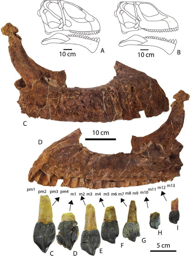
2   R. ROYO-TORRES ET AL.

                                                                                                                                 Downloaded from https://academic.oup.com/zoolinnean/advance-article/doi/10.1093/zoolinnean/zlaa091/5900936 by guest on 02 October 2020

Figure 1. Reconstructions of the skull of Turiasaurus (A) and Losillasaurus (B) in the lateral view. Left premaxilla, maxilla
and teeth (MAP-6005) of Losillasaurus giganteus (San Lorenzo specimen) in labial or lateral (C) and lingual or medial (D)
views. Right premaxillary tooth (MAP-6013) (E); and right maxillary teeth (MAP-6014) (F), (MAP-6015) (G), (MAP-6016)
(H), (MAP-6017) (I), (MAP-6018) (J), (MAP-6019) (K) in lingual views.

                               © 2020 The Linnean Society of London, Zoological Journal of the Linnean Society, 2020, XX, 1–27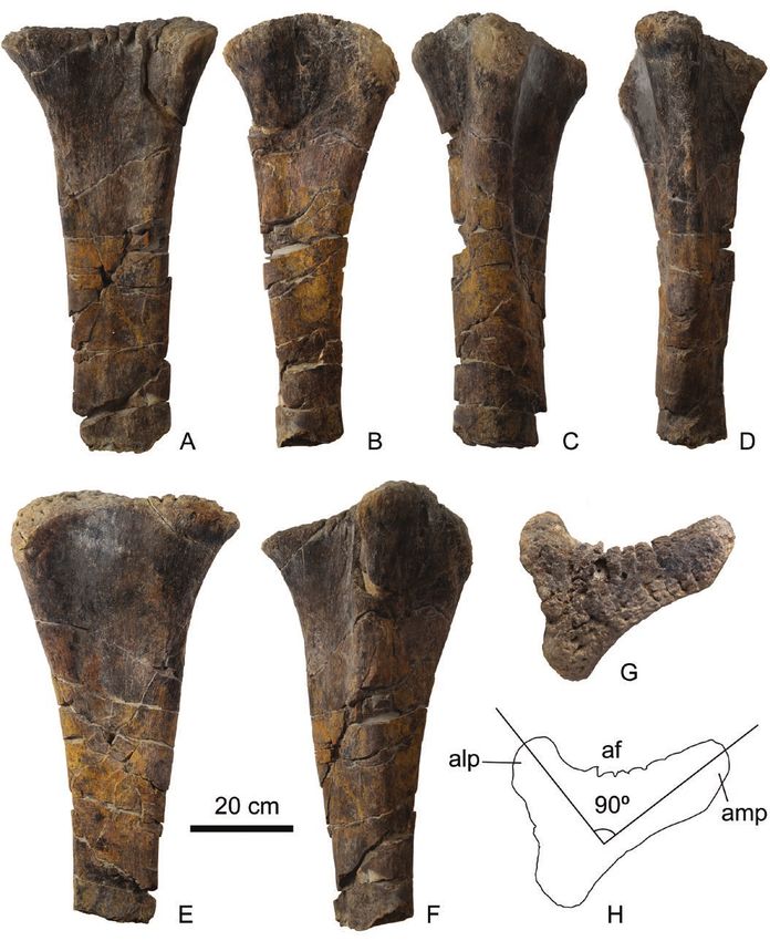
EVOLUTION OF TURIASAUR DINOSAURS                3

                                                                                                                          Downloaded from https://academic.oup.com/zoolinnean/advance-article/doi/10.1093/zoolinnean/zlaa091/5900936 by guest on 02 October 2020

Figure 2. Left dentary and teeth (MAP-6008) of Losillasaurus giganteus (San Lorenzo specimen) in lingual view (A); left
dentary teeth (MAP-6045) (d1), (MAP-6031) (d2), (MAP-6035) (d3), (MAP-6037) (d4), (MAP-6036) (d7), (MAP-6038) (d8),
(MAP-6039) (d9), (MAP-6040) (d10), (MAP-6020) (d13), (MAP-6021) (d14) in lingual views. Right dentary and teeth (MAP-
6009) of Losillasaurus giganteus (San Lorenzo specimen) in lingual view (B). Right dentary teeth (MAP-6032) (d2) and
(MAP-6033) (d3) of Losillasaurus giganteus (San Lorenzo specimen) in lingual view.

© 2020 The Linnean Society of London, Zoological Journal of the Linnean Society, 2020, XX, 1–27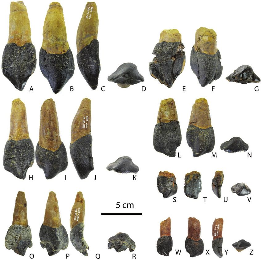
4    R. ROYO-TORRES ET AL.

Portugal, has been described with a tooth and forelimb         Mierasaurus, Moabosaurus, Tendaguria, Turiasaurus,
dated as Late Kimmeridgian (Mateus et al., 2014). In           Zby, (Royo-Torres et al., 2017; Mannion et al., 2019) and
the European domain, Schwarz et al. (2020) proposed            probably Amanzia (Schwarz et al., 2020), the relative
the taxa Amanzia greppini (von Huene, 1922) from               phylogenetic position between them is still debated.
Switzerland as a possible member of Turiasauria.               We investigated their position via an updated version
Recent studies also suggest a wider palaeogeographic           of the largest available data matrix for sauropods

                                                                                                                                Downloaded from https://academic.oup.com/zoolinnean/advance-article/doi/10.1093/zoolinnean/zlaa091/5900936 by guest on 02 October 2020
distribution and stratigraphic range for Turiasauria           (Mannion et al., 2019; Schwarz et al., 2020), which
extending to North America (Royo-Torres et al.,                included all turiasaur taxa and the new material
2017) with Mierasaurus Royo-Torres et al., 2017 and            from San Lorenzo referred to here to Losillasaurus. In
Moabosaurus Britt et al., 2017, and Africa (Mannion            addition, in order to test the potential close relationship
et al., 2019) with Tendaguria Wild, 1991. Our work             of taxa from the Middle Jurassic of Madagascar (Läng,
describes a new turiasaur specimen from Spain, defines         2008) (see Systematic Palaeontology section) with the
a new genus and species as a result of the review of           Turiasauria clade (Mocho et al., 2016), we included
historical material from Madagascar, and updates the           these Madagascar taxa in the phylogenetic analyses.
characters to support the Turiasauria clade.                   The final dataset comprises 118 taxa scored for 542
                                                               characters with some modifications for Losillasaurus
                                                               and Turiasaurus due to the new elements included in
           MATERIAL AND METHODS                                these taxa (see Supporting Information, File S1). We
                                                               provide our full data matrix in MESQUITE and TNT
We have been working on the excavation of the new
                                                               file formats (Supporting Information, Files S5, S6).
specimen from the San Lorenzo site in Riodeva (Teruel,
                                                               Characters 11, 14, 15, 27, 40, 51, 104, 122, 147, 148,
Spain) since 2010. We also provided special supervision
                                                               195, 205, 259, 297, 426, 435, 472 and 510 were treated
of the laboratory preparation and conditioning of the
                                                               as ordered multistate characters, as in Mannion et al.
exhibition fossils for the Dinópolis Museum (Teruel)
                                                               (2019). Following previous versions of this data matrix,
in the Dinosaur Hall. We visited every institution with
                                                               and the preliminary analyses, several fragmentary and
identified or purported turiasaurs in Spain, Portugal,
                                                               unstable taxa were identified and excluded a priori
France, the UK, Germany, the USA and Argentina. The
                                                               (Mannion et al., 2017; Mannion et al., 2019; Schwarz
specimens studied are housed in different museums (see
                                                               et al., 2020) (for information about codification see
Supporting Information, File S1) and all were studied
                                                               Supporting Information, File S1).
by us first-hand. We prepared the measurements (see
Supporting Information, Files S2–S4) and pictures.
   Material from the San Lorenzo site (Riodeva,
Teruel, Spain) in the Villar del Arzobispo Formation
(Kimmeridgian–Tithonian) is described in this work. It                                  RESULTS
was initially only known by a caudal vertebra (Royo-                  Description and comparison of a new
Torres et al., 2009) until a partial skeleton was recovered           giant specimen of Spanish Turiasaur
after excavation work in 2010 and 2011 (Cobos et al.,               from the Villar del Arzobispo formation
2011). It consists of a cranial and postcranial skeleton                   (Kimmeridgian–Tithonian)
(for a list of bones see Supporting Information, File S1).
Another specimen identified in this work is a complete         Premaxilla–maxilla
anterior caudal vertebra (SHN (JJS) 180) from Baleal           An articulated right premaxilla and maxilla (MAP-
(Peniche, Portugal), Praia de Amoreira-Porto Novo              6005, see institutional abbreviations in Supporting
Formation dated to the Upper Kimmeridgian–Lower                Information, File S1) are preserved, although the
Tithonian (Mocho et al., 2017b). The third specimen            ascending process of the premaxilla is broken just
studied in this work are remains (for a list of bones          after its base (Fig. 1C, D). The ascending process
see Supporting Information, File S1) located at the            in lateral view is seen to be slightly concave, as in
Ankinganivalaka municipality on the right bank of              Mamenchisaurus Young, 1954 (Ouyang & Ye, 2002),
a meander of the Loza River in the Middle Isalo III            and it probably has a large dorsal projection. It is
Formation (Bathonian) (Lemoine, 1906; Besairie &               a robust premaxilla with a subrectangular shape
Collignon, 1972; Läng, 2008; Mannion, 2010).                   in lateral view and has four functional teeth. The
                                                               anterior margin gives a short ‘muzzle’ without a step,
                                                               and the contact with the maxilla is straight. The
    Analytical protocol for the phylogenetic                   external surface has an anteroventrally orientated
                        analyses                               vascular groove originating from the contact with
While there is a consensus regarding the monophyly             the maxilla. The maxilla is rectangular in shape and
of the Turiasauria clade, which includes Losillasaurus,        long anteroposteriorly with 13 dental alveoli. The

                              © 2020 The Linnean Society of London, Zoological Journal of the Linnean Society, 2020, XX, 1–27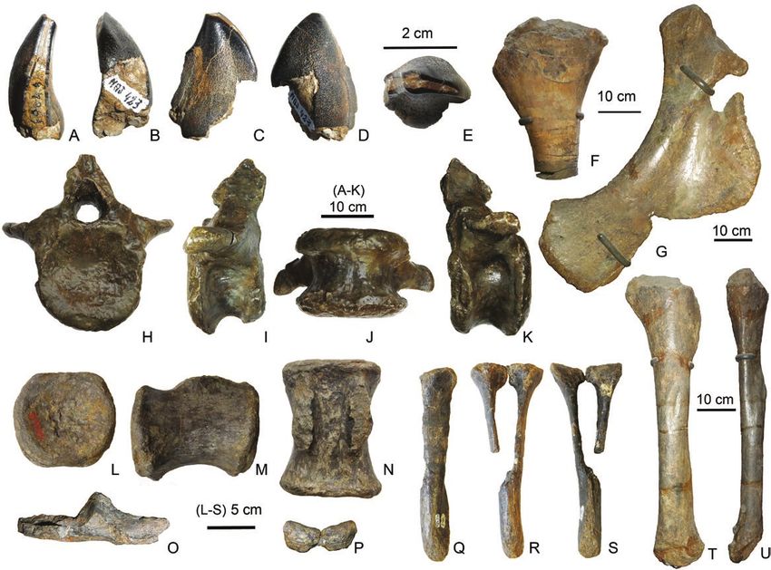
EVOLUTION OF TURIASAUR DINOSAURS                 5

maxillary tooth row ends anterior to the preserved               As in other sauropods such as Camarasaurus Cope,
portion of the antorbital fenestra, a feature also               1877 (Wiersma & Sander, 2017), the tooth crowns
present in diplodocoids (Upchurch, 1998; Wilson,                 gradually decrease in size, being larger mesially
2002, 2005). The posterior end of the body is a                  and smaller distally. In the mesialmost region, the
little less robust than the anterior end. The dorsal             crown apex is apically oriented to the base of the
ascending process is flat and thin, different to that            crown. These are the premaxillary teeth (Figs 1, 3)

                                                                                                                           Downloaded from https://academic.oup.com/zoolinnean/advance-article/doi/10.1093/zoolinnean/zlaa091/5900936 by guest on 02 October 2020
of Turiasaurus, and slopes strongly posterodorsally              and anteriormost teeth in the dentary (Figs 2, 4). In
to likely contact the upper end of the lacrimal. The             the case of the dentary, from d3 to d15 (Fig. 2) and
preserved margins of the antorbital fenestra infer               the maxillary teeth, the apices move to the more
a small fenestra, smaller than that of Turiasaurus               distal region and become apicodistally oriented.
(Fig. 1). The lateral surface of the premaxilla and              This leads to a more distally shifted apex with an
maxilla has dorsoventrally elongated grooves.                    asymmetric appearance in the mesiodistal view. In
A derived feature only described in diplodocoids                 general, every tooth has a convex labial crown face
(Apatosaurus Marsh, 1877, Diplodocus Marsh, 1878,                and concave lingual face. This asymmetric feature
Dicraeosaurus Janensch, 1914 and Nigersaurus                     makes it possible to assign isolated teeth to the
Sereno et al., 1999) and Nemegtosaurus Nowinski,                 right or left side of the skull. However, we need the
1971 (Wilson, 2005; Tshopp et al., 2015; Mannion                 help of another feature to assign isolated teeth to
et al., 2019). It is considered a synapomorphy of                either the maxillary or dentary. When looking at the
Dicraeosauridae (Mannion et al., 2012; Tshopp et al.,            teeth in mesial or distal view, the crown is curved
2015). This feature may be a diagnostic character                lingually in both the premaxillary and maxillary
for this turiasaur genus, because it is absent in the            teeth, and the crown is directed towards the labial
Turiasaurus and Mierasaurus skulls. The tooth                    in the dentary teeth (Figs 3, 4). This anatomical
formula is four premaxillary and 13 maxillary teeth.             difference allows us to set the teeth in the skull, and
                                                                 this criterion may be applicable to other groups of
                                                                 sauropods. Furthermore, it has been confirmed in
Dentary                                                          Turiasaurus and in a thus far unpublished specimen
The left and right dentaries (MAP-6008 and MAP-                  referred to Camarasaurus from Colorado (CPT-4445
6009) have been preserved (Fig. 2). They consist of              to CPT-4460). These features were also described in
thick vertical plates of bone, the posterior two-thirds          Camarasaurus (Carey & Madsen, 1972; Wiersma &
of which lie in a parasagittal plane, while the anterior         Sander, 2017), Diplodocus, Europasaurus Mateus
portion curves slightly to meet its partner at the               et al., 2006 (Wiersma & Sander, 2017), Giraffatitan
symphysis. The anterior end of the dentary is bent               Paul, 1988, Nemegtosaurus and Quaesitosaurus
downwards so that the long-axis of the symphysis                 Kurzanov & Bannikov, 1983 (Upchurch, 1998;
forms an angle of 110º to the long-axis of the mandible.         Upchurch et al., 2004a). The tooth distribution in
The cross-sectional shape of symphysis is oblong and             the skull of the San Lorenzo specimen consists of
the anteroventral margin of the dentary is gently                four alveoli in the premaxillae, 13 in the maxillae
rounded in shape. Each alveolus has two or three                 and 15 in each dentary. In contrast to neosauropods,
replacement teeth. The dentary has a longitudinal                which are described as homodont (McIntosh et al.,
groove going from the lingual position in the chin to            1996; Chatterjee & Zheng, 2005; Wiersma & Sander,
the posterior end on the ventral surface. There are 15           2017), turiasaurs have a heterodont dentition, such
alveoli for the left dentary teeth, which decrease in            as Turiasaurus (Royo-Torres & Upchurch, 2012) and
size as they progress posteriorly.                               Mierasaurus (Royo-Torres et al., 2017). Previous
                                                                 studies described three morphotypes for the heart-
                                                                 shaped teeth of this clade (Mocho et al., 2016, 2017a).
Dentition                                                        However, thanks to the teeth in the skull of the
The San Lorenzo specimen has several teeth in                    San Lorenzo specimen, we can identify the correct
anatomical position (Figs 1, 2; see Supporting                   anatomical position for each morphotype in the skull.
Information, File S3 for measurements): two                      This leads to the identification of four types of tooth
unerupted teeth in the right premaxillary–maxillary              morphology: (1) anterior dentary teeth, (2) middle and
bones, seven teeth in the right dentary and two teeth            posterior dentary teeth, (3) premaxillary teeth and
in the left dentary. Thirty isolated teeth have been             (4) maxillary teeth. In general, each tooth crown in
referred to the same specimen. Regarding morphology,             this specimen has a labiolingually compressed heart/
namely the shape and size of the teeth in articulation           spoon-shaped morphology and presents enamel with
with the skull, each isolated tooth has been placed              a wrinkled texture. This texture is produced by a set
in the left or right dentaries or maxillae/premaxillae.          of small scales. The teeth are different to those of

© 2020 The Linnean Society of London, Zoological Journal of the Linnean Society, 2020, XX, 1–27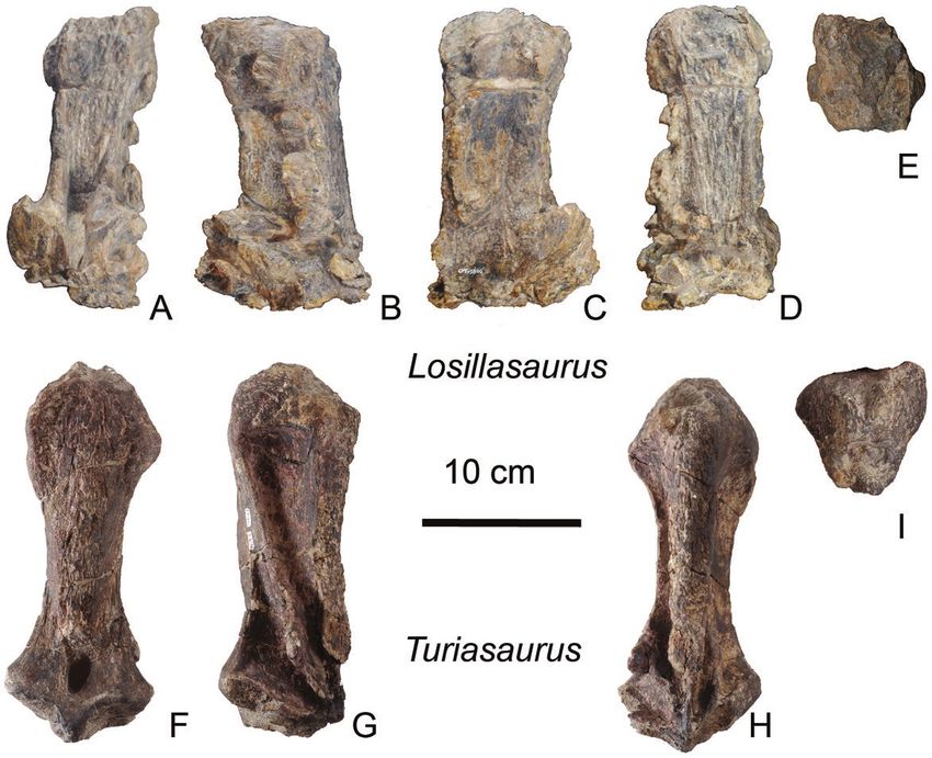
6   R. ROYO-TORRES ET AL.

Figure 3. Right premaxillary and maxillary teeth of Losillasaurus giganteus (San Lorenzo specimen): premaxillary teeth           Downloaded from https://academic.oup.com/zoolinnean/advance-article/doi/10.1093/zoolinnean/zlaa091/5900936 by guest on 02 October 2020
(MAP-6013) in lingual (A), labial (B), mesial (C) and apical (D) views; maxillary teeth (MAP-6014) in lingual (E), labial (F)
and apical (G) views; maxillary teeth (MAP-6015) in lingual (H), labial (I), mesial (J) and apical (K) views; maxillary teeth
(MAP-6016) in lingual (L), labial (M) and apical (N) views; maxillary teeth (MAP-6017) in lingual (O), labial (P), mesial (Q)
and apical (R) views; maxillary teeth (MAP-6018) in labial (S), lingual (T), mesial (U) and apical (V) views; and maxillary
teeth (MAP-6019) in lingual (W), labial (X), mesial (Y) and apical (Z) views.

Turiasaurus, which are acicular and whose wrinkled              crown is slightly apicomesially projected and the
texture features more complex scales. The wrinkles              teeth reach the maximum mesiodistal width near the
are most prominent on the labial surface, similar to            base of the apex. On the labial face, the teeth display
the teeth of Turiasaurus (Royo-Torres & Upchurch,               an apicobasal bulge bounded by two shallow grooves
2012), namely on the bulge area and near the base               (Fig. 6) with the same orientation. The lingual face has
of the crown (Mocho et al., 2017a). In general, the             a low apicobasal ridge, which may extend along the

                               © 2020 The Linnean Society of London, Zoological Journal of the Linnean Society, 2020, XX, 1–27
EVOLUTION OF TURIASAUR DINOSAURS                         7

                                                                                                                                     Downloaded from https://academic.oup.com/zoolinnean/advance-article/doi/10.1093/zoolinnean/zlaa091/5900936 by guest on 02 October 2020
Figure 4. Dentary teeth of Losillasaurus giganteus (San Lorenzo specimen): right dentary teeth (MAP-6032) in lingual
(A), labial (B), mesial (C), distal (D) and apical (E) views; left dentary teeth (MAP-6035) in lingual (F), labial (G), distal
(H), mesial (I) and apical (E) views; left dentary teeth (MAP-6036) in lingual (K), labial (L), distal (M), mesial (N) and
apical (O) views; left dentary teeth (MAP-6038) in lingual (P), labial (Q), distal (R), mesial (S) and apical (T) views; left
dentary teeth (MAP-6039) in lingual (U), labial (V), distal (W) and apical (X) views; right dentary teeth (MAP-6034) in
lingual (Y), labial (Z), distal (A’) and apical (B’) views; and left dentary teeth (MAP-6021) in labial (C’), lingual (D’), distal
(E’) and apical (F’) views.

entire apicobasal length. It occupies the central part             sauropod teeth (i.e. Amygdalodon Cabrera, 1947;
of the lingual concavity. The lingual crest is variable            Carballido & Pol, 2010). Unworn crowns typically
and sometimes reaches the base of the crown where                  lack serrations, except in the d2 (MAP-6032), where
it develops a flat-to-convex mesiodistal platform. The             a couple of rounded denticles are seen in the mesial
mesial and distal edges of the crown are not parallel              apex boundary. The roots are slightly labiolingually
and diverge from the base of the tooth. The transition             compressed cones that are mesiodistally narrower
between the root and crown is well defined in every                than the base of the crown. They have several smooth
tooth. The teeth exhibit an asymmetrical ‘D’-shaped                apicobasal grooves: three in the dentary teeth (Fig. 5),
cross-section with a convex labial face and flat-to-               two in the labial face and one in the lingual face.
smooth concave lingual face. The asymmetrical apex                 They are similar to the roots of the dentary teeth
deflects distally and could bear mesial, distal and                of Turiasaurus. Some dentary teeth have up to four
apical facets, depending on the development of wear.               grooves, i.e. MAP-6033, although the usual amount
Except for in the premaxillary teeth (see below), the              is three (Fig. 5 B, C; MAP-6035). The roots of the
mesial edge of the apex is convex, while the distal edge           premaxillary and maxillary teeth usually have six
is concave in the labial and lingual views. Crown-to-              low apicobasal grooves (unknown in Turiasaurus):
crown occlusion produced ‘V’-shaped wear facets. At                four grooves in the labial face and two in the lingual
the tip of the apex, the surface is smoother as in other           face, namely MAP 6013 (Fig. 5A).

© 2020 The Linnean Society of London, Zoological Journal of the Linnean Society, 2020, XX, 1–27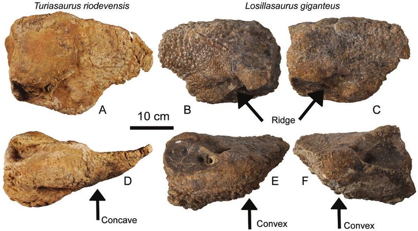
8   R. ROYO-TORRES ET AL.

                                                                                                                               Downloaded from https://academic.oup.com/zoolinnean/advance-article/doi/10.1093/zoolinnean/zlaa091/5900936 by guest on 02 October 2020
Figure 5. Longitudinal grooves in the right premaxillary teeth (MAP-6013) in the labial view (A) and left dentary teeth
(MAP-6035) in labial (B) and lingual (C) views of Losillasaurus giganteus (San Lorenzo specimen).

Premaxillary teeth                                            heart and rounded square, and they are labiolingually
The four premaxillary teeth (MAP-6013) are bent               more compressed and shorter than the other teeth.
lingually and their crowns are curved in the same             The maxillary teeth have intermediate SI values of
direction (Figs 1, 3). They are the most robust teeth,        1.3–1.07. The lingual face is apicobasally concave and
labiolingually longer compared to the other teeth (see        flat-to-concave mesiodistally with a platform at the
Supporting Information, File S3) and have a symmetric         base of the crown. On the labial face, the maxillary
heart-shape appearance. The mesial and distal edges           teeth of the San Lorenzo specimen display an
of the apex are both slightly concave in the labial and       apicobasal bulge bounded by two shallow grooves with
lingual views. The SI values of this morphotype are           the same orientation, while Turiasaurus has only one
close to 1.33 (Pm4, MAP-6013). The lingual face is            labial groove on the mesial edge (Fig. 6).
apicobasally concave and flat-to-concave mesiodistally
with a platform at the base of the crown.
                                                              Dentary teeth
                                                              The 15 dentary teeth (Figs 2, 4; Supporting
Maxillary teeth                                               Information, File S3) decrease in size as they progress
There are 13 maxillary teeth (Figs 1, 3; Supporting           posteriorly. The teeth curve labially and the crown is
Information, File S3). They are bent lingually and the        also directed labially. The first and second mesialmost
asymmetric crown is also curved lingually. They have          dentary teeth differ from the middle and distalmost
a well-defined heart-shaped outline, an apicomesially         dentary teeth. They have heart-shaped crowns, which
projected crown and a pronounced curvature of the             are more apicobasally elongated and labiolingually
apex. The maxillary teeth are diagnostic. The apex is         compressed than the posterior dentary teeth. They
shorter (apex/crown height ratio 0.3–0.24) than the           also have higher SI values: 1.73 in d2, and ranging
apex of Turiasaurus (CPT-3941, apex/crown height              from 1.18 to 1.53 in d3 to d15. In the mesialmost teeth,
ratio 0.4) (Fig. 6). In addition, the teeth of the San        the mesial and distal edges at the base of the crown
Lorenzo specimen have a ‘secondary apex’ on the               are closely parallel and straighter than in the other
distal edge in the transition between the apex and            dentary teeth. In d1 and d2, the apex is particularly
base of the crown (Figs 1, 3). This ‘secondary apex’ is       long and presents a slight distal deflection, which is
considered a potential autapomorphy and is unknown            not as pronounced as in the posterior dentary teeth.
in any other sauropod. The mesial and distal edges of         The distal edge of the apex is concave, while the mesial
the main apex are straight and concave, respectively.         edge is convex-to-straight. The middle and distalmost
The shape of the maxillary teeth is between that of a         dentary teeth have a well-defined heart-shaped outline,

                             © 2020 The Linnean Society of London, Zoological Journal of the Linnean Society, 2020, XX, 1–27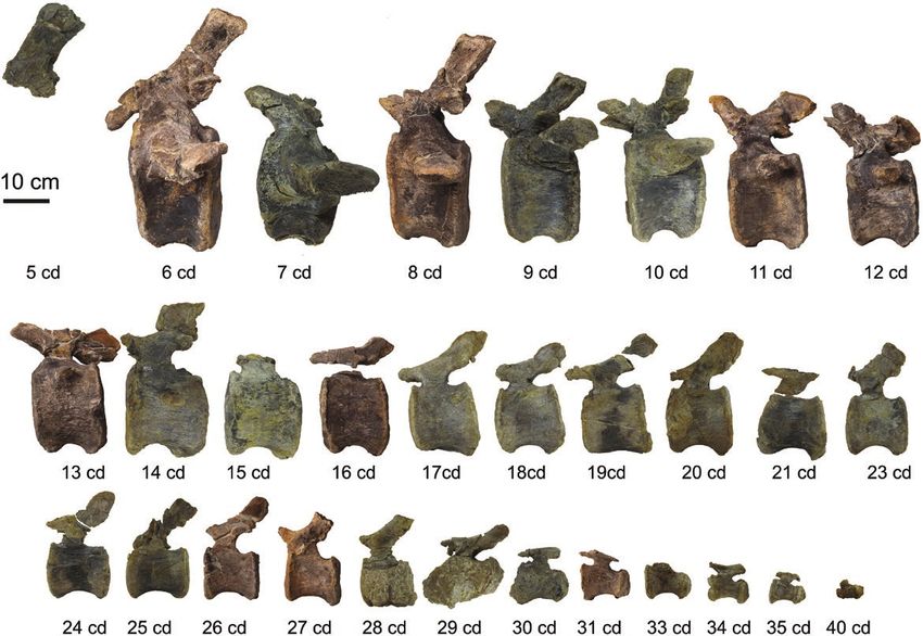
EVOLUTION OF TURIASAUR DINOSAURS                       9

                                                                                                                                 Downloaded from https://academic.oup.com/zoolinnean/advance-article/doi/10.1093/zoolinnean/zlaa091/5900936 by guest on 02 October 2020
Figure 6. Comparison between Turiasaurus and Losillasaurus teeth: right premaxillary teeth (MAP-6013) in labial (A)
and apical (B) views of Losillasaurus giganteus (San Lorenzo specimen); right maxillary teeth (MAP-6019) in labial (C)
and apical (D) views of Losillasaurus giganteus (San Lorenzo specimen); left maxillary teeth (CPT-1262) in labial (E) and
apical (F) views of Turiasaurus riodevensis; left maxillary teeth (CPT-3941) in labial (G) and apical (H) views of Turiasaurus
riodevensis.

an apicomesially projected crown, and a pronounced               They represent the fourth to 38th vertebrae in the caudal
curvature of the apex.                                           series. In addition, the last preserved caudal vertebra
                                                                 is interpreted as a distal caudal vertebra, likely located
                                                                 between the 40th and 50th caudal vertebrae. The length
Dorsal vertebra and rib                                          for the caudal centra is approximately the same in the
Only one dorsal vertebral fragment (MAP-6047) is                 first 15 preserved caudal vertebrae, and the caudal
preserved in the San Lorenzo specimen. It comprises              transverse processes persist through 15 vertebrae.
the neural spine, transverse lateral processes, one              According to the holotype specimen of Losillasaurus,
prezygapophysis and hyposphene. This fragment likely             for which it is possible to study the first five caudal
belongs to a middle dorsal vertebra compared to the              vertebrae (Casanovas et al., 2001), the first complete
dorsal vertebrae of Turiasaurus. It lacks the anchor             caudal vertebra from the San Lorenzo specimen
morphology seen in Turiasaurus (Fig. 7). A dorsal rib            should be the sixth. Both the fifth (type specimen) and
(MAP-6048) was found, which is probably related to a             sixth vertebrae (San Lorenzo specimen) are similar
posterior vertebra, although it may be related to the            in morphology, but differ slightly in articulation: the
preserved fragment. It displays a long capitulum and             fifth is more convex posteriorly than the sixth caudal
short tuberculum with solid bone in its interior. The            vertebra. The sixth caudal centrum is flat and slightly
neck lacks pneumatic cavities or foramina, but there is          convex on the posterior surface, displaying variable
one in the proximal surface of the tuberculum.                   concavity in the central area of the articulation,
                                                                 which becomes barely perceptible from the sixth
                                                                 caudal vertebra onwards. Thus, the anterior caudal
Caudal vertebrae                                                 articular surfaces are slightly procoelous. This feature
In total, 35 caudal vertebrae belonging to this specimen         is differentiated from the strong convex posterior
(CPT-1846a and b, MAP-6050 to MAP-6083) have been                articulations of titanosaurs and mamenchisaurids
recovered (Figs 8–10; Supporting Information, File S3).          such as Wamweracaudia Mannion et al., 2019 (HMN

© 2020 The Linnean Society of London, Zoological Journal of the Linnean Society, 2020, XX, 1–27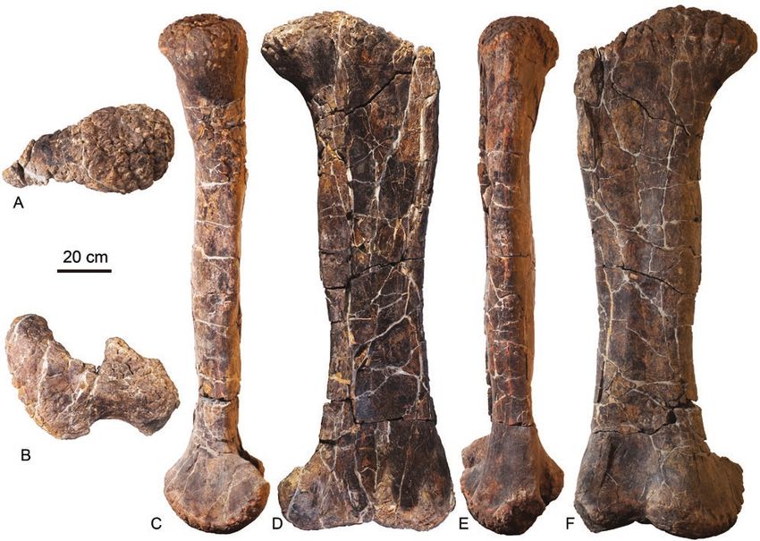
10    R. ROYO-TORRES ET AL.

                                                                                                                                 Downloaded from https://academic.oup.com/zoolinnean/advance-article/doi/10.1093/zoolinnean/zlaa091/5900936 by guest on 02 October 2020
Figure 7. Dorsal spines (MAP-6047) of Losillasaurus giganteus (San Lorenzo specimen) in anterior (A), dorsal (B) and
posterior views. Dorsal spines (MCNV Lo-11) of Losillasaurus giganteus (paratype) in posterior view (D). Dorsal spines
(MCNV Lo-17b) of Losillasaurus giganteus (paratype) in posterior view (E). Dorsal spine (CPT-2688) of Turiasaurus
riodevensis (paratype) in posterior (F) and anterior (G) views. Dorsal spine (CPT-1633) of Turiasaurus riodevensis (referred
to material) in posterior (H) and anterior (I) views.

MB.R.2901.1–30) (Mannion et al., 2019). The middle              the transverse processes are directed laterally or
caudal vertebrae are amphicoelous, with the anterior            anterolaterally. In the middle caudal vertebrae (from
cup moderately deeper than the posterior one. The               the 11th to 14th caudal vertebrae) of the San Lorenzo
posterior and distal caudal vertebrae are procoelous            specimen (RD-28), they do not reach the posterior
with the posterior surface completely covered by the            margin of the centrum, and it is thus different from the
convexity. The latter differs from the opisthocoelous           condition for basal Titanosauriformes (D’Emic, 2012).
condition seen in Turiasaurus (Royo-Torres et al.,              The lateral processes are rectangular and compressed
2006). Rugosity is present in the dorsal half of the            dorsoventrally in the dorsal and ventral views. The
posterior articulation surface between the sixth                neural arch in the anterior and middle caudal vertebrae
and eighth caudal vertebrae, and a small concavity              is located on the anterior half of the centrum. The
appears posteriorly in the same area. There is a                caudal neural spine is transversely compressed and
horizontal notch in a similar position in the 23 rd to          has a dorsoventrally short spinopostzygapophyseal
27th caudal vertebrae. The caudal bone texture is solid         fossa. This character is similar to that found in basal
in all vertebrae, meaning they lack pneumatic fossae            eusauropods and basal macronarians. They have
and foramina. The transverse processes extend from              a relatively short neural spine for anterior caudal
the neural arch to lateral face of the centrum in the           vertebrae, resembling the condition present in
anterior caudal vertebrae, with a triangular distal             Giraffatitan (Janensch, 1950). In the anterior caudal
taper in the anterior and posterior views. They are             vertebrae, the spinoprezygapophyseal lamina is present
directed posteriorly and dorsally. This characteristic          at the base of the spine and disappears into the lateral
differs in the caudal vertebrae of Turiasaurus, where           surface of the neural spine. The anterior surface of the

                               © 2020 The Linnean Society of London, Zoological Journal of the Linnean Society, 2020, XX, 1–27
EVOLUTION OF TURIASAUR DINOSAURS 11
Downloaded from https://academic.oup.com/zoolinnean/advance-article/doi/10.1093/zoolinnean/zlaa091/5900936 by guest on 02 October 2020
Figure 8. Caudal vertebrae in posterior views of Losillasaurus giganteus (San Lorenzo specimen): 4th (MAP-6050), 6th
(MAP-6052), 7th (MAP-1846b), 8th (MAP-6053), 9th (MAP-6054), 10th (MAP-6055), 11th (MAP-6056), 12th (MAP-6057), 13th
(MAP-6058), 14th (MAP-6059), 15th (MAP-6062), 16th (MAP-6061), 17th (MAP-6060), 18th (MAP-6063), 19th (MAP-6064), 20th
(MAP-6066), 21st (MAP-6065), 23rd (MAP-6068), 24th (MAP-6069), 25th (MAP-6070), 26th (MAP-6071), 27th (MAP-6072), 28th
(MAP-6073), 29th (MAP-6074), 30th (MAP-6075), 31st (MAP-6076), 33rd (MAP-6078), 34th (MAP-6079), 35th (MAP-6080) and
40th (MAP-6083).
spine is rugose and lacks a transversely constricted and related to the pronounced lateral deformation of the
deep prespinal lamina. This is similar to the posterior neural spine in the first and second caudal vertebrae
face, where the surface is also rugose and lacks a of the Losillasaurus type specimen. This character
postspinal lamina and the spinopostzygapophyseal is missing in the San Lorenzo specimen from the
fossa. The neural spine is larger anteroposteriorly than 11th caudal vertebrae onwards. This character is not
transversely. One important character is the presence developed in the specimen from Puntal de Santa
of a rugose ridge at the base of the spine between the Cruz referred to as Turiasaurus riodevensis and is
prezygapophyses. It is oriented perpendicularly to the considered a new autapomorphy for Losillasaurus
spine and does not continue towards the dorsal spine, giganteus. This character is also present in the isolated
differentiating it from the prespinal lamina. This vertebra (SHN 180) from the Upper Jurassic Praia de
ridge is present in the Losillasaurus type specimen Amoreira-Porto Novo Formation in Baleal (Peniche,
and in Turiasaurus (Puntal de Santa Cruz specimen, Portugal) (Mocho et al., 2017b). The caudal vertebrae
RD-13; Royo-Torres et al., 2009). It is thus considered a of the San Lorenzo specimen lack a hyposphene
possible synapomorphy for Turiasauria. The character similar to the Losillasaurus type specimen and SHN
is possibly present between the second to ninth caudal 180. The postzygapophyses are joined at the base of
vertebrae and disappears from the tenth one onwards. the neural spine by a horizontal lamina in the anterior
In other sauropods, such as Tastavinsaurus, a thin caudal vertebrae, but separated from the tenth caudal
lamina appears in the same position, but in the fossa that vertebra onwards (Fig. 8).
exists between the prezygapophyses. Finally, another
important feature is the presence of a dorsoventral
ridge on the anterolateral surface of the spine from Chevrons
the third to tenth caudal vertebrae (Figs 9, 10). In total, 21 chevrons (MAP-6085 to MAP-6110) have
The character is also evident in the Losillasaurus been recovered for this specimen (Fig. 11; Supporting
type specimen in the third to fifth caudal vertebrae. Information, File S3). They represent the anterior,
The absence in the first two caudal vertebrae can be middle and posterior chevrons in a continuous series,
© 2020 The Linnean Society of London, Zoological Journal of the Linnean Society, 2020, XX, 1–2712 R. ROYO-TORRES ET AL.
Downloaded from https://academic.oup.com/zoolinnean/advance-article/doi/10.1093/zoolinnean/zlaa091/5900936 by guest on 02 October 2020
Figure 9. Caudal vertebrae in lateral views of Losillasaurus giganteus (San Lorenzo specimen): 5 th (MAP-1846a), 6th
(MAP-6052), 7th (MAP-1846b), 8th (MAP-6053), 9th (MAP-6054), 10th (MAP-6055), 11th (MAP-6056), 12th (MAP-6057), 13th
(MAP-6058), 14th (MAP-6059), 15th (MAP-6062), 16th (MAP-6061), 17th (MAP-6060), 18th (MAP-6063), 19th (MAP-6064), 20th
(MAP-6066), 21st (MAP-6065), 23rd (MAP-6068), 24th (MAP-6069), 25th (MAP-6070), 26th (MAP-6071), 27th (MAP-6072), 28th
(MAP-6073), 29th (MAP-6074), 30th (MAP-6075), 31st (MAP-6076), 33rd (MAP-6078), 34th (MAP-6079), 35th (MAP-6080) and
40th (MAP-6083).
showing three different morphological types. The Young, 1939 or Tazoudasaurus Allain et al., 2004, in
first chevron is likely formed by two separated left which all chevrons are closed (Otero et al., 2012). In
and right distal blades with an inverted ‘U’-shaped Camarasaurus, the presence of this bridge seems
morphology (Sereno et al., 1999). The remaining to depend on the ontogeny, as some chevrons are
anterior chevrons display two dorsal rami that are open while others are closed (Ikejiri et al., 2005).
dorsally bridged and ventrally fused. The distal The ratio of the dorsoventral length of the haemal
end corresponds to a single blade structure and is canal to the total length of chevron in the first ten
‘Y’-shaped in the anterior and posterior views. The anterior chevrons is less than 0.30 (0.27). Thus, this
relative length of the main stem of the ‘Y’ gradually condition is primitive with respect to Europasaurus
shortens until forming a ‘V’ shape while moving and Titanosauriformes, which have haemal canals
backwards along the tail. The third type is represented representing 50% of the chevron length (Wilson,
by a forked structure in the middle and posterior 2002; D’Emic, 2012). However, the extension of the
chevrons, such as in non-macronarian eusauropods haemal canal in the San Lorenzo specimen is greater
(Otero et al., 2012). The proximal ends of the anterior than that of Mamenchisaurus (Ouyang & Ye, 2002),
and middle chevrons are dorsally bridged by a bar Apatosaurus (Upchurch et al., 2004b), and deeply
of bone, while the posterior chevrons are dorsally nested Titanosauriformes, such as Tamabatitanis
open. This feature is similar in basal neosauropods (Saegusa & Ikeda, 2014). This ratio is 0.40 in the
such as diplodocoids. However, it is different in more middle chevrons (relative position, 14th) and greater
derived sauropods like Mamenchisaurus, Omeisaurus than 0.50 in the posterior ones (relative position, 17th).
© 2020 The Linnean Society of London, Zoological Journal of the Linnean Society, 2020, XX, 1–27EVOLUTION OF TURIASAUR DINOSAURS 13
Downloaded from https://academic.oup.com/zoolinnean/advance-article/doi/10.1093/zoolinnean/zlaa091/5900936 by guest on 02 October 2020
Figure 10. Comparison of the caudal spines of Losillasaurus giganteus (CPT-1846, San Lorenzo specimen) in posterior (A),
right lateral (B), left lateral (C), anterior (D) and dorsal (E) views and Turiasaurus riodevensis (CPT-1611, Puntal de Santa
Cruz) in posterior (F), right lateral (G), anterior (H) and dorsal (I) views.
Forelimb titanosauriforms, as it has with a long amp (Upchurch
The left ulna (MAP-6111) is the only well-preserved et al., 2015; Mannion et al., 2017). The articular
bone from the forelimb. The ulna (Fig. 12; Supporting surface of the amp process is slightly concave along
Information, File S3) is a relatively slender element. its length and transversely flat. Both processes define
In proximal view, the ulna is triradiate, as in every a slightly anterior fossa with dorsoventrally oriented
sauropod (McIntosh, 1990; Upchurch et al., 2004a), ridges that receives the proximal end of the radius
with the anteromedial process (amp) and anterolateral (Fig. 12), as in other eusauropods (Wilson & Sereno,
(alp) processes meeting each other at approximately 90º 1998). The olecranon region, where the anteromedial
(Fig. 12). The amp is longer than the alp (ratio = 1.48), and anterolateral proximal processes meet, is low
which is similar to the condition in most sauropods (see and shorter than the amp and alp processes. Thus,
table 2 in Upchurch et al., 2015). The ulna (MAP-6111) the San Lorenzo specimen possesses the reduced
differs from the subequal proximal processes seen in derived olecranon that occurs in other turiasaurs such
Mamenchisaurus (Ouyang & Ye, 2002), Omeisaurus as Zby (Mateus et al., 2014) and Mierasaurus (Royo-
(He et al., 1988; Läng, 2008), Shunosaurus Dong et al. Torres et al., 2017) and in most sauropods except
1983 (Läng, 2008) and diplodocoids (Wilson, 2002). some titanosaurs, where a more prominent olecranon
The San Lorenzo specimen is different to from some is reacquired (Upchurch, 1995, 1998; Wilson, 2002).
© 2020 The Linnean Society of London, Zoological Journal of the Linnean Society, 2020, XX, 1–2714 R. ROYO-TORRES ET AL.
Downloaded from https://academic.oup.com/zoolinnean/advance-article/doi/10.1093/zoolinnean/zlaa091/5900936 by guest on 02 October 2020
Figure 11. Chevrons of Losillasaurus giganteus (San Lorenzo specimen) in posterior view: from anterior to posterior,
chevron 1 to chevron 9 (MAP-6085 to MAP-66093), chevron 12 (MAP-6096), chevron 14 to 19 (MAP 6098 to MAP-6103),
chevron 21 (MAP 6015) and chevron 22 (MAP-6016).
Passing distally along the shaft of the ulna, the amp Femur
and alp processes and radial fossa gradually decrease The femur of the San Lorenzo specimen (MAP-6113;
in prominence, disappearing at around mid-height in Fig. 13; Supporting Information, File S3) is straight
a similar way to those in Turiasaurus (Royo-Torres in all views. An articular head at the proximal end
et al., 2006) and Zby (Mateus et al., 2014). The posterior projects dorsomedially in the anterior view. The head
surface of the proximal half of the ulna is strongly is not separated from the greater trochanter by a
concave mediolaterally (Fig. 12) and bounded by the constriction, a feature usually present in sauropods
distal extension of the amp process and a ridge formed (McIntosh, 1990; Upchurch et al., 2004a). The head is
along the proximal half of the posterolateral margin. rounded, subspherical, tapers laterally and is narrow
Therefore, this deep fossa rivals the radial fossa in in the greater trochanter. The shaft is oval in cross-
depth. This strong concavity is probably a feature section, transversely. In the anterior and posterior
in turiasaurs such as Losillasaurus, Mierasaurus, views, the section has the same length and tapers
Moabosaurus, Turiasaurus and Zby (Royo-Torres et al., slightly in the middle. The femur lacks a ‘lateral bulge’
2017). In other sauropods, this concavity is only slightly in its lateral surface in the proximal third, a feature
developed, except in several somphospondylans, where that allows the exclusion of the San Lorenzo specimen
it has also been described (Upchurch et al., 2015). The from the Titanosauriformes’ clade (Salgado et al., 1997;
anterior surface of the distal half of the ulna has a Wilson & Sereno, 1998). The lateral margin of the
deep vertical groove and ridge as seen in Turiasaurus proximal end is straight relative to the lateral margin
(CPT-1197) and Losillasaurus (MCNV). This character of the midshaft and lacks a medially deflected lateral
was erroneously interpreted as a synapomorphy for margin (Royo-Torres et al., 2012; Mannion et al.,
Turiasauria (Royo-Torres et al., 2006; but amended in 2013), which is similar to the case of non-macronarian
Royo-Torres et al., 2017), as it was interpreted as being eusauropods. The fourth trochanter is located on
present on the posterior surface of the distal half of the the posteromedial margin of the shaft at midlength,
ulna. Thus, character number 413 used by Mannion where it is reduced to a low ridge. The distal end is
et al. (2017, 2019) should be deleted (Royo-Torres divided into two condyles: the tibial condyle is twice
et al., 2017). It is codified here with a ‘?’ for each taxa as big as the fibular one. This condition is different
in the phylogenetic analyses. The articular surface of in other turiasaurs, such as Mierasaurus (DBGI 39),
the ulna in Turiasaurus is damaged, and the proximal and could be an autapomorphy. Usually, the tibial
profile of the ulna is shaped between a ‘T’ and ‘Y’. condyle is larger than the fibular one in all sauropods,
© 2020 The Linnean Society of London, Zoological Journal of the Linnean Society, 2020, XX, 1–27EVOLUTION OF TURIASAUR DINOSAURS 15
Downloaded from https://academic.oup.com/zoolinnean/advance-article/doi/10.1093/zoolinnean/zlaa091/5900936 by guest on 02 October 2020
Figure 12. Left ulna (MAP-6111) of Losillasaurus giganteus (San Lorenzo specimen) in anterior (A), lateral (B), posterior
(C), anteromedial (D), medial (E), anterolateral (F) and dorsal (G, H) views.
such as Brachiosaurus Riggs, 1903 and Giraffatitan intercondylar groove is shallow on the distal part
(HMN St 291, Janensch, 1961), but smaller than in of the anterior face. However, the tibial and fibular
the San Lorenzo specimen. In the posterior margin condyles display a deep intercondylar groove on their
there is a notch lateral to the fibular condyle, as is posterior faces. The articular condyles are rotated so
usual in most dinosaurs (Upchurch, 1995, 1998). The that their long-axes are directed anterolaterally at an
© 2020 The Linnean Society of London, Zoological Journal of the Linnean Society, 2020, XX, 1–2716 R. ROYO-TORRES ET AL.
Downloaded from https://academic.oup.com/zoolinnean/advance-article/doi/10.1093/zoolinnean/zlaa091/5900936 by guest on 02 October 2020
Figure 13. Right femur (MAP-6113) of Losillasaurus giganteus (San Lorenzo specimen) in dorsal (A), ventral (B), medial
(C), posterior (D), lateral (E) and anterior (F) views.
angle of approximately 20º to the parasagittal plane, most eusauropods, but absent in most diplodocoids and
as also occurs in Apatosaurus (Upchurch et al., 2004b) somphospondylans (Mannion et al., 2013). The area
whose long axis of the transverse section of the shaft for the ‘tuberculum fibularis’ is rugose and there is a
is horizontal. small vertical ridge, different to the ridge in the tibia
of Suuwassea Harris & Dodson, 2004 (Harris, 2007).
The distal end is less expanded than the proximal
Tibia end and has a rough surface with a heart-shaped
The tibia (MAP-6115; Fig. 14; Supporting Information, outline in the ventral view. Both malleoli are laterally
File S3) is straight. The tibia to femur length ratio is oriented, and the anterior one is larger and square in
0.65 and, therefore, within the typical range of those shape. It also displays the articular surface for the
in sauropods (McIntosh, 1990; Upchurch et al., 2004a). ascending process. The posterior malleolus is smaller
The proximal articular surface is between subcircular and rounded. It is described as a posterior ventral
and transversely compressed, a plesiomorphic process. The posterior condyle projects more distally
condition for Sauropoda (Wilson, 2002; Tschopp et al., and meets the posterior side of the ascending process
2015). The anterolateral corner of the proximal end of the astragalus. The ratio between the mediolateral
displays a rounded triangular cnemial process, which and anteroposterior widths is close to 1.
is an anteriorly projecting and vertically elongate
ridge. The lateral margin of the proximal end bulges
laterally, forming a vertical groove between its anterior Fibula
face and posterior face of the cnemial crest. A small T h e f i b u l a ( M A P - 6 1 1 6 ; F i g. 1 5 ; S u p p o r t i n g
ridge posterior to the cnemial crest is present at the top Information, File S3) is a straight bone with
of this groove. This crest is interpreted as the second an expanded proximal end. The distal end is
cnemial crest described by Bonaparte et al. (2000) not preserved. The proximal end of the fibula
for the tibia (SMNS 12144) of Janenschia. In the San is anteroposteriorly elongated and transversely
Lorenzo specimen, this crest is markedly pointed and flattened and rectangular in the dorsal view. The
parallel to the cnemial crest. This feature is present in lateral surface is convex and the medial one concave.
© 2020 The Linnean Society of London, Zoological Journal of the Linnean Society, 2020, XX, 1–27EVOLUTION OF TURIASAUR DINOSAURS 17
Downloaded from https://academic.oup.com/zoolinnean/advance-article/doi/10.1093/zoolinnean/zlaa091/5900936 by guest on 02 October 2020
Figure 14. Right tibia (MAP-6115) of Losillasaurus giganteus (San Lorenzo specimen) in dorsal (A), ventral (B), medial (C),
posterior (D), anterior (E) and lateral (F) views.
The latter surface displays a triangular muscle posterior margin of the main body of the astragalus.
scar. The anterior margin has a sharp triangular The astragalus is wedge-shaped in proximal view and
expansion in the lateral and medial views with a becomes medially narrow in anterior view. The anterior
medially oriented vertical ridge. The proximal end edge is straight-to-convex transversely in the proximal
is twice the anteroposterior length of the diaphysis, view. Laterally, there is a ventral shelf underlying
differentiating it from that in Turiasaurus and the distal end of the fibula. The lateral surface of
Camarasaurus. The lateral trochanter is a concave the astragalus has a central concavity, which is not
surface, different to the flat and rugose surface present deflected posteriorly. The posterior astragalar fossa
in Turiasaurus. The lateral muscle insertion scar shares is deep and bears two main foramina separated by a
the plesiomorphic oval shape of those in Omeisaurus subvertical ridge, as in Lapparentosaurus Bonaparte,
and Shunosaurus. This shape is different from that in a 1986 (Läng, 2008) and Oceanotitan Mocho et al., 2019.
trochanter associated with one or two vertical elongate This subvertical ridge is another important difference
ridges, as occurs in some Titanosauriformes (D’Emic, from the astragalus of Turiasaurus (Fig. 16). The
2012; Mannion et al., 2019). posterodistal edge of the astragalus, ventral to the
ascending process, is laterally projected, resulting
in a pronounced and flat tongue-like structure. The
Astragalus laterally facing articular surface for the fibula is
The astragalus (left: MAP-6117; right: MA-6118) is concave, well-marked and occupies the dorsal part of
wider transversely than anteroposteriorly (Fig. 16; the lateral surface of the astragalus.
Supporting Information, File S3), typical of Sauropoda
(McIntosh, 1990; Upchurch et al., 2004a). Its lateral
end is broad and tapers medially to a blunt point, Taxonomic assignation of the San Lorenzo
but is more rounded than in the case of Turiasaurus specimen
(Fig. 16). The ventral surface is rugose and convex, Following the description of the San Lorenzo
both anteroposteriorly and transversely. The specimen, it is considered to possess characters that
ascending process is flat and runs two-thirds of the differ from Turiasaurus, Zby and others that have
© 2020 The Linnean Society of London, Zoological Journal of the Linnean Society, 2020, XX, 1–2718 R. ROYO-TORRES ET AL.
Downloaded from https://academic.oup.com/zoolinnean/advance-article/doi/10.1093/zoolinnean/zlaa091/5900936 by guest on 02 October 2020
Figure 15. Right fibula (MAP-6116) of Losillasaurus giganteus (San Lorenzo specimen) in medial (A), dorsal (B), anterior
(C), lateral (D) and posterior (E) views.
only been previously described in Losillasaurus. Phylogenetic analyses
Some are new and must be included in a revised We performed the analyses using extended implied
diagnosis (see below) for this genus. Both the weighting (Goloboff, 2014), with the default settings
Losillasaurus type and San Lorenzo specimen in TNT. The pruned data matrix was analysed
share the following new autapomorphies, which are using the Search in TNT v.1.5 (Goloboff et al., 2008;
not present in any other sauropod: the presence of Goloboff & Catalano, 2016). Searches were conducted
a dorsoventral ridge in the anterolateral surface by employing sectorial searches, drift and tree fusing.
of the spine at least between the fourth and tenth Consensus was stabilized five times before using the
caudal vertebrae. This character has been confirmed resultant trees as the starting trees for a ‘Traditional
as absent in the Turiasaurus specimen from the Search’ using Tree Bisection-Reconstruction. The
Puntal de Santa Cruz site (Royo-Torres et al., 2009), first analysis includes the San Lorenzo specimen
in which an anterior caudal spine (CPT-1649) has as separated OTU from Losillasaurus type and the
been identified (Fig. 10). Another character seen new taxa from the Middle Jurassic of Madagascar
in the Losillasaurus holotype and San Lorenzo (119 OTUs). This produced 135 MPTs with a length
specimen is a spine with a shallow dorsal groove of 239.99 steps and a well-resolved strict consensus
projected anteroposteriorly. This groove is bigger (Supporting Information, File S1, Fig. S1). The
in the anterior caudal vertebrae, especially in the San Lorenzo specimen and Losillasaurus type are
first and second caudal vertebrae (Losillasaurus sister-taxa of Turiasaurus. They are all recovered as
type specimen) and then shallow from the third members of the Turiasauria clade and the new taxa
to 30th caudal vertebrae (Losillasaurus type and from the Middle Jurassic of Madagascar is given as a
San Lorenzo specimens). Based on the anatomical sister-taxon of European turiasaurs. We ran another
descriptions and comparisons, it is concluded that analysis, which positioned the Losillasaurus type and
the San Lorenzo specimen should be classified as San Lorenzo specimen in the same OTU (118 OTUs).
Losillasaurus giganteus, a hypothesis also supported The result (Fig. 18) is similar to the previous analysis
by the phylogenetic analyses performed (see below and produced 135 MPTs with a 239.7 step length. We
and Supporting Information, File S1). also performed the analyses using conventional equal
© 2020 The Linnean Society of London, Zoological Journal of the Linnean Society, 2020, XX, 1–27EVOLUTION OF TURIASAUR DINOSAURS 19
Downloaded from https://academic.oup.com/zoolinnean/advance-article/doi/10.1093/zoolinnean/zlaa091/5900936 by guest on 02 October 2020
Figure 16. Comparison between astragali. Left astragalus (CPT-1244) in dorsal (A) and posterior (D) views of Turiasaurus
riodevensis (paratype) and right (B, E) (MAP-6119) and left (C, F) astragalus (MAP-6117) of Losillasaurus giganteus (San
Lorenzo specimen) in dorsal (B, C) and posterior (E, F) views.
weights parsimony (Goloboff, 2014). The results of the et al., 2001; Royo-Torres et al., 2006). Dated as
analysis with 99 999 MPTs (overflow) and 2591 step Kimmeridgian according to the data of Campos-Soto
length show a polytomy for Eusauropoda (Supporting et al. (2019).
Information, File S1, Fig. S2).
Referred material: The material referred to
Systematic palaeontology Losillasaurus comes from the same specimen from
where the holotype and paratype were selected. It
Sauropoda Marsh, 1878 consists of a skull fragment and partial postcranial
skeleton (see Supporting Information, File S1). A second
Eusauropoda Upchurch, 1995
specimen, described in this paper, is from the San
Turiasauria Royo-Torres et al., 2006 Lorenzo site (Riodeva, Teruel, Spain) in the Villar del
Arzobispo Formation. It consists of a partial skull with
Losillasaurus Casanovas et al., 2001 teeth and partial postcranial skeleton (see the previous
description section and Supporting Information, File
Figures: See Casanovas et al., 2001: figs 1–7; Figs 1–16; S1). A third specimen referred to as Losillasaurus is
Supporting Information, File S3. a complete anterior caudal vertebra (SHN 180) found
in Baleal (Peniche municipality, Portugal) in the Praia
Type species: Losillasaurus giganteus Casanovas de Amoreira-Porto Novo Formation, dated as Upper
et al. (2001). Kimmeridgian–Lower Tithonian (Manuppella et al.,
1999; Mocho et al., 2017b).
Holotype: Anterior caudal vertebra housed in the
Museo de Ciencias Naturales de Valencia (MCNV).
Revised diagnosis: Losillasaurus is diagnosed by
Paratype: Two anterior caudal vertebrae housed in eight autapomorphies: (1) (new) lateral surface of
the Museo de Ciencias Naturales de Valencia (MCNV). the premaxilla and maxilla with dorsoventrally
elongated grooves convergent with diplodocids and
Type locality and horizon: La Cañada site (Valencia), Nemegtosaurus (Mannion et al., 2019); (2) (new) a
Vi l l a r d e l A r z o b i s p o Fo r m a t i o n ( C a s a n o v a s maxillary tooth with a secondary apex on the distal
© 2020 The Linnean Society of London, Zoological Journal of the Linnean Society, 2020, XX, 1–2720 R. ROYO-TORRES ET AL.
Downloaded from https://academic.oup.com/zoolinnean/advance-article/doi/10.1093/zoolinnean/zlaa091/5900936 by guest on 02 October 2020
Figure 17. Narindasaurus thevenini: right premaxillary-maxillary teeth (MNHN MAJ 423) in mesial (A), distal (B), lingual
(C), labial (D) and apical (E) views; right tibia (MNHN MAJ 428) in medial view (F); left pubis (MNHN MAJ 430) in lateral
view (G); anterior caudal vertebrae (MNHN MAJ 424) in anterior (H), right lateral (I), ventral (J) and left lateral (K) views;
posterior caudal vertebrae (MNHN MAJ 426) in posterior (L), right lateral (M) and dorsal (N) views; distal chevron (MNHN
MAJ 429) in right lateral view (O); middle-anterior chevron (MNHN MAJ 425) in dorsal (P), right lateral (Q), posterior (R)
and anterior (S) views; and right fibula in lateral (T) and anterior (U) views from the same specimen.
edge; (3) markedly curved neural spines of the proximal condyle of the femur is twice as large as the fibula
caudal vertebrae that in the first and second caudal condyle.
vertebrae produce a pronounced cutlass-like shape
in the lateral view (Casanovas et al., 2001; Upchurch Additional comments: The character, anteroposterior
et al., 2004a); (4) (new) presence of a dorsoventral length at the base of the neural spines of the proximal
ridge in the anterolateral surface of the spine at caudal vertebrae being approximately half the height
least between the fourth and tenthcaudal vertebrae of the spine (ratio = 0.5), was included in the original
(it is not clearly present in the first three caudal diagnosis (Casanovas et al., 2001) and accepted in
vertebrae and disappears in the 11th caudal vertebra); Upchurch et al. (2004a). However, this is not considered
(5) (new) caudal neural spines with a shallow dorsal valid as the spines are compressed by taphonomic
groove with directed anteroposteriorly, bigger in the deformation.
anteriormost caudal vertebrae, especially in the first
and second bones and shallow from the third to the
30th caudal vertebrae; (6) (new) the long-axis of the Narindasaurus thevenini gen. & sp. nov.
obturator foramen is perpendicular to the long-axis
of the pubis; (7) (new) the lateral trochanter of the Figures: See Thevenin (1907): pl. 1, figs 1, 1a–1c, 6–6a,
fibula is concave and rugose; and (8) (new) the tibial 7–7a, 9, 9a.; pl. 2, figs 5–8; Läng (2008): figs II-24,
© 2020 The Linnean Society of London, Zoological Journal of the Linnean Society, 2020, XX, 1–27You can also read