The Suboccipital Carrefour: Cervical and Vertebral Arterial Anastomosis
←
→
Page content transcription
If your browser does not render page correctly, please read the page content below
AJNR Am J Neuroradiol 19:925–931, May 1998

                   The Suboccipital Carrefour: Cervical and
                       Vertebral Arterial Anastomosis
                               Michael Ayad, Fernando Viñuela, and Eduardo H. Rubinstein

             PURPOSE: Our objective was to anatomically define the anastomoses between cervical and
          carotid arterial distributions (the carrefour) in the rabbit and to assess the contribution of
          these collaterals to cortical blood flow (CBF) during cerebral ischemia.
             METHODS: Angiography was carried out in six rabbits with basilar artery occlusion using
          selective contrast injection into the right subclavian, external carotid, and internal carotid
          arteries. Anastomoses were corroborated with methacrylate vascular casts prepared in five
          additional rabbits. CBF was measured in eight rabbits by H2 clearance after basilar artery
          occlusion and again after bilateral common carotid artery occlusion. Cortical DC potential was
          measured during ischemia in these rabbits and in another 19 rabbits after additional occlusion
          of the cervical collateral arteries.
             RESULTS: A network of anastomoses between superficial and ascending cervical, superior
          intercostal, vertebral, and occipital arteries was found by angiography and corrosion casts.
          Additional communications in the ophthalmic, ethmoidal, and cerebellar arterial distributions
          are described. These pathways were found to supply a mean of 15 6 7 mL/100 g per minute
          residual CBF during three-vessel ischemia, or 24% of the preischemic CBF. Ischemic depolar-
          ization of DC potential occurred in seven of the eight rabbits with collateral CBF at a mean
          latency of 2.64 6 0.59 minutes and at 1.71 6 0.09 minutes in those without.
             CONCLUSION: The suboccipital collateral network of the rabbit resembles that of humans
          and can contribute significantly to CBF during ischemia. The results suggest that this model
          may be useful for evaluating methods of optimizing hemodynamic control of the anastomoses
          in situations such as those encountered during endovascular therapy.

   The suboccipital carrefour or “knot” refers to a                      moses are clearly dependent on the pressure gradient
network of anastomoses interconnecting the cervical,                     across the channels (6), thus explaining the variability
vertebral, occipital, and carotid arteries that has been                 of their opacification on routine angiography (7).
well described in humans (1). The functional signifi-                       The present study was undertaken to characterize
cance of this arterial network stems from its re-                        the arterial network described above in the rabbit.
cruitment during pathologic conditions of arterial                       This animal possesses a well-developed circle of Wil-
occlusion or in the presence of local vascular malfor-                   lis and brain blood flow, which, like that of humans, is
mations. After stenosis of the common carotid artery,                    not confounded by the presence of a rete mirabile. A
the suboccipital carrefour enlarges to provide collat-                   protocol involving proximal basilar artery occlusion
eral blood flow from subclavian to external carotid                      and selective injection of the subclavian artery with
territories, ultimately supplying the brain by way of                    contrast material was used to optimize the pressure
intraorbital ophthalmic anastomoses (2, 3). A re-                        gradient and thus to visualize the collateral system
versed pattern of flow in the system occurs after                        during angiography. To correlate the angiographic
proximal subclavian artery occlusion, contributing to
                                                                         results with three-dimensional anatomy, methacrylate
the so-called subclavian steal phenomenon (4, 5). The
                                                                         arterial casts were prepared of the craniocervical vas-
extent and directionality of flow through the anasto-
                                                                         culature. Second, the potential contribution of the
                                                                         suboccipital collateral system to brain blood flow dur-
  Received August 29, 1997; accepted October 12.                         ing ischemia was measured by using the hydrogen
  From the Departments of Anesthesiology (E.H.R.), Physiology
(M.A., E.H.R.), and Radiological Sciences (F.V.), UCLA School of         clearance method (8). Finally, a functional correlate
Medicine, Los Angeles.                                                   of this collateral blood flow was determined by mea-
  Address reprint requests to Eduardo H. Rubinstein, MD, PhD,            suring the latency for the onset of ischemic depolar-
Department of Anesthesiology, UCLA School of Medicine, Los
Angeles, CA 90024.
                                                                         ization of cortical DC potential. This index was re-
                                                                         corded in rabbits with basilar and bilateral carotid
© American Society of Neuroradiology                                     artery occlusion alone (and thus preserved suboccip-

                                                                   925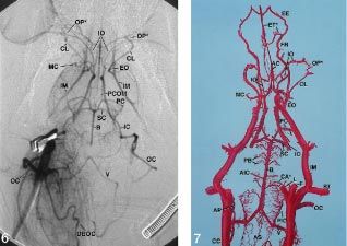
926      AYAD                                                                                                   AJNR: 19, May 1998

ital collateral blood flow) as compared with that in                  in two animals with a plastic cannula directed toward the heart
rabbits with additional blockade of the collateral                    (inner diameter, 3.5 mm; outer diameter, 4.9 mm). In one of
blood flow.                                                           these rabbits, the basilar artery and both common carotid
                                                                      arteries were occluded before injection of contrast material.
                           Methods                                    The remaining two rabbits first underwent occlusion of the
                                                                      basilar artery and both common carotid arteries followed by
   Experiments were carried out on male New Zealand white             placement of a centrally directed 5F catheter in the right
rabbits weighing between 2.8 and 4.0 kg. The protocols were           carotid and a snare around the brachiocephalic trunk. The
approved by the Chancellor’s Animal Research Committee and            internal carotid arteries were occluded bilaterally in one of the
were in compliance with NIH guidelines for the care and use of        two rabbits. Filling of the suboccipital collateral network was
laboratory animals. All rabbits underwent induction of anes-          achieved by central injection into the carotid catheter following
thesia with 5% halothane in an enclosed chamber followed by           snare occlusion of the brachiocephalic trunk.
tracheotomy, intubation, and mechanical ventilation. Paralysis           After completion of the vessel preparations described above,
was maintained with intravenous pancuronium bromide (0.3              each rabbit was killed with an intravenous injection of 3 mEq
mg/kg per hour). Halothane was then reduced to 0.8% for the           KCl. The craniocervical vasculature was then perfused with 60
duration of the experiment and respiratory rate was adjusted to       to 120 mL of normal saline containing 10 U/mL heparin and 50
maintain an end-tidal PCO2 of 26 mm Hg. Esophageal temper-            mg/mL papaverine until the effluent from an opened external
ature was maintained at 38.0°C with radiant heat throughout           jugular vein was clear. Methyl methacrylate monomer, catalyst,
the experiment. The femoral artery and vein were catheterized         coloring agent, and promoter were prepared in a ratio of
for measurement of arterial blood pressure and fluid or drug          200:30:12:1 (Batson’s #17 Anatomical Corrosion Compound,
administration, respectively.                                         Polysciences, Warrington, Pa), and approximately 30 mL was
                                                                      injected under hand pressure into either the aortic or carotid
                            Angiography                               catheter. The thorax was then hemisected and the upper body
   To isolate the vertebral arteries from the circle of Willis, we    immersed in cold water for 3 hours to facilitate curing of the
approached the proximal basilar artery via retropharyngeal            acrylic resin. After the polymerization was complete, the spec-
craniotomy and occluded it with bipolar electrocautery for a          imens were placed in 35% KOH to dissolve soft tissues and
distance of 2 mm beginning at the vertebrobasilar junction.           bone.
Both internal and external carotid arteries were carefully ex-
posed at their bifurcation, and 4-0 silk ligatures were placed
loosely around the two arteries on the right for selective occlu-         Measurement of Collateral Blood Flow and DC Potential
sion with an aneurysmal clip during angiography. On the left             In eight rabbits, nylon snares were placed around both com-
side, the external carotid artery was ligated at its origin to        mon carotid arteries following basilar artery occlusion, and the
prevent reflux of contrast material from that artery into the         animals were placed prone in a stereotactic head frame. Bilat-
brain circulation. At the midpoint between the carotid bifurca-       eral parietal craniotomies 5 mm in diameter were made just
tion and aorta, the right common carotid artery was cannulated        caudal to the coronal suture and lateral to the midline. The
with two 5F catheters, each extending for 1.5 cm in either            dura mater of the left hemisphere was pierced and a Teflon-
direction. The rostrally directed catheter was used for injecting     coated platinum-iridium (Pt-Ir) wire electrode (0.005-inch di-
contrast material into the occipital, internal, and external ca-      ameter bare; 0.007-inch diameter coated) was advanced 1 mm
rotid arteries. The aortic arch was then exposed through a            into the cerebral cortex using a Narishige (Stoelting; Wood
sternotomy at the second intercostal space. Nylon snares were         Dale, IL) micromanipulator. Current between the Pt-Ir elec-
placed around the left subclavian artery and brachicephalic           trode and an indifferent silver-silver chloride (Ag-AgCl) elec-
trunk, sparing the left common carotid artery. Occlusion of the       trode placed on the lumbar trapezius muscle was measured
brachiocephalic trunk permitted selective perfusion of the right      with a polarographic amplifier polarized to 1400 mV. For
subclavian artery when contrast material was injected into the        determination of cortical blood flow (CBF), hydrogen was
caudally directed right carotid catheter. The left subclavian         added to the inspired gas mixture (FIH2 5 0.1) and maintained
artery was occluded during angiography to induce a subclavian         until the hydrogen current reached a stable plateau for at least
steal and thus to increase retrograde filling of the cervical         2 minutes, signifying saturation of cerebral tissue with H2.
collateral vessels on that side with contrast agent. Similarly, the   Inflow of H2 was then stopped and the desaturation slope
left common carotid artery was transiently occluded with a            recorded. In each animal, CBF was first measured under base-
bulldog during angiography to induce cerebral ischemia and to         line conditions; that is, basilar artery occlusion alone. Blood
improve the pressure gradient for anterograde filling of anas-        flow was then remeasured during bilateral carotid occlusion. In
tomotic vessels to the brain circulation.                             the calculation of CBF, the first 1.5 minutes of each desatura-
   Six rabbits underwent angiography. In each rabbit, angio-          tion was deleted in order to avoid recirculation artifacts.
grams were taken during separate injections of contrast mate-         The remaining data were normalized, plotted, and fitted by
rial into the right external carotid, right internal carotid, and     computer with an exponential equation. The half-time for
right subclavian arteries; each injection was replicated at least     washout in minutes (t1/2) was derived from the exponential
once. Anastomotic arteries between the subclavian, external,          equation, and CBF was then calculated according to the
and internal carotid territories were readily identifiable by their   expression CBF 5 (100) ln(2/t1/2), giving values in mL/100 g
presence on arteriograms taken from selective injection of two        per minute (8).
different arteries; the larger anastomoses were often visible on         To measure cortical DC potential, a second Ag-AgCl elec-
each of the three different series. All arteriograms were taken       trode mounted within a glass pipette containing Ringer’s solu-
in the supine position using Omnipaque contrast medium. The           tion was placed on the pial surface of the right parietal cortex
amount of contrast agent used varied with the artery injected         via a cotton bridge. The potential difference between the active
approximately as follows: external carotid artery, 4 mL; internal     and indifferent electrodes was recorded on a Neuroprobe DC
carotid artery, 2.5 mL; subclavian artery, 6 mL. Digitally sub-       amplifier (A-M Systems; Everett, WA) with input impedance
tracted arteriograms were taken in an angiography room using          of 1011 ohms. DC potential was also recorded in 19 rabbits that,
a GE AdvantX system at a frame rate of 7.5 per second.                in addition to undergoing occlusion of the basilar and bilateral
                                                                      carotid arteries, underwent ligation of the subclavian vessels
                                                                      that supply inflow to the suboccipital collateral network. In
                   Vascular Cast Preparation                          these animals, the proximal subclavian, vertebral, internal tho-
  Anatomic casts of the cervical and cerebral vasculature were        racic, superior intercostal, and superficial cervical arteries were
prepared in five rabbits. The abdominal aorta was catheterized        ligated bilaterally before the induction of cerebral ischemia by
AJNR: 19, May 1998 SUBOCCIPITAL CARREFOUR 927
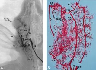
FIG 1. Angiogram of the right
subclavian artery vasculature
in the rabbit. Contrast material
was injected in the right com-
mon carotid artery during
proximal occlusion of the bra-
chiocephalic trunk. (Defini-
tions for abbreviations used in
all figures are in box.)
FIG 2. Methacrylate corro-
sion cast of the proximal aortic
arch obtained by retrograde
perfusion of the right common
carotid artery with resin. Note
the ascending branch of the
superior intercostal artery,
which anastomoses with fine
branches of the superficial cer-
vical and muscular branches of
the vertebral arteries.
snare occlusion of the brachiocephalic trunk. The latter set of
experiments was carried out as part of a separate in-depth Key to Abbreviations of Arteries
electrophysiological study.
AA anterior auricular
Statistical Analysis AC anterior cerebral
ACV ascending cervical
The results of CBF and latency of DC potential onset are
AIC anterior inferior cerebellar
expressed as mean 6 SEM. Comparisons of DC onset between
AO aorta
animals with collateral CBF and those without were carried out
using factorial analysis of variance and Scheffe’s test for mul- AP ascending pharyngeal
tiple comparisons. P values less than .05 were considered sta- AS anterior spinal
tistically significant. AX axillary
B basilar
Results BC brachiocephalic
CA* cerebellar anastomoses
Figure 1 is an angiogram depicting the primary CC common carotid
branches of the subclavian artery in the rabbit. A CL ciliary
typical pattern seen in a corrosion cast specimen of DBOC descending branch of occipital
the aortic arch is shown in Figure 2 for comparison. EC external carotid
The vertebral artery, the most proximal branch of the EE external ethmoidal
subclavian artery, courses dorsomedially through the ET* ethmoidal rete
transverse foramena of the first six cervical vertebrae, EO external ophthalmic
giving off muscular branches at each intervertebral F facial
FR frontal
level. The superficial cervical artery arises anterior to
IC internal carotid
the vertebral origin. Traveling in the ventrolateral IM internal maxillary
neck, it often gives off an ascending cervical branch, IO internal ophthalmic
as seen in Figure 1. Both the superficial and ascend- IT internal thoracic
ing cervical arteries fuse at various points with mus- L lingual
cular branches of the vertebral artery. The superior LT lateral thoracic
intercostal artery and the internal thoracic artery usu- MB muscular branches of vertebral
ally arise from a common trunk on the inferior aspect MC middle cerebral
of the subclavian just distal to the vertebral origin. OC occipital
The internal thoracic artery descends along the ven- OP* ophthalmic anastomosis
PA posterior auricular
tral thoracic wall anastomosing with intercostal arter-
PB perforating branches of basilar
ies arising from the descending aorta. The intercostal PC posterior cerebral
arteries are not seen in Figure 1 because of an unfa- PIC posterior inferior cerebellar
vorable pressure gradient during contrast injection, SB subclavian
because they were under pressure from the aorta. SC superior cerebellar
However, the internal thoracic-intercostal anastomo- SCV superficial cervical
ses down to the T7– 8 costal level were present on SI superior intercostal
both vascular casts obtained by perfusion of the ab- SS subscapular
dominal aorta. The superior intercostal artery (some- ST superficial temporal
times referred to as the costocervical artery) bifur- TA thoracoacromial
TF transverse facial
cates the descending artery, giving off branches that
TS transverse scapular
supply the lateral aspect of the first three ribs. The V vertebral
ascending superior intercostal branch courses as high VOC* vertebral-occipital anastomosis
as the C-3 vertebral level, ramifying extensively with928 AYAD AJNR: 19, May 1998 FIG 3. Serial frames of an angiogram taken by the same methods as Figure 1 show filling of the cerebral vasculature solely by collateral input. The proximal basilar artery and both external carotid ar- teries have been occluded. A, Taken 1.72 seconds after the begin- ning of contrast injection. B, At 2.13 seconds, note filling of the basilar artery via cerebellar collaterals. C, At 3.19 seconds, large vertebral-oc- cipital anastomoses permit filling of the circle of Willis by way of the common ori- gins of the internal carotid and occipital arteries. D, At 7.06 seconds, note the ophthalmic anastomoses between the internal and ex- ternal carotid arterial territories. The exten- sive course of descending branches of the occipital artery is also evident. branches of the superficial cervical artery as well as rabbit, the diameter of the vertebral-occipital anasto- with muscular branches of the vertebral artery, as mosis approximates the caliber of the vertebral artery shown in Figure 2. The transverse scapular artery has itself. Because of the common origins of the occipital wide variability in its site of origin from the subcla- and internal carotid arteries in this rabbit, later im- vian, but in this series of experiments it was found to ages (Fig 3C and D) show anterograde filling of the arise most frequently several millimeters distal to the circle of Willis via the internal carotid artery. In ad- superficial cervical artery on the superior aspect of dition, a contribution to the circle of Willis from the the subclavian artery. In two instances, the superficial external carotid distribution can be appreciated. The cervical artery was found to arise from a proximally ophthalmic anastomosis between the ciliary branch of located transverse scapular artery. The transverse scap- the internal maxillary artery and the internal ophthal- ular artery itself passes over the scapula and was not mic branch of the internal carotid artery is clearly found to anastomose appreciably with the superior in- visible in Figure 3D. tercostal, superficial cervical, or vertebral arteries. In contrast to Figure 3, the occipital artery may The serial angiograms in Figure 3 illustrate the arise from a variety of different origins in the rabbit. function of the cervical vasculature as a collateral Figure 4 shows the occipital artery originating near pathway to the cerebral circulation. Direct inflow to the trifurcation of the external carotid artery, where it the circle of Willis was blocked by prior occlusion of forms an anastomotic ring with the superficial tem- the proximal basilar artery and both common carotid poral artery and gives off numerous descending arteries. Selective perfusion of the right subclavian branches. While the vertebral occipital anastomosis artery in Figure 3A shows early filling of its primary constitutes the principal collateral pathway from the branches, including a common origin of the superfi- cervical vasculature to the carotid territory, the exten- cial cervical and transverse scapular arteries and ret- sive ramifications of the descending branch of the rograde filling of the contralateral vertebral artery. In occipital artery communicate with other cervical ves- Figure 3B, 0.41 seconds later, even though its origin is sels as well. This can best be seen in the vascular cast occluded, the basilar artery begins to fill via anasto- shown in Figure 5. A large vertebral-occipital anasto- moses between the posterior and anterior cerebellar mosis is present in this specimen. The tortuous occip- arteries arising from the vertebral and basilar arteries, ital artery and its descending branches form an anas- respectively. Muscular branches of the vertebral ar- tomotic plexus with the ascending cervical artery and, tery are also apparent, the largest of which, at the C-1 to a lesser extent, with the superficial cervical artery level, becomes the prominent anastomosis between from which the ascending cervical artery arises. Mus- the vertebral and occipital arteries. In this particular cular branches of the vertebral artery contribute to
AJNR: 19, May 1998                                                         SUBOCCIPITAL CARREFOUR                         929

                                                                               FIG 4. Selective injection of contrast ma-
                                                                               terial into the occipital and internal maxil-
                                                                               lary arteries (facial, lingual, and internal ca-
                                                                               rotid arteries have been ligated). Note the
                                                                               distal origin of the occipital artery (as com-
                                                                               pared with Fig 3) and the anastomotic ring
                                                                               formed between the occipital and superfi-
                                                                               cial temporal arteries.

                                                                               FIG 5. Anterior oblique view. Corrosion
                                                                               cast of the suboccipital carrefour prepared
                                                                               by perfusion of the right subclavian artery
                                                                               with resin (internal and external carotid ar-
                                                                               teries have been ligated). The right com-
                                                                               mon carotid artery has been rotated later-
                                                                               ally to provide exposure of the extensive
                                                                               anastomotic network between the anterior
                                                                               and superficial cervical arteries, the mus-
                                                                               cular branches of the vertebral artery, and
                                                                               the descending branches of the occipital
                                                                               artery. The anterior spinal artery, terminat-
                                                                               ing in the distal vertebral arteries, is also
                                                                               apparent in this specimen.

the carrefour at multiple levels as well. Although not      bifurcation fuses in the midline with its contralateral
shown in this cast, the ascending branch of the supe-       counterpart and gives off small branches that join the
rior intercostal artery contributes to a variable extent    ethmoidal arteries anteriorly. The lateral branch of
from one rabbit to another.                                 this internal ophthalmic artery joins the ciliary artery,
   The anastomoses between the internal and external        arising from the external ophthalmic branch of the
carotid arteries, which permit suboccipital collateral      internal maxillary artery, to make up the ophthalmic
inflow to or outflow from the brain (depending on the       anastomosis. As seen in Figure 7, the short external
pressure gradient), are shown in Figures 6 and 7.           ophthalmic artery also gives off the frontal artery,
Figure 6 shows angiographic filling of the internal         which travels rostromedially. The frontal artery gives
maxillary arteries after selective injection of contrast    rise to one or more external ethmoidal arteries, which
material into the right internal carotid artery. The        meet in the midline with the contralateral external
internal ophthalmic artery, taking origin from the          ethmoidal vessel(s). The anterior cerebral arteries
internal carotid artery shortly before joining the circle   fuse in the midline to form a common trunk that
of Willis, travels for a short distance and branches to     travels a short distance before anastomosing with the
give off two to three parallel vessels that supply the      ethmoidal arteries and midline branch of the internal
retina and sclera. The more distal vessel of the group      ophthalmic artery to make up the ethmoidal rete.
then bifurcates at almost 180°. The medial arm of this         Two other sources of collateral blood flow may be

                                                                               FIG 6. Selective injection of contrast ma-
                                                                               terial into the right internal carotid artery.
                                                                               The ophthalmic anastomosis between the
                                                                               ciliary branch of the external and internal
                                                                               ophthalmic arteries is clearly displayed.

                                                                               FIG 7. Dorsal view of the corrosion cast
                                                                               of the internal maxillary arteries and circle
                                                                               of Willis displays the ophthalmic anasto-
                                                                               moses, ethmoidal rete, and cerebellar col-
                                                                               lateral circle (descriptions are in text).
930 AYAD AJNR: 19, May 1998
appreciated from Figure 7. The cerebellar collateral anatomy that has previously been identified in the rab-
circle, between the anterior and posterior inferior bit; however, the literature contains some discrepancies.
cerebellar arteries, is apparent in this cast, which may Bensley (13) described the transversa colli artery as
allow blood or contrast material to bypass the verte- arising from the subclavian artery and coursing toward
brobasilar junction, as in the angiograms in Figure 3. the neck of the first rib. This depiction was not docu-
The anterior spinal artery, seen in Figures 5 and 7, mented by other authors (14, 15), and in the specimens
provides a potential small accessory pathway from the reported here, the artery was a branch of the superior
aorta to the vertebral artery, since it also receives intercostal artery, which did not contribute to the cervi-
radicular input from the posterior intercostal and cal collateral plexus. Similarly, the thyroidea ima artery,
lumbar arteries. Small communications between de- arising from the brachiocephalic trunk to supply the
scending thyroid arterial branches of the common trachea and infrahyoid musculature, has been inconsis-
carotid artery and tracheal branches derived from the tently observed by different authors (13–16) and was not
superior intercostal artery were also found in two of the found in the corrosion casts studied here. The ascending
corrosion casts, but were not seen angiographically. pharyngeal artery is a small vessel sometimes present in
Cortical blood flow measured by the H2 clearance the rabbit. Arising medially from the carotid bifurcation,
method in eight rabbits with occlusion of the basilar it has been observed by some authors (17, 18) but not by
artery alone was 62.2 6 6.9 mL/100 g per minute. others (19–21). The ascending pharyngeal artery was
Repeat measurements taken after occlusion of bilat- identified in two vascular cast specimens in the present
eral common carotid arteries gave a mean CBF of study (eg, Fig 7), but in neither case did the artery
15.0 6 7.0 mL/100 g per minute, or 24% of the anastomose with the occipital artery, as it often does in
baseline flow. This residual flow represents collateral humans (1, 12).
CBF that can be accounted for by the pathways dis- While the collective cervical collateral pathway re-
cussed in the previous section. ported here has not previously been examined in the
The latency for ischemic depolarization of DC po- rabbit, some of the individual anastomoses have been
tential was measured in eight rabbits during bilateral described. The vertebral-occipital anastomosis was
common carotid artery occlusion after prior cautery briefly mentioned in two accounts (17, 20) but refuted
of the basilar artery alone. Ischemic depolarization by Jeppsson and Olin (18). Their failure to observe
occurred in seven of the eight rabbits; the mean la- the communication angiographically was most likely
tency of ischemic depolarization was 2.64 6 0.59
due to the absence of a pressure gradient during
minutes. The rabbit that did not elicit ischemic depo-
contrast injection in their preparation, as is often the
larization had a baseline CBF of 74.4 mL and isch-
case in humans (6). Wide variability in the origin of
emic CBF of 58.5 mL/100 g per minute. The com-
the occipital artery of the rabbit has been reported by
pleteness of the occlusions in this animal were
several authors (18, 19, 21). The anastomoses be-
verified and ischemia was replicated, giving compara-
ble CBF results. Therefore, this unusually high resid- tween superficial temporal and occipital arteries and
ual CBF value was not attributable to inadvertent between descending branches of the occipital artery
patency of the common carotid arteries or vertebro- and ascending and superficial cervical arteries have
basilar junction. If the CBF values in this rabbit were not, to our knowledge, been previously described.
excluded from the overall analysis, the mean baseline Ophthalmic (17–20) and ethmoidal (19, 20) anasto-
and ischemic CBF of the remaining animals were 61.0 moses between the internal and external carotid cir-
and 8.8 mL/100 g per minute, respectively. Ischemia culations in the rabbit have previously been docu-
without residual flow was produced in 19 rabbits with mented in varying degrees of detail. The collateral
prior bilateral occlusion of the vertebral, superficial cer- circle between the anterior and posterior inferior cer-
vical, superior intercostal, and internal thoracic arteries. ebellar arteries and perforating branches of the basi-
All of these animals exhibited ischemic depolarization, lar artery found here was also present in the corrosion
with a mean latency of 1.71 6 0.09 minutes. The differ- casts of Freisenhausen (22).
ence in ischemic depolarization latency between the The anatomic, CBF, and DC potential data all
seven rabbits with residual flow compared with those suggest that the suboccipital collateral system may
without it was statistically significant at P 5 .03. exhibit significant variability from one rabbit to the
next. On the other hand, its contribution to CBF
under ischemic conditions does not appear to be
Discussion trivial, and, in fact, in some animals it was consider-
Whereas the suboccipital collateral system has able. Overall it represented 24% of the preischemic
been extensively described in humans (1– 4, 9 –12), its CBF values in this series. The preischemic flow of 62
presence in experimental animals has not been nearly mL/100 g per minute obtained with basilar occlusion
so well appreciated. This study has found a surprising alone is within the normal range of CBF for this
degree of similarity between the cervical collateral net- species (19). An unknown but probably small compo-
work of the rabbit and that of humans, suggesting that nent of the residual CBF may be attributed to flow in
the rabbit may be a useful model for further studies of the cerebellar collateral anastomoses (Figs 3A and 7)
the carrefour’s physiological function in various occlu- across the vertebrobasilar junction. Another small
sive states. potential source of inflow into the suboccipital plexus
By and large, our findings correspond to the basic may have come from anterograde flow from the an-AJNR: 19, May 1998 SUBOCCIPITAL CARREFOUR 931
terior spinal artery into the vertebral artery supplied References
by thoracolumbar radicular arteries.
1. Lasjaunias P, Berenstein A. Surgical Neuroangiography, 1: Functional
Apart from the animal with aberrantly high resid- Anatomy of Craniofacial Arteries. New York: Springer; 1987:283–292
ual CBF, the collateral flow in the remaining seven 2. Miyachi S, Negoro M, Sugita K. The occipital-vertebral anastomo-
rabbits was below the threshold for ischemic depolar- sis as a collateral pathway: hemodynamic patterns. Surg Neurol
1989;32:350 –355
ization of DC potential. However, the latency was 3. Zülch KJ. Some basic patterns of the collateral circulation of the
significantly prolonged relative to rabbits without col- cerebral arteries. In: Zülch KJ, ed. Cerebral Circulation and Stroke.
lateral blood flow. This delay in ischemic depolariza- New York: Springer; 1971:1106 –1122
tion suggests that the collateral flow may be of suffi- 4. Bosniak MA. Cervical arterial pathways associated brachioce-
phalic occlusive disease. AJR Am J Roentgenol 1964;91:1232–1244
cient volume to alter the outcome from a transient 5. Herring M. The subclavian steal syndrome: a review. Am Surg
ischemic episode (23, 24). Moreover, this blood flow 1977;43:220 –228
represents the capacity of preexisting collaterals dur- 6. Mosmans PCM, Jonkman EJ. The significance of the collateral
vascular system of the brain in shunt and steal syndromes. Clin
ing an acute event, before there is time for adapta- Neurol Neurosurg 1980;82:145–156
tion. Symon and Russell (25) found that primates 7. Seeger JF, Gabrielson TO, Latchaw RE. Some technique-depen-
could tolerate bilateral occlusion of both the vertebral dent patterns of collateral flow during cerebral angiography. Neu-
roradiology 1974;8:149 –155
and carotid arteries without apparent brain damage 8. Young W. H2 clearance measurement of blood flow: a review of
provided that the ligations were carried out sequen- technique and polarographic principles. Stroke 1980;11:552–564
tially over several months. They concluded that the 9. Fields WS, Bruetman ME, Weibel J. Collateral circulation of the
progressive enlargement of cervical collateral anasto- brain. Mongraph Surg Sci 1965;2:183–259
10. Peeters FLM. Collateral circulation between the external and in-
moses accounted for this remarkable tolerance. ternal carotid arteries. Diagn Imaging Clin Med 1981;50:91–98
The clinical significance of the suboccipital carre- 11. Schechter MM. The occipital-vertebral anastomosis. J Neurosurg
four in endovascular therapy has been recognized by 1964;21:758 –762
12. Pelz DM, Fox AJ, Vinuela F, et al. The ascending pharyngeal
neuroradiologists both for its potential benefits and artery: a collateral pathway in complete occlusion of the internal
for its hazards. During embolization of arteriovenous carotid artery. AJNR Am J Neuroradiol 1987;8:177–178
malformations supplied by occipital or cervical arter- 13. Bensley BA. Practical Anatomy of the Rabbit. 3rd ed. Philadelphia:
ies, there is a potential risk of an embolic agent Blakiston; 1921
14. Baldwin FM. Notes on the branches of the aorta (arcus aortae) and
escaping into the cerebral circulation via the cranio- the subclavian artery of the rabbit. Anat Rec 1920;19:173–183
cervical anastomoses. Several reports have described 15. Angell-James JE. Variations in the vasculature of the aortic arch
this complication (26 –28). Direction of flow in the and its major branches in the rabbit. Acta Anat 1974;87:283–300
16. Bugge J. Arterial supply of the cervical viscera in the rabbit. Acta
carrefour during this procedure is thought to depend Anat 1967;68:216 –227
on many factors, including rate and force of injection, 17. Daniel PM, Dawes JDK, Pritchard MML. Studies of the carotid
local tissue ischemia caused by obliteration of feeding rete and its associated arteries. Phil Trans R Soc London (Series B)
1953;237:173–215
vessels, PCO2, and systemic arterial pressure (26, 29). 18. Jeppsson PG, Olin T. Cerebral angiography in the rabbit: an
Another situation in which the cervical collateral sys- investigation of vascular anatomy and variations in circulatory
tem may be problematic is after external or common pattern with conditions of injection. Lund Univ Arsskkr N F Avd
carotid artery ligation to control operative bleeding 1960;56:1–56
19. Scremin OU, Sonnenschein RR, Rubinstein EH. Cerebrovascular
during head and neck surgery (30). In some cases, anatomy and blood flow measurements in the rabbit. J Cereb Blood
ligation of the occipital or cervical arteries has been Flow Metab 1982;2:55– 66
necessary to adequately reduce bleeding. These situ- 20. Chungcharoen D, DeBurgh Daly M, Neil E, Schweitzer A. The effect
of carotid occlusion upon the intrasinusal pressure with reference to
ations underscore the importance of understanding vascular communications between the carotid and vertebral circula-
the variables influencing hemodynamics of this arte- tions in the dog, cat and rabbit. J Physiol 1952;117:56–76
rial system in various circumstances. 21. Baldwin FM. Variations in the carotid arteries of the rabbit. Anat
Rec 1919;16:309 –315
22. Freisenhausen H-D. Gefassanordnung und Kapillardichte im Ge-
Conclusion hirn des Kaninchens. Acta Anat 1965;62:539 –562
23. Astrup J, Siesjo BK, Symon L. Thresholds in cerebral ischemia: the
A network of anastomoses between ascending cer- ischemic penumbra. Stroke 1981;12:723–725
vical, superficial cervical, superior intercostal, verte- 24. Ayad M, Verity MA, Rubinstein EH. Lidocaine delays cortical
bral, and occipital arteries has been described in the ischemic depolarization: relationship to electrophysiologic recov-
ery and neuropathology. J Neurosurg Anesthesiol 1994;6:98 –110
rabbit by means of angiography and methacrylate 25. Symon L, Russell RWR. The development of cerebral collateral
vascular casts. Measurements of blood flow and DC circulation following occlusion of vessels in the neck: an experi-
potential indicate that the contribution of this net- mental study in baboons. J Neurol Sci 1971;13:197–208
26. Spetzler RF, Modic M, Bonstelle C. Spontaneous opening of large
work to CBF during ischemia may be variable but occipital-vertebral artery anastomosis during embolization. J Neu-
significant. The results suggest that the arterial system of rosurg 1980;3:849 – 850
the rabbit may be a useful model by which to study the 27. Ahn HS, Kerber CW, Deeb ZL. Extra- to intracranial arterial
anastomoses in therapeutic embolization: recognition and role.
hemodynamics of collateral blood flow to the brain. Of AJNR Am J Neuroradiol 1980;1:71–75
particular interest would be the hemodynamic response 28. Bitoh S, Hasegawa H, Obashi J, Maruno M. Sudden appearance of
of the collaterals to pharmacologic manipulation, occipital-vertebral arterial anastomoses during therapeutic embo-
chronic ischemia, PCO2, and hypertension. lization. Surg Neurol 1985;24:160 –164
29. Russell EJ. Functional angiography of the head and neck. AJNR
Am J Neuroradiol 1986;7:927–936
Acknowledgments 30. Takeuchi Y, Numata T, Konno A, Suzuki H, Hino T, Kaneko T.
Hemodynamic changes in the head and neck after ligation of the
We thank John Roberts and Christopher Carungi for their unilateral carotid arteries: a study using color Doppler imaging.
technical assistance during angiography. Ann Otol Rhinol Laryngol 1994;103:41– 45You can also read