Case Report Case Reports of a New Method for Differential Diagnosis of Calcified Carotid Artery Atheroma
←
→
Page content transcription
If your browser does not render page correctly, please read the page content below
Hindawi
Case Reports in Dentistry
Volume 2021, Article ID 8874087, 5 pages
https://doi.org/10.1155/2021/8874087

Case Report
Case Reports of a New Method for Differential Diagnosis of
Calcified Carotid Artery Atheroma

          Guilherme Augusto Alves de Oliveira ,1 Cleiterson Rezende de Sá ,2
          Omar Ribeiro Santos Junior ,2 Rafael Pereira da Mata Santos ,1
          and Flávio Ricardo Manzi1
          1
              Department of Dentistry of the Pontifical Catholic University of Minas Gerais (PUC-MG), Belo Horizonte, Minas Gerais, Brazil
          2
              Preventive Cardiology Department of Núcleo Cardiológico Integrado Ltda., Mateus Leme, Minas Gerais, Brazil

          Correspondence should be addressed to Rafael Pereira da Mata Santos; rpmsantos@sga.pucminas.br

          Received 30 April 2020; Revised 4 November 2020; Accepted 14 December 2020; Published 5 January 2021

          Academic Editor: Jiiang H. Jeng

          Copyright © 2021 Guilherme Augusto Alves de Oliveira et al. This is an open access article distributed under the Creative
          Commons Attribution License, which permits unrestricted use, distribution, and reproduction in any medium, provided the
          original work is properly cited.

          Introduction. Early diagnosis of calcified atheromas may decrease morbidity and mortality caused by brain and cardiovascular
          diseases, in which atherosclerosis is the main etiological factor of these pathologies. Dental examinations with the aim of
          detecting this pathology have been in progress since 1981, such as panoramic radiography, considered the most widely studied
          method for this diagnosis. However, some limitations of this exam have been reported with reference to inability to visualize the
          cervical region and difficulty of establishing a precise diagnosis because of many structures and calcifications that have similar
          radiographic characteristics. Case Report. The present study to describe a dental radiographic technique for establishing the
          differential diagnosis of calcified atheromas regarding other calcifications and reporting 3 clinical cases that demonstrate its
          effectiveness in different clinical situations. Discussion. Manzi Projection can promote a differential diagnosis of calcified
          atheromas in dental practice and consequently subsidize the clinician for referring the patient to the physician.

1. Introduction                                                          examinations, e.g., the panoramic radiograph. In this exami-
                                                                         nation, widely used in dental practice, calcified atheroma
Atherosclerosis may be considered the main cause of the                  may be visualized as circled, irregular or heterogeneous, uni-
global death rate due to its association with the etiology of            lateral or bilateral radiopaque masses in the neck soft tissue
cardiovascular and cerebrovascular diseases. Clinically, it is           region, adjacent to the C3-C4 intervertebral space, distinct
characterized by the formation of atheromatous plaques                   from the radiopaque structures of this region, next to the
between the tunica intima and tunica media of the arterial               hyoid bone [3, 4].
wall [1]. The bifurcation region of the carotid artery repre-                 Many other calcifications and structures represented in
sents one of the main sites for the development of atheroma,             the cervical region of the panoramic radiograph may make
favored by the presence of a turbulent flow that generates a              it difficult to diagnose calcified atheromas, as they may pres-
low intensity yet constant stress on the arterial wall, causing          ent similar radiographic characteristics, such as triticeous
endothelial lesions that will trigger the formation of these             cartilage calcification, sialoliths, phleboliths, hyoid bone, cal-
lipoprotein plates [2].                                                  cified lymph nodes, and among others. Thus, complementary
    These atheromas may undergo a dystrophic calcification                examinations are necessary for establishing a differential
process, enabling these plates to be identified in radiographic           diagnosis [5, 6].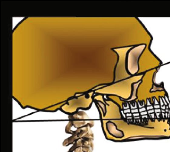
2 Case Reports in Dentistry
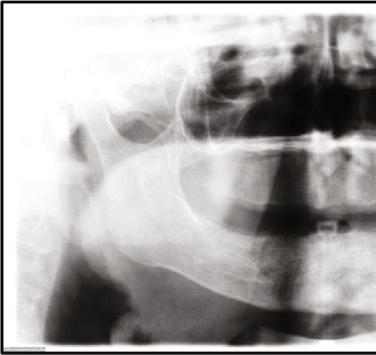
Frankfurt Plane
+150°–30°
X-Ray
Figure 2: Panoramic radiograph showing radiopaque image
Exposure factor circumscribed on the left side of the neck soft tissue region
adjacent to the C3 and C4 intervertebral space (arrow).
Figure 1: Manzi Projection description.
Notably, Intra-arterial Angiography is the gold standard
for the detection of these atheromatous plaques and the ste-
nosis they cause; however, because it is an invasive method
with an estimated risk morbidity and mortality of 2%, nonin-
vasive examinations are chosen in clinical practice, in which
the Doppler Ultrasound is outstanding, as it has an accuracy
rate of up to 90% when compared with Intra-arterial Angiog-
raphy [7–9].
The purpose of this study is to describe a dental radio-
graphic technique for establishing the differential diagnosis
of calcified atheromas relative to other calcifications with
similar radiographic characteristics. Furthermore, the inten-
tion is to report three clinical cases that demonstrate the
diagnostic possibilities of this technique.
2. Technical Report
The Manzi Projection is performed using the cephalometric
unit of the panoramic radiographic equipment, in which
the technique is described as follows: anteroposterior inci-
dence, Frankfurt plane with vertical inclination between
+15 and 30°, angle of the vertical and horizontal X-ray central
beam at 0°, and reduction of radiation exposure factors, usu-
ally at 50% (Figure 1).
Figure 3: Manzi Projection showing radiopaque image
3. Case Reports circumscribed on both sides of the neck soft tissue region, in
which the left side is adjacent to the C3 and C4 intervertebral
3.1. Clinical Case 1. The patient, a 78-year-old woman, sys-
space (arrow) and the right side is adjacent to the C4 (arrow).
temically presenting the following risk factors related to ath-
eroma formation: diabetes, dyslipidemia, arterial
hypertension, and tobacco use. The patient was asked to have
a panoramic radiograph taken for dental treatment. The unit of the Kodak Ceph 9000 3D (Carestream Health, Inc.,
examination was performed with the Kodak Ceph 9000 3D Rochester, NY, USA) equipment, by which the presence of
(Carestream Health, Inc.) equipment, in which the presence bilateral radiopaque masses were detected in the patient,
of radiopaque mass adjacent to the C3-C4 intervertebral showing that the one on the right side was located adjacent
space on the left side of the patient was detected, compatible to the C4 vertebra (Figure 3).
with calcified atheroma (Figure 2). As these calcifications were also presented in the Manzi
The Manzi Projection was then performed on the patient Projection, the patient was referred for cardiac monitoring,
to establish a differential diagnosis between calcified athero- in which the Doppler Ultrasound was requested to establish
mas and other calcifications with similar imaginological the degree of arterial stenosis. The presence of calcified pla-
characteristics. This was performed with the cephalometric que in the carotid bulb (bulbar or medulla oblongata) region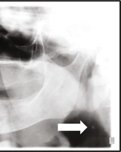
Case Reports in Dentistry                                                                                                         3

of the right and left carotid arteries was detected in this
examination, causing between 30 and 49% stenosis.

3.2. Clinical Case 2. The patient, a 56-year-old man, system-
ically presenting the following risk factors for atherosclerosis:
dyslipidemia, arterial hypertension, active periodontal dis-
ease, and being a self-declared ex-tobacco user. The pano-
ramic radiograph for dental treatment was requested, in
which the presence of bilateral radiopaque masses adjacent
to the cervical region was detected (Figure 4).
     The patient was asked to undergo the Manzi Projection
examination, which demonstrated the presence of radi-
opaque masses compatible with calcified atheromas on                 Figure 4: Panoramic radiograph showing the presence of bilateral
the patient’s right side, in which the presence of a second         radiopaque masses (arrows).
plaque, in a more inferior position than that of the one
demonstrated by the panoramic radiograph, and the
absence of calcified plaque on the left of the patient were
verified (Figure 5).
     The patient was referred to the responsible cardiologist,
who confirmed, by Doppler Ultrasound, the presence of cal-
cified plaques in the right carotid artery, located in the
carotid bulb and common carotid artery region, causing
between 30 and 49% stenosis. On the left side, the presence
of calcium free plaque was also verified, causing between 30
and 49% stenosis. However, the calcification visualized on
the left side of the panoramic radiograph was not a calcified
atheroma.

3.3. Clinical Case 3. The patient, a 90-year-old woman, sys-
temically presenting the following risk factors for atheroscle-
rosis: dyslipidemia and arterial hypertension. She was
submitted to panoramic radiograph examination for dental
treatment in which the presence of a calcified plaque was ver-
ified and visualized in the cervical region on the patient’s left
side (Figure 6).
    The Manzi Projection for the differential diagnosis of
other calcifications and structures was performed in the den-
tal office, in which the presence of radiopaque masses adja-          Figure 5: Manzi Projection showing the presence of two calcified
cent to the cervical region was verified, on both sides              plaques adjacent to the cervical region (arrows).
(bilateral). Relevant to highlight is that the localization of
the calcified structure (demonstrated by the panoramic
radiograph) was superior position to that of the structures         although highly accurate, is an invasive method with an esti-
visualized on the Manzi Projection and that these structures        mated rate of morbidity and mortality of 2% [7–9]
did not appear to be prominently in the cervical region, the            Other calcifications in the soft tissues can be observed
region of the carotid artery itself (Figure 7).                     radiographically in the cervical region. Carotid atheroma
    The patient was referred for cardiac monitoring, in which       must be distinguished from those other radiopacities that
the Doppler Ultrasound was requested. Thus, on examina-             lie in proximity to the artery. Based on the location, the
tion, the presence of bilateral calcified plaques in the carotid     calcified triticeous cartilage is the most likely calcification to
bulb and common carotid artery regions was verified, caus-           be confused with a carotid atheroma. However, as a matter
ing over 70% stenosis on the right side and between 50 and          of distinguishing, there are some main features that can be
69% stenosis on the left side.                                      observed. Calcified triticeous cartilages are well delimited
                                                                    and regular radiopacities, while carotid atheroma is visualized
4. Discussion                                                       as irregular, heterogeneous, verticolinear radiopacities. Also,
                                                                    in a panoramic radiograph, the carotid atheroma appears
Atherosclerosis is usually asymptomatic, and it is diagnosed        more laterally than a calcified triticeous cartilage [10, 11].
by means of medical examinations, such as Doppler Ultra-                Intending to provide an early diagnosis of these calcified
sound, considered the most widely used examination for this         atheromatous plaques, Friedlander and Lande, as far back as
purpose, because it is a noninvasive method with excellent          1981, analyzed the possibility of identifying these calcifica-
accuracy. It differs from Intra-arterial Angiography which,          tions by means of using panoramic radiography. Thirty-five
4                                                                                                              Case Reports in Dentistry

                                                                      played without being superimposed by other calcified struc-
                                                                      tures, and enlargement of the vertebral visualization enables
                                                                      an analysis of the cervical region. This is important because
                                                                      around 11% of the carotid bifurcations, the sites most fre-
                                                                      quently affected by atheromatous plaques are found inferior
                                                                      to the C3-C4 intervertebral space, where visualization in the
                                                                      panoramic radiograph is most unlikely [14]. The patient
                                                                      undergoes the examination with the Frankfurt plane (that
                                                                      starts at the superior portion of the acoustic meatus and is
                                                                      tangential to the infraorbital foramen) leaning slightly in
                                                                      the superior direction (between 15 and 30°), which dimin-
                                                                      ishes the possibility of mandibular superimposition of the
Figure 6: Panoramic radiograph showing the presence of
                                                                      cervical region. Because it is an anteroposterior technique,
radiopaque mass circumscribed on the left side of the cervical
region (arrow).                                                       the region of the carotid bifurcation is found closer to the
                                                                      radiographic sensor which, according to the geometric prin-
                                                                      ciples of radiological image formation, may clearly distin-
                                                                      guish calcified atheromas, in case they exist.

                                                                      5. Conclusion
                                                                      The Manzi Projection can promote differential diagnosis of
                                                                      calcified atheroma in relation to other cervical calcifications
                                                                      in dental practice; however, further studies are necessary to
                                                                      establish its accuracy.

                                                                      Additional Points
                                                                      Human and Animal Rights. No animals were used in this
                                                                      research. All research procedures followed were in accor-
                                                                      dance with the ethical standards of the committee responsi-
                                                                      ble for human experimentation (institutional and national)
                                                                      and with the Helsinki Declaration of 1975, as revised in
                                                                      2008          (http://www.wma.net/en/20activities/10ethics/
                                                                      10helsinki/).

                                                                      Consent
                                                                      Written and informed consent was obtained from the
                                                                      patient. Informed consent form was submitted to the patients
                                                                      enrolled in this study.

                                                                      Conflicts of Interest
Figure 7: Manzi Projection showing the presence of bilateral          The authors declare no conflict of interest, financial or
radiopaque masses, adjacent to the cervical region (arrows).          otherwise.
Notice that these structures are inferior to the calcified structure
region demonstrated by the panoramic radiograph.
                                                                      Acknowledgments
years have passed, and this method is still being studied and         The authors would like to thank CAPES (Coordination for
compared with other medical examinations for establishing             the Improvement of Higher Education Personnel) for their
its sensitivity, specificity, and accuracy. Although the pano-         financial support.
ramic radiograph does not present similar rates when com-
pared with Doppler Ultrasound, its findings must not be
disregarded, as the literature infers an association between
                                                                      References
the presence of calcified plaques in the cervical region (pano-         [1] D. Mozaffarian, E. J. Benjamin, A. S. Go et al., “Heart disease
ramic) and carotid calcifications [10–13].                                  and stroke statistics -2015 update: a report from the American
     The Manzi Projection represents a dental radiographic                 Heart Association,” Circulation, vol. 131, pp. 29–322, 2015.
technique that may assist with the differential diagnosis of            [2] M. F. Linton, P. G. Yancey, S. S. Davies et al., “The role of lipids
these calcified atheromatous plaques in the carotid because,                and lipoproteins in atherosclerosis,” in Endotext, L. J. Groot, P.
in this examination, the course of the carotid artery is dis-              Beck-Peccoz, G. Chrousos, K. Dungan, A. Grossman, and J. M.
Case Reports in Dentistry 5
Hershman, Eds., pp. 1–174, South Dartmouth (MA): NCBI
Bookshelf, 2015.
[3] A. H. Friedlander, “Panoramic radiography: the differential
diagnosis of carotid artery atheromas,” Special Care in Den-
tistry, vol. 15, no. 6, pp. 223–227, 1995.
[4] A. H. Friedlander and I. K. Friedlander, “Panoramic dental
radiography: an aid in detecting individuals prone to stroke,”
Brazilian Dental Journal, vol. 181, no. 1, pp. 23–26, 1996.
[5] L. C. Carter, K. Tsimidis, and J. Fabiano, “Carotid calcifica-
tions on panoramic radiography identify an asymptomatic
male patient at risk for stroke: a case report,” Oral Surgery,
Oral Medicine, Oral Pathology, Oral Radiology, and Endodon-
tics, vol. 85, no. 1, pp. 119–122, 1998.
[6] F. R. Manzi, F. N. Bóscolo, S. M. de Almeida, and F. H. Neto,
“Panoramic radiography as an auxiliary in detecting patients
at risk for cerebrovascular accident (CVA): a case report,”
Journal of Oral Science, vol. 45, no. 3, pp. 177–180, 2003.
[7] H. J. M. Barnett, D. W. Taylor, M. Eliasziw et al., “Benefit of
carotid endarterectomy in patients with symptomatic moder-
ate or severe stenosis,” The New England Journal of Medicine,
vol. 339, no. 20, pp. 1415–1425, 1998.
[8] European Carotid Surgery Trialists Colaborative Group, “Ran-
domised trial of endarterectomy for recently symptomatic
carotid stenosis: final results of the MRC European Carotid
Surgery Trial (ECST),” The Lancet, vol. 351, no. 9113,
pp. 1379–1387, 1998.
[9] E. G. Grant, C. B. Benson, G. L. Moneta et al., “Carotid artery
stenosis: gray-scale and Doppler US diagnosis—Society of
Radiologists in Ultrasound Consensus Conference,” Radiol-
ogy, vol. 229, no. 2, pp. 340–346, 2003.
[10] R. S. Kamikawa, M. F. Pereira, Â. Fernandes, and M. I. Meurer,
“Study of the localization of radiopacities similar to calcified
carotid atheroma by means of panoramic radiography,” Oral
Surgery, Oral Medicine, Oral Pathology, Oral Radiology, and
Endodontology, vol. 101, no. 3, pp. 374–378, 2006.
[11] L. C. Carter, “Discrimination between calcified triticeous carti-
lage and calcified carotid atheroma on panoramic radiogra-
phy,” Oral Surgery, Oral Medicine, Oral Pathology, Oral
Radiology, and Endodontology, vol. 90, no. 1, pp. 108–110,
2000.
[12] A. H. Friedlander and A. Lande, “Panoramic radiographic
identification of carotid arterial plaques,” Oral Surgery, Oral
Medicine, and Oral Pathology, vol. 52, no. 1, pp. 102–104,
1981.
[13] S. J. Yoon, W. Yoon, O. S. Kim, J. S. Lee, and B. C. Kang, “Diag-
nostic accuracy of panoramic radiography in the detection of
calcified carotid artery,” Dento Maxillo Facial Radiology,
vol. 37, no. 2, pp. 104–107, 2008.
[14] J. Griniatsos, S. Damaskos, N. Tsekouras, C. Klonaris, and
S. Georgopoulos, “Correlation of calcified carotid plaques
detected by panoramic radiograph with risk factors for stroke
development,” Oral Surgery, Oral Medicine, Oral Pathology,
Oral Radiology, and Endodontics, vol. 108, no. 4, pp. 600–
603, 2009.You can also read