COVID-19 and spontaneous esophageal perforation: a case report
←
→
Page content transcription
If your browser does not render page correctly, please read the page content below
Case Report
                                                                                                                                       Page 1 of 5

COVID-19 and spontaneous esophageal perforation: a case report
Seema S. Rao1^, Katy Marino2, Matthew A. Steliga2, Jason L. Muesse2
1
    Department of Surgery, University of Arkansas for Medical Sciences, Little Rock, Arkansas, USA; 2Division of Thoracic Surgery, Department of
Surgery, University of Arkansas for Medical Sciences, Little Rock, Arkansas, USA
Correspondence to: Jason L. Muesse, MD. 4301 W Markham Street, Slot 725, Little Rock, Arkansas, 72205, USA. Email: JMuesse@uams.edu.

                   Abstract: Pulmonary manifestations of the novel coronavirus, COVID-19, have been discussed heavily
                   in the literature, however, there have been minimal reports regarding extra-pulmonary manifestations
                   of the disease to date. In particular, there has been no literature to date discussing the pathophysiology
                   or incidence of esophageal perforation in the COVID-19 patient. This case report describes a 65-year-
                   old COVID-19 positive male presenting with a case of spontaneous esophageal perforation. The patient
                   underwent esophagogastroduodenoscopy (EGD) with stent placement followed by thoracoscopic evacuation
                   of gastric contents from the pleural spaces and mediastinal drainage. His clinical course was unique in
                   that his esophageal perforation management was complicated by logistical and technical challenges due to
                   COVID-19 infection. Several precautions were required before, during and after each test or intervention
                   performed on the patient. This created a challenging set of circumstances which had not been dealt with in
                   the past. Nevertheless, after a two-week hospital stay, the patient was discharged in stable condition with
                   plans for outpatient follow-up and removal of stent. This case report provides an unusual presentation of
                   esophageal perforation in a patient with concurrent COVID-19 infection while highlighting the special
                   techniques required to diagnose and treat the patient.

                   Keywords: Esophagus; esophageal perforation; esophageal stent; COVID-19; case report

                   Received: 12 July 2020; Accepted: 19 May 2021.
                   doi: 10.21037/aoe-20-60
                   View this article at: https://dx.doi.org/10.21037/aoe-20-60

Introduction                                                                     Case presentation

While COVID-19 has been classified as a predominantly                            This is a 65-year-old male with history of previous distal
respiratory illness, some patients have had involvement of                       esophageal perforation from vomiting (Boerhaave’s
other organ systems including neurological, gastrointestinal                     type) treated with a fully covered esophageal stent and
and hematological symptoms (1). Pneumomediastinum,                               gastrojejunostomy tube in 2017. The esophageal stent
thoracic empyema as well as mediastinal lymphadenopathy                          was removed a few weeks after placement and healing
have been reported in COVID-19 patients (2,3). There                             confirmed with an esophagram. In April 2020, he presented
has been no association described in the literature between                      to the emergency department with intractable nausea and
COVID-19 and esophageal perforation. This case report                            vomiting. This had started two days earlier with an episode
describes an esophageal perforation in a patient with                            of hematemesis followed by dyspnea and intermittent fevers.
COVID-19. We present the following case in accordance                            There was no history of foreign body ingestion or onset of
with the CARE reporting checklist (available at https://                         symptoms after a specific meal. His only home medications
dx.doi.org/10.21037/aoe-20-60).                                                  were a proton-pump inhibitor and sucralfate. An initial

^ ORCID: 0000-0002-8155-2813.

© Annals of Esophagus. All rights reserved.                                              Ann Esophagus 2021 | https://dx.doi.org/10.21037/aoe-20-60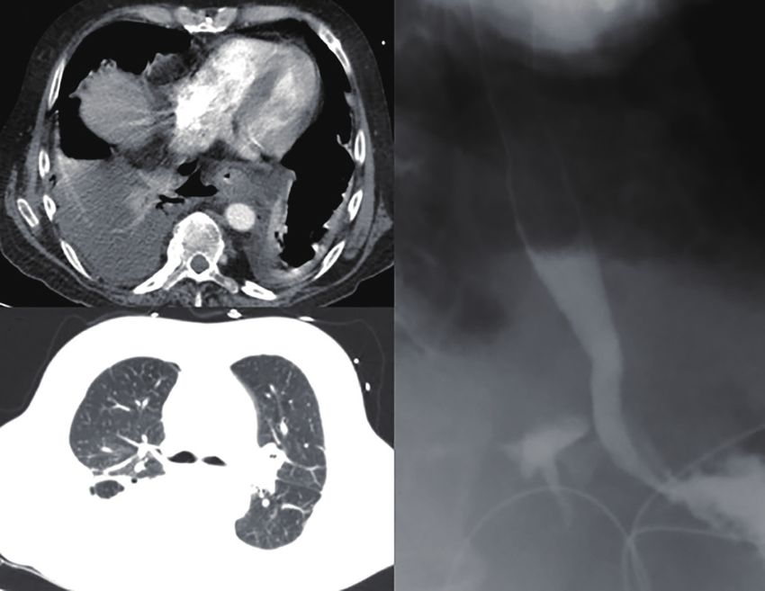
Page 2 of 5                                                                                                    Annals of Esophagus, 2021

                    A                                                    B

                    C

Figure 1 (A) Esophagram upon presentation revealing leak of contrast material into right posterior chest; (B) initial computed tomography
(CT) scan showing bilateral effusions; (C) initial CT scan showing lack of classic radiological findings of COVID-19.

chest X-ray was obtained which demonstrated bilateral                    sounds bilaterally, a soft abdomen and well healed midline
lower lobe effusions with no other radiolucent material to               laparotomy incision and gastrostomy site. A CT scan with
suggest a foreign body. Laboratory abnormalities included                IV and oral contrast showed severe esophageal edema
leukocytosis of 17,400, acute kidney injury (creatinine of               at the distal third with a complex pleural fluid collection
1.6 mg/dL), hypokalemia (potassium of 2.7 mmol/L) and                    in the posterior mediastinum suspicious for perforation
lactic acidosis of 2.4 mmol/L. D dimer was slightly elevated             (Figure 1A). A water soluble contrast esophagram
and anion gap was 16. A CT scan performed to evaluate for                demonstrated obvious leakage of contrast material
pulmonary embolism in the emergency department revealed                  confirming esophageal perforation (Figure 1B). He met
extra-luminal air posterior and to the right of the esophagus            screening criteria for COVID-19, including fevers, malaise
concerning for a perforation and bilateral effusions. He was             and cough, therefore a COVID-19 test was performed via
transferred to our institution for a higher level of care.               nasopharyngeal swab in the intensive care unit upon arrival.
   On arrival, his vitals were: pulse 65 beats per minute,               The rapid qualitative RT-PCR test resulted as positive.
blood pressure 93/56 mmHg, SpO2 100% and temperature                     His CT scan did not have any of the classic findings of
of 36.5 ℃. On review of systems he endorsed some                         COVID-19 (Figure 1C).
dyspnea and malaise. He was retired and reported living                     He was taken emergently to the operating room. The
a relatively isolated life in a small rural town. He had a               esophagogastroduodenoscopy (EGD) revealed a 2–3 mm
history of alcoholism in the past, but currently consumed                perforation at 39 cm from the incisors at the 4 o’clock
2–3 alcoholic drinks per day and did not use tobacco. He                 right posterolateral position, with the gastroesophageal
reported a lessened desire to consume alcohol in the days                junction at 40 cm from the incisors (Figure 2). There was
prior to presentation with no heavy alcohol abuse leading                no previous stent or signs of a foreign body noted that
up to the vomiting event. He had diminished breath                       could have caused a traumatic erosion. Because of the

© Annals of Esophagus. All rights reserved.                                        Ann Esophagus 2021 | https://dx.doi.org/10.21037/aoe-20-60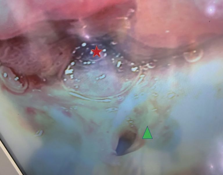
Annals of Esophagus, 2021 Page 3 of 5
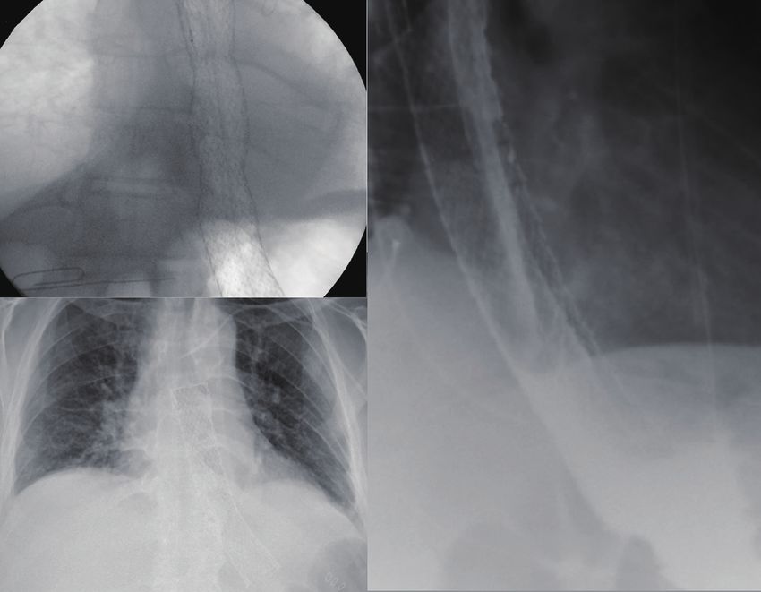
perforation having occurred almost 48 hours prior and stent was deployed successfully with fluoroscopic guidance
due to the relatively small size of the defect, it was felt (Figure 3A,B). A gastrojejunostomy tube was endoscopically
that an esophageal stent was the best option to address the placed through the patient’s previous gastrostomy site.
perforation. An EndoMaxx (Merit Medical, South Jordan, Bilateral thoracoscopic evacuation of gastric contents and
UT, USA) 23×150 millimeter fully covered esophageal mediastinal drainage was performed, left followed by right.
There was extensive contamination of gastric contents into
the pleural spaces, more significant on the right. A LigaSure
Anterior device (Medtronic, Dublin, Ireland) was used to open the
pleura around the esophagus near the site of the perforation
to allow drainage of the mediastinum. Nasogastric tube
decompression of the stomach along with gastric gravity
decompression via gastrojejunostomy tube was continued
until post-operative day 8, when a repeat esophagram
was performed to evaluate for leak, which was negative
(Figure 3C). He was initiated on a clear liquid diet and then
advanced to full liquids. His chest tubes were removed on
post-operative days 10 and 11 sequentially following further
demonstration of clinical stability.
Figure 2 Esophagogastroduodenoscopy; star denotes Infectious disease service was consulted and suggested
gastroesophageal junction, triangle represents perforation at right using clinical markers to track COVID-19 infection
posterior esophagus. including IL-6 and CRP (4). His CRP initially drawn on
A C
B
Figure 3 Clockwise from left. (A) Stent placement under fluoroscopic guidance, paperclip denotes gastroesophageal junction; (B) chest X-ray
on day 13 with stent and no radiographic evidence of COVID infection; (C) esophagram on day 8.
© Annals of Esophagus. All rights reserved. Ann Esophagus 2021 | https://dx.doi.org/10.21037/aoe-20-60Page 4 of 5 Annals of Esophagus, 2021
post-operative day 4 was 105.10 mg/L and IL-6 on post- esophagram given that our fluoroscopy suite lacked negative
operative day 5 was elevated to 145.7 pg/mL. These trended pressure air clearance. To combat this, we taped off the
downward to a value of 12.90 mg/L and 26.1 pg/mL doorways, wore N95 equipment for airborne precautions
respectively, on post-operative day 7 when a repeat throughout the procedure and terminally cleaned the room
COVID-19 test resulted as negative suggesting clinical afterwards. Assessment and management of esophageal
resolution of COVID-19 infection. perforations is time sensitive due to rapid progression of
He remained on the COVID-19 isolation floor mediastinal sepsis. Although the patient had COVID-19,
throughout his hospital stay. Our patient refused alcohol he was able to undergo diagnosis and treatment in an
withdrawal prophylaxis and never showed signs of alcohol expeditious manner, and no health care providers involved
withdrawal. He was maintained on antibiotics and anti- in the case subsequently contracted COVID-19.
fungal agents for 13 days and was discharged home with oral This case report depicts an esophageal perforation in a
antibiotic and anti-fungal treatment on post-operative day patient with COVID-19. It is important to note that this
13 in good condition with plans to remove the esophageal patient had a prior history of esophageal perforation as well
stent and repeat his esophagram in 4 weeks. as risk factors for recurrent esophageal perforation, however,
All procedures performed in studies involving human little is known about extra-pulmonary manifestations of
participants were in accordance with the ethical standards of COVID-19. While there has been no conclusive evidence
the institutional and/or national research committee(s) and linking esophageal perforations as a complication or
with the Helsinki Declaration (as revised in 2013). Written presentation of COVID-19, more research and observation
informed consent was obtained from the patient. of COVID-19 patients is required to determine whether
any association exists.
Discussion
Acknowledgments
To our knowledge, this is the first case report describing
spontaneous esophageal perforation in a patient who Funding: None.
concurrently presented with COVID-19.
The patient’s chest X-ray, as well as CT scan did not
Footnote
reveal any of the classic radiologic findings of COVID-19
(Figure 1C). It has been noted that the radiological findings Reporting Checklist: The authors have completed the
of ground glass opacities are more commonly located in CARE reporting checklist. Available at https://dx.doi.
the lower lobes in a subpleural distribution (5), which org/10.21037/aoe-20-60
may have been obscured by the bilateral effusions in our
patient caused by the esophageal perforation. Bronchoscopy Peer Review File: Available at https://dx.doi.org/10.21037/
performed at the time of surgery demonstrated extensive aoe-20-60
mucous production bilaterally. This suggests our patient
may have had pulmonary signs of COVID-19, which were Conflicts of Interest: All authors have completed the ICMJE
likely subclinical and not the primary manifestation of the uniform disclosure form (available at https://dx.doi.
disease. org/10.21037/aoe-20-60). The authors have no conflicts of
Most Boerhaave’s perforations are seen preferentially interest to declare.
on the left side, due to structural weakness at that portion
of the esophagus (6). Spontaneous recurrences are rare (7). Ethical Statement: The authors are accountable for all
Given his history of emesis, we anticipated the perforation aspects of the work in ensuring that questions related
on the left posterior location, but clearly spontaneous to the accuracy or integrity of any part of the work are
perforations can occur on either side, which emphasizes the appropriately investigated and resolved. All procedures
importance of pre-operative imaging. performed in studies involving human participants were in
It is also important to note challenges in caring for accordance with the ethical standards of the institutional
patients with COVID-19 and esophageal pathology. We and/or national research committee(s) and with the Helsinki
administered fluid conservatively to prevent pulmonary Declaration (as revised in 2013). Written informed consent
edema. We also faced challenges with obtaining an was obtained from the patient.
© Annals of Esophagus. All rights reserved. Ann Esophagus 2021 | https://dx.doi.org/10.21037/aoe-20-60Annals of Esophagus, 2021 Page 5 of 5
Open Access Statement: This is an Open Access article 3. Valette X, du Cheyron D, Goursaud S. Mediastinal
distributed in accordance with the Creative Commons lymphadenopathy in patients with severe COVID-19.
Attribution-NonCommercial-NoDerivs 4.0 International Lancet Infect Dis 2020;20:1230.
License (CC BY-NC-ND 4.0), which permits the non- 4. Zhu Z, Cai T, Fan L, et al. Clinical value of immune-
commercial replication and distribution of the article with inflammatory parameters to assess the severity of
the strict proviso that no changes or edits are made and the coronavirus disease 2019. Int J Infect Dis 2020;95:332-9.
original work is properly cited (including links to both the 5. Guan CS, Lv ZB, Yan S, et al. Imaging Features of
formal publication through the relevant DOI and the license). Coronavirus disease 2019 (COVID-19): Evaluation on
See: https://creativecommons.org/licenses/by-nc-nd/4.0/. Thin-Section CT. Acad Radiol 2020;27:609-13.
6. Pate JW, Walker WA, Cole FH Jr, et al. Spontaneous
rupture of the esophagus: a 30-year experience. Ann
References
Thorac Surg 1989;47:689-92.
1. Behzad S, Aghaghazvini L, Radmard AR, et al. 7. Naitoh H, Fukuchi M, Kiriyama S, et al. Recurrent,
Extrapulmonary manifestations of COVID-19: Radiologic spontaneous esophageal ruptures associated with
and clinical overview. Clin Imaging 2020;66:35-41. antiphospholipid antibody syndrome: report of a case. Int
2. Zhou C, Gao C, Xie Y, et al. COVID-19 with spontaneous Surg 2014;99:842-5.
pneumomediastinum. Lancet Infect Dis 2020;20:510.
doi: 10.21037/aoe-20-60
Cite this article as: Rao SS, Marino K, Steliga M, Muesse JL.
COVID-19 and spontaneous esophageal perforation: a case
report. Ann Esophagus 2021.
© Annals of Esophagus. All rights reserved. Ann Esophagus 2021 | https://dx.doi.org/10.21037/aoe-20-60You can also read