Minimally invasive treatment of gynecomastia by ultrasound-guided vacuum-assisted excision: report of a case series
←
→
Page content transcription
If your browser does not render page correctly, please read the page content below
CASE REPORT
                                               https://doi.org/10.29289/2594539420200069

     Minimally invasive treatment of gynecomastia by
      ultrasound-guided vacuum-assisted excision:
                  report of a case series
            Henrique Lima Couto1,2 , Carolina Nazareth Valadares1,2,3* , Osmar Pellegrini Junior4 ,
     Tereza Cristina Ferreira de Oliveira1 , Patricia Martins Gomes El Bacha1 , Shirley das Graças Ferreira1

    ABSTRACT
Introduction: Gynecomastia (GM) is a benign proliferation of glandular breast tissue in men. Some cases need surgical intervention.
Traditional open surgery by semicircular inferior periareolar incision is the most common surgical approach. In order to obtain
better esthetic results, some alternatives to open surgery have been proposed, such as liposuction, endoscopic mastectomy,
and vacuum-assisted excision (VAE). Objective: To describe the technical surgical approach of ultrasound-guided VAE of GM and
its results from a case series. Method: This is an evaluation of seven GM cases submitted to ultrasound-guided VAE with a 10G
needle using the ENCOR ® BD whole circumference automated breast biopsy system in Redimasto – Redimama, a Brazilian breast
center. The result was considered good or satisfactory when it showed minimal remaining gland, good symmetry, no retraction,
necrosis, hypertrophic scar, or displacement of the nipple-areola complex. All patients answered a questionnaire to evaluate
their satisfaction and perception of the procedure. Results: Seven (7) patients with Simon grade 1 and 2 bilateral GM underwent
ultrasound-guided VAE. No case of displacement, necrosis, or retraction of the nipple-areola complex, post-procedure bleeding,
infection, skin necrosis, or asymmetry was detected. No patient reported decrease or change in nipple sensation or erection.
All patients had bruises and hematomas that spontaneously resolved within 30 days. All results were considered good or excellent
by patients and surgeons. Conclusion: Minimally invasive ultrasound-guided VAE is an excellent alternative for the treatment of
GM. It is better indicated for Simon grade 1 and 2 GM, with good and excellent esthetic results, small scar, and low rates of nipple
and areolar complications. It allows an outpatient procedure with low morbidity (local anesthesia) and fast recovery.
KEYWORDS: gynecomastia; mammary ultrasonography; interventional ultrasound; needle bipsy.

                                                                           GM typically results from an absolute or relative deficiency
INTRODUCTION                                                           of androgen action or excessive estrogen action in the breast tis-
Gynecomastia (GM) is a benign proliferation of glandular breast        sue2. No treatment is necessary for asymptomatic adolescents or
tissue in men1. It is the most common male breast disorder,            men, but it is required when GM is progressive, painful, or causes
accounting for nearly 60% of them. It can be unilateral or, most       cosmetic discomfort. It usually resolves by itself or by removing
often, bilateral. GM is a common condition with a prevalence           the underlying cause, such as medication, anabolic-androgenic
of 32% to 65%, depending on age, and can affect up to 70% of           steroid abuse, or treatment of systemic diseases3. Medical ther-
all pubescent boys2. A man’s lifespan has three peaks: the first       apy can also be prescribed for patients with a recent diagnosis —
occurs during infancy, the second during puberty, and the third        within two years —, but is less effective for long-standing GM.
in middle-aged and older men1,2 . GM in infancy and puberty            Some cases need surgical intervention. According to Simon, GM
resolves spontaneously in most cases. Proper investigation is          can be classified into grades4 (Table 1).
highly recommended among adults and older adults to exclude                Traditional open surgery by semicircular inferior periareolar
underlying diseases1.                                                  incision is the most common surgical approach, but it may cause

1
 Redimasto, Redimama – Belo Horizonte (MG), Brazil.
2
 Universidade Federal de Minas Gerais – Belo Horizonte (MG), Brazil.
3
  Santa Casa de Belo Horizonte – Belo Horizonte (MG), Brazil.
4
  Hospital da Força Aérea – Brasília (DF), Brazil.
*Corresponding author: carolinanvaladares@gmail.com
Conflict of interests: nothing to declare.
Received on: 11/03/2020. Accepted on: 11/18/2020

                                                       Mastology 2021;31:e20200069                                                  1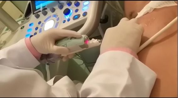
Couto HL, Valadares CN, Pellegrini Junior O, Oliveira TCF, El Bacha PMG, Ferreira SG
significant morbidities, such as asymmetry, poor scarring, and for the patient’s weight. No sedation was necessary. After the
nipple-areola complex retraction or necrosis5-7. In order to obtain 10G needle was introduced and positioned via ultrasound,
better esthetic results, some alternatives to open surgery have the automated vacuum device was activated (Figures 1 and 2).
been proposed, such as liposuction, endoscopic mastectomy, and The number of fragments extracted from each breast varied
vacuum-assisted excision (VAE)7-9. according to the surgeon’s judgment of each case, taking into
In the last few years, the use of vacuum-assisted devices, account the amount of breast tissue during clinical examina-
originally created to diagnose breast lesions by radiologically- tion, mammography, and breast ultrasound before surgery, as
guided procedures, has shown to be promising in the surgical well as the real-time breast ultrasound evaluation during the
management of GM8-12. procedure. The vacuum method for dense breasts with fine
precision was used for all cases. The resection performed left
a 1-cm thick gland behind the nipple, just like the standard
OBJECTIVE surgical procedure. At the end of the VAE of the GM, vacuum
To describe the technical surgical approach of ultrasound-guided and manual suction of the residual cavity were performed to
VAE of GM and its results from a case series. avoid or reduce the incidence of postoperative hematomas and
bruises. Only one patient had the surgical cavity marked with a
metal clip. Mammographic images were obtained one and six
METHOD months after VAE to evaluate the removal of the glandular tissue
The study consists of seven GM cases evaluated from December (Figure 3). Patients wore a thoracic compression belt for at least
1, 2018, to December 1, 2019. The patients underwent ultrasound- 30 days. Follow-up was scheduled at 7 days, 14 days, 1 month,
guided VAE with a 10G needle using the ENCOR® BD whole cir- 2 months, and 6 months after the procedure, and consisted of
cumference automated breast biopsy system in Redimasto — clinical examination, pictures, and survey of the patient’s and
Redimama, a Brazilian breast center. Before the procedure, all breast surgeon’s satisfaction. The result was considered good or
patients were submitted to a clinical evaluation with full his- satisfactory when it showed minimal remaining gland, good
tory and physical examination by a breast surgeon, as well as symmetry, no retraction, necrosis, hypertrophic scar, or dis-
mammography, breast ultrasound, and blood tests. All patients placement of the nipple-areola complex. All patients answered
signed an informed consent form for the VAE procedure. All pro- a questionnaire to evaluate their satisfaction and perception
cedures were performed by breast surgeons experts in ultra- of the procedure.
sound-guided VAE. The procedures took place in the breast
center, in an outpatient approach, through a 3 mm incision
in each breast, with local anesthesia, using 2% lidocaine and RESULTS
bupivacaine when necessary, according to the maximum dose Seven patients with Simon grade 1 and 2 bilateral GM under-
went ultrasound-guided VAE. One of them had undergone pre-
Table 1. Simon grade of gynecomastia. vious traditional open surgical treatment of GM with unsatis-
Grade 1 small breast without excess skin factory results, and all patients expressed their wish to have an
Grade 2 moderate breast without excess skin excision with less morbidity, small scars, and good esthetic out-
come. The mean age was 27.5 years (ranging from 19 to 34 years).
Grade 3 moderate breast with excess skin
The average procedure time was 28 minutes (ranging from 23
Grade 4 large breast with excess skin
to 54 minutes). The main complaint and indication for the pro-
cedure was the esthetic appearance of GM, followed by physi-
cal deformity. One patient had an areola fissure caused by the
vacuum suction during the procedure, which was promptly
sutured and did not affect the final esthetic result. At follow-
up, all patients and breast surgeons reported excellent or good
satisfaction (Figures 4 and 5), and at the six-month review, no
patient presented recurrence or asked for another intervention
or open surgery. No patient had postoperative seroma, bleeding,
or hemorrhage or needed to be taken to the operating room at
any time, during or after the surgical procedure and follow-up.
All procedures were performed in an outpatient setting, with
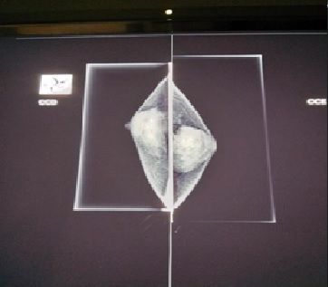
Figure 1. Ultrasound-guided vacuum-assisted excision of gyne- local anesthesia and no sedation. Histological evaluation revealed
comastia: surgical approach. benign GM in all patients. No case of displacement, necrosis, or
2 Mastology 2021;31:e20200069
Minimally invasive treatment of gynecomastia
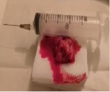
Figure 2. Ultrasound-guided vacuum-assisted excision of gynecomastia: surgical specimen.
Figure 3. Mammograms before and six months after ultrasound-guided vacuum-assisted excision of gynecomastia.
Mastology 2021;31:e20200069 3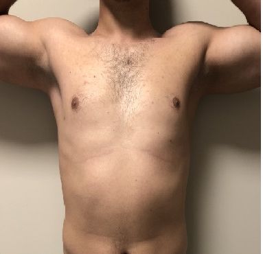
Couto HL, Valadares CN, Pellegrini Junior O, Oliveira TCF, El Bacha PMG, Ferreira SG
retraction of the nipple-areola complex was detected. None of procedure for this condition. The results are usually satisfac-
the individuals investigated presented postoperative bleed- tory, but postoperative complications are common, including
ing, infection, skin necrosis, or asymmetry. No patient reported areola deformity or retraction; “saucer-shaped defect” (from
decrease or change in nipple sensation or erection. All patients over-resection of breast tissue); seroma; poor scarring, such
had bruises and hematomas that spontaneously resolved within as retraction, hypertrophic scar, or keloid formation; wound
30 days of VAE, with excellent or good cosmetic results and no dehiscence; and nipple retraction, necrosis, or altered sensa-
skin sequelae. The individuals investigated were able to return tion. The side effects of standard surgery have been a long-
to their life activities in 2 days and to physical work in 14 days. standing concern. In 1987, Courtiss et al. published an article
Physical activities were allowed two weeks after the procedure. reporting that 101 out of 159 patients presented high com-
All results were considered good or excellent by patients and plication rates after traditional excision for the treatment of
surgeons (Table 213 and Figure 3). GM, including under-resection (21.9%), “saucer-shaped defect”
(18.7%), poor scarring (18.7%), hematoma (16.1%), and seroma
(9.4%) 6 . In order to decrease morbidity and improve esthetic
DISCUSSION results, the GM treatment should improve with new surgical
The main goal of treating GM is to remove the excess of breast techniques and minimally invasive procedures.
tissue, achieving the best symmetry with minimal scarring More recently, some groups have described an endoscope-
and good or excellent esthetic results. Different from subcu- assisted subcutaneous mastectomy 5 , with a smaller inci-
taneous mastectomy for cancer treatment, the purpose of sion. However, this technique did not eliminate the potential
GM surgery is not to excise all breast tissue in an oncologic complication of having a scar on a visible part of the chest
fashion. GM surgery aims to remove enough breast tissue to or axillae, and the risk of nipple-areola complex complica-
obtain a good cosmetic result and avoid clinical recurrence. tions remains 8 .
The open surgical approach is still the standard procedure for In 2010, the Royal College of Surgeons of England pub-
persistent GM after one or two years, especially when associ- lished the first article about a vacuum-assisted biopsy device
ated with psychological distress, unsatisfactory body image, associated with liposuction to provide a minimally invasive
and avoidance of activities in which the chest is exposed approach for GM, with excellent results8. The group suggested
(sports and swimming) 4. For years, subcutaneous mastectomy that ultrasound guidance could be positive in those cases.
through a semicircular inferior areolar incision, associated One year later, the Chinese experience with a vacuum-assisted
or not with liposuction, has been the gold-standard surgical biopsy device was also published9. Recently, the indications
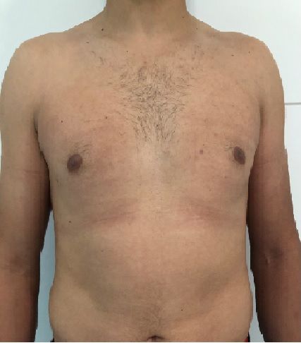
Figure 4. 34-year-old man with Simon grade 2 gynecomastia.
4 Mastology 2021;31:e20200069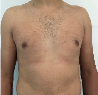
Minimally invasive treatment of gynecomastia

for VAE have expanded to more severe Simon grades of GM,                         The benefits of VAE are similar to those of minimally
with the procedure performed in the operating room under                     invasive procedures in general — reduced morbidity, better
general anesthesia10.                                                        esthetic results, fewer recovery days, and no hospitalization
    A recent prospective series compared VAE of GM with open                 time or cost 8 . The results from this series corroborate the
traditional surgery. The VAE group had significantly smaller scar            findings of other series and studies. Depending on the GM
sizes (0.40 ± 0.08 cm vs. 5.34 ± 0.38 cm, p < 0.01), shorter healing         grade, the VAE can be performed with local anesthesia, with
time (3.67 ± 0.71 days vs. 7.90 ± 0.92 days, p < 0.01) and hospital-         or without sedation. With the evolution of vacuum-assisted
ization (2.60 ± 0.62 vs. 7.17 ± 0.83 days, p < 0.01), as well as higher      devices, better vacuum aspiration, and multiple fragments
postoperative satisfaction (4.70 ± 0.60 scores vs. 3.20 ± 0.55 scores,       collected in an automated circular approach with one-step
p < 0.01). The incidence rate of bruises was significantly higher            needle insertion, it is possible to remove a considerable amount
in the VAE group compared to the open surgical group (47% vs.                of breast tissue in a few minutes, reducing the odds of infec-
17%, p = 0.013 and 54% vs. 20%, p = 0.007), respectively11.                  tion or complication. A study reported a median time of 50

Figure 5. Same patient six months after ultrasound-guided vacuum-assisted excision of gynecomastia.

                                                           Mastology 2021;31:e20200069                                                  5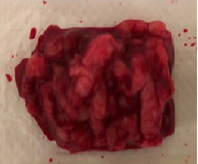
Couto HL, Valadares CN, Pellegrini Junior O, Oliveira TCF, El Bacha PMG, Ferreira SG

Table 2. Satisfaction evaluation: adaptation of the consultation satisfaction questionnaire.
 n=7                                            Esthetic discomfort         Physical deformity          Medical indication
 Patient complaint                                         5                           2                          0
 n=7                                                  Excellent                     Good                       Regular          Bad
 Final esthetic result (6 months) – patient                5                           2                          0               0
 Final esthetic result (6 months) –
                                                           4                           3                          0               0
 surgeon
 n=7                                                      yes                         no
 Would the patient repeat or recommend
                                                           7                           0
 the procedure for someone?
 Was the procedure well tolerated?                         7                           0
 Complications n = 7
 Seroma                                                    0
 Bruises                                                   7
 Anesthesia scar                                           0
 Bleeding                                                  0
 Areola fissure                                            1
 Displacement, necrosis, or retraction of
                                                           0
 the nipple-areola complex.
 Decrease or change in nipple sensation
                                                           0
 or erection
Source: Mazzarone13.

minutes using an 8G needle with a semi-automated device 8 ,                    grade 1 and 2 GM, with good and excellent esthetic results
while in this series, the median time was 25 minutes using                     and low rates of nipple and areolar complications. It allows
a 10G needle with a whole circumference automated device.                      an outpatient procedure with low morbidity (local anesthesia)
The patients’ procedure tolerance was high, even with just                     and fast recovery. Hematomas and bruises are always present
local anesthesia . Automated devices allow faster, safe, and                   due to the nature of the approach. Breast surgeons can obtain
outpatient procedures that preclude hospitalization and have                   satisfactory cosmetic results with little morbidity and postop-
the potential of saving costs.                                                 erative complications, such as nipple retraction or necrosis.
    Doubts related to long-time recurrence remain and require                  Ultrasound-guided VAE has become a valuable approach for
more studies for clarification. Longer follow-up will be neces-                the surgical management of Simon grade 1 and 2 GM, with
sary to evaluate this issue better. Nevertheless, the amount                   or without liposuction according to necessity. Trials compar-
of breast tissue excised described by the literature and this                  ing VAE of GM with open surgery should also evaluate clini-
series is not different from the traditional open surgical                     cally relevant recurrence throughout the years to establish
specimen. Mammographic images gradually change over                            the safety of these surgical approaches over time.
time. After six months, it is possible to estimate the amount
of tissue resected, but, like in benign surgeries, the degree of
architectural distortion is high, especially due to large hema-                AUTHORS’ CONTRIBUTION
tomas and bruises, which fade with time. This finding indi-                    C.V.: Investigation, Methodology, Project Administration,
cates that the best moment for a mammographic evaluation                       Writing — Review and Editing.
of the amount of breast resected should probably be after one                  H.L.: Investigation, Methodology, Project Administration,
year of the procedure.                                                         Supervision, Validation, Writing — Review and Editing.
                                                                               T.O.: Writing — Review and Editing, Formal Analysis.
                                                                               P.B.: Methodology, Writing — Review and Editing.
CONCLUSION                                                                     S.F.: Data Curation, Validation, Writing — Review and Editing.
Minimally invasive ultrasound-guided VAE is an excellent alter-                O.J.: Investigation, Visua lization, Writing — Orig ina l
native for the treatment of GM. It is better indicated for Simon               Draft, Validation.

   6                                                        Mastology 2021;31:e20200069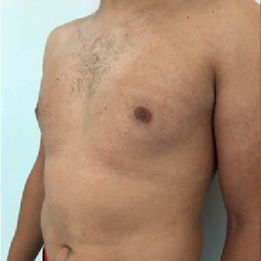
Minimally invasive treatment of gynecomastia

     REFERENCES
1. Kanakis GA, Nordkap L, Bang AK, Calogero AE, Bártfai G,                   8. Qutob O, Elahi B, Garimella V, Ihsan N, Drew PJ. Minimally
   Corona G, et al. EAA clinical practice guidelines-gynecomastia               invasive excision of gynaecomastia—a novel and effective
   evaluation and management. Andrology. 2019;7(6):778-93.                      surgical technique. Ann R Coll Surg Engl. 2010;92(3):198-200.
   https://doi.org/10.1111/andr.12636                                           https://doi.org/10.1308/003588410x12628812458815
2. Narula HS, Carlson HE. Gynaecomastia: pathophysiology,                    9. He Q, Zheng L, Zhuang D, Fan Z, Xi C, Zhou P. Surgical
   diagnosis and treatment. Nat Rev Endrocrinol. 2014;10(11):684-               treatment of gynecomastia by vacuum-assisted biopsy device.
   98. https://doi.org/10.1038/nrendo.2014.139                                  J Laparoendosc Adv Surg Tech A. 2011;21(5):431-4. https://doi.
3. Vojvodic M, Xu FZ, Cai R, Roy M, Fielding JC. Anabolic-androgenic            org/10.1089/lap.2011.0019
   Steroid Use Among Gynecomastia Patients: Prevalence and                   10. Yao Y, Yang Y, Liu J, Wang Y, Zhao Y. Vacuum-assisted
   Relevance to Surgical Management. Ann Plast Surg. 2019;83(3):258-             minimally invasive surgery. An innovative method for the
   63. https://doi.org/10.1097/SAP.0000000000001850                              operative treatment of gynecomastia. Surgery. 2019;166(5):934-
4. Simon BE, Hoffman S, Kahn S. Classification and surgical                      9. https://doi.org/10.1016/j.surg.2019.04.032
   correction of gynecomastia. Plast Reconstr Surg. 1973;51(1):48-           11. Wang Y, Wang J, Liu L, Liang W, Qin Y, Zheng Z, et al. Comparison
   52. https://doi.org/10.1097/00006534-197301000-00009                          of curative effects between mammotome-assisted minimally
5. Varlet F, Raia-Barjat T, Bustangi N, Vermersch S, Scalabre A.                 invasive resection (MAMIR) and traditional open surgery
   Treatment of Gynecomastia by Endoscopic Subcutaneous                          for gynecomastia in Chinese patients: A prospective clinical
   Mastectomy in Adolescents. J Laparoendosc Adv Surg Tech A.                    study. Breast J. 2019;25(6):1084-9. https://doi.org/10.1111/
   2019;29(8):1073-6. https://doi.org/10.1089/lap.2019.0256                      tbj.13424
6. Courtiss EH. Gynecomastia: analysis of 159 patients and current           12. Iwuagwu O, Drew P. Vacuum-assisted biopsy device-diagnostic
   recommendations for treatment. Plast Reconstr Surg. 1987;79(5):740-           and therapeutic applications in breast surgery. Breast.
   53. https://doi.org/10.1097/00006534-198705000-00010                          2004;13(6):483-7. https://doi.org/10.1016/j.breast.2004.06.004
7.   Colombo-Benkmann M, Buse B, Stern J, Herfarth C. Indications for        13. Mazzarone F. Avaliação da satisfação do resultado de
     and results of surgical therapy for male gynecomastia. Am J Surg.           cirurgia plástica [dissertation]. Rio de Janeiro: Fundação
     1999;178(1):60-3. https://doi.org/10.1016/s0002-9610(99)00108-7             Cesgranrio; 2013.

                                                                                                © 2021 Brazilian Society of Mastology
                                         This is an open access article distributed under the terms of the Creative Commons license.

                                                          Mastology 2021;31:e20200069                                                        7
You can also read