Detection of Cavernous transformation of the portal vein in a 22-year-old male with upper gastrointestinal bleeding and anemia by Abdominal Duplex ...
←
→
Page content transcription
If your browser does not render page correctly, please read the page content below
Posted on Authorea 18 Jul 2021 | The copyright holder is the author/funder. All rights reserved. No reuse without permission. | https://doi.org/10.22541/au.162664946.62190736/v1 | This a preprint and has not been peer reviewed. Data may be preliminary.
Detection of Cavernous transformation of the portal vein in a
22-year-old male with upper gastrointestinal bleeding and anemia
by Abdominal Duplex Ultrasonography. A Case Report.
Joseph Arkorful1 , Theophilus Adu - Bredu2 , and Obed Otoo1
1
Kwame Nkrumah University of Science and Technology
2
Komfo Anokye Teaching Hospital
July 18, 2021
Abstract
Cavernous transformation of the portal vein (CTPV) is a rare condition characterized by the formation of multiple tortuous
tiny blood vessels within and around a portal vein which has been occluded by thrombus. Early diagnosis of CTPV is relevant
for timely intervention which could save the liver from non-reversible damage
Title: Detection of Cavernous transformation of the portal vein in a 22-year-old male with upper gastroin-
testinal bleeding and anemia by Abdominal Duplex Ultrasonography. A Case Report.
Running Head : Sonographic detection of Cavernous transformation of the portal vein
Authors : Joseph Arkorful1,2 , Theophilus Kofi Adu – Bredu2,3 , Obed Otoo2 .
1. University Hospital, Kwame Nkrumah University of Science and Technology, Kumasi, Ghana
2. Department of Medical Diagnostics, College of Health Sciences, Kwame Nkrumah University of Science
and Technology, Kumasi, Ghana
3. Department of Obstetrics and Gynecology, Komfo Anokye Teaching Hospital, Kumasi, Ghana
Corresponding Author : Joseph Arkorful. Postal Address: P.O. Box UP 1717, K.N.U.S.T – Kumasi. Tel:
+233540604576 Email: josepharkorful@hotmail.com
ABSTRACT
Cavernous transformation of the portal vein (CTPV) is a rare condition characterized by the formation of
multiple tortuous tiny blood vessels within and around a portal vein which has been occluded by thrombus.
Early diagnosis of CTPV is relevant for timely intervention which could save the liver from end stage disease.
However, the diagnosis is often difficult due to its rarity and diverse clinical symptoms. Ultrasonography
could serve as a reliable modality in the early diagnosis of this condition to improve patients’ outcomes.
This case report narrates the role of Abdominal Duplex ultrasonography in the diagnosis of CTPV in a
22-year-old male presenting with upper gastrointestinal (GI) bleeding and anemia.
KEY CLINICAL MESSAGE
CTPV; a rare condition can be missed clinically due to its non-specific symptoms. CTPV should hence
be part of the differential diagnosis for patients who present with upper GI bleeding and anemia; and
the utilization of Abdominal Duplex ultrasound encouraged since it helps in detection of CTPV to ensure
adequate patientcare.
1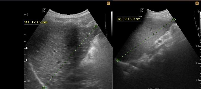
KEYWORDS:
Cavernous transformation of the Portal vein, Duplex Ultrasonography, Portal hypertension, Upper gastroin-
Posted on Authorea 18 Jul 2021 | The copyright holder is the author/funder. All rights reserved. No reuse without permission. | https://doi.org/10.22541/au.162664946.62190736/v1 | This a preprint and has not been peer reviewed. Data may be preliminary.
testinal bleeding, Portal cavernoma.
INTRODUCTION
Cavernous transformation of the portal vein (CTPV) also known as portal cavernoma is a very rare condition
of the liver which is usually characterized by the formation of multiple tortuous tiny blood vessels within
and around a portal vein which has been partially or completely occluded by a thrombus 1–3 .
Liver cirrhosis, hepatoma, splenectomy, liver transplant and pancreatitis are known causes of portal vein
thrombosis that subsequently leads to CTPV in adults 1 . However, CTPV have been mostly recorded
in individuals with long-term thrombosis of the portal vein without evidence of primary liver disease1–3 .
Cases of CTPV have also been recorded in individuals with hematological diseases 4 and those with primary
systemic vascular abnormalities which have resulted in chronic portal vein thrombosis 1,2,5 .
The initial diagnosis of CTPV by clinicians may be challenging due to its diverse causes and clinical pre-
sentations 4,6 . Common clinical manifestations are however those associated with portal hypertension as a
result of chronic portal vein thrombosis; such as upper gastrointestinal bleeding (secondary to esophageal
varices), anemia, splenomegaly, and ascites 3 .
Abdominal Duplex Ultrasonography is a useful first-line imaging tool for both adult and pediatric patients
with clinical presentations pointing towards the suspicion of liver disease or portal hypertension; because it
allows for examination of abdominal organs and their vascular architecture in a real-time, efficient and safe
manner7 . Computed Tomography Angiography (CTA) and Magnetic Resonance Angiography (MRA) may
be used for the confirmation of CTPV when an initial diagnosis is made with ultrasonography.
We therefore report on a case of a 22-year-old male in whom a diagnosis of cavernous transformation of the
portal vein was made with Duplex ultrasonography who presented clinically with anemia and hematemesis.
CASE REPORT
A 22-year-old male presented to the emergency room of our hospital with slight dizziness, four episodes
of vomiting mixed with blood and passage of dark stool over a 48-hour period. The patient had no prior
history of chronic alcoholism, liver cirrhosis or hepatoma. According to the patient, he has had an episode
of vomiting of blood and abdominal pain a year ago and was treated and discharged on account of bleeding
peptic ulcer disease. This current episode of vomiting of blood with slight dizziness occurred again and was
brought to our hospital by his parents for further management.
Upon physical examination, patient appeared pale with no evidence of respiratory distress. There was no
complaints of cough, chills, headache or fever. No obvious abdominal mass palpated.
Vital signs: Blood pressure: 143/96mmHg, Heart rate: 100beats/minute, Temperature: 36.9o C, Respiratory
rate: 15 breaths/minute, Oxygen saturation: 97%.
Based on the patient’s clinical presentation, an initial diagnosis of bleeding peptic ulcer disease with differ-
ential diagnosis of Mallory-Weiss tear was made.
Laboratory investigation was then initiated and highlighted as follows:
There was evidence of severe anemia (low hemoglobin and red blood cells (RBC)), low platelets, low hema-
tocrit (HCT) and low mean corpuscular volume (MCV) upon full blood count investigation
Findings:
Hemoglobin: 5g/dl (Reference range: 13.0 – 17.0g/dl); RBC: 3.09 (Reference range: 4.2 – 6.0
6/ul);P latelets:86(Ref erencerange:150.0−−50010 6/ul); HCT : 17.3%(Ref erencerange : 39.0 − −52.0%); M CV : 56f L(Ref erencerange : 76.0 − −100
10
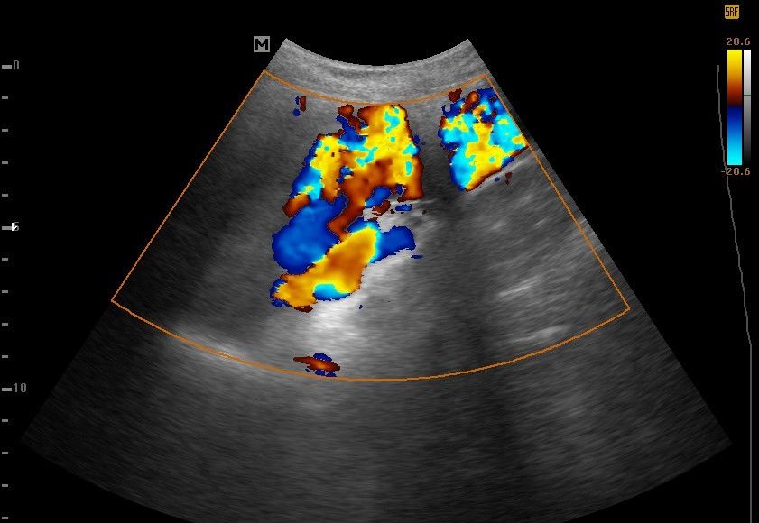
2FIGURE 1. Sonogram demonstrating an occluded portal vein (arrow head) with noticeable surrounding
formed tortuous blood vessels (arrows) at the porta hepatis
Posted on Authorea 18 Jul 2021 | The copyright holder is the author/funder. All rights reserved. No reuse without permission. | https://doi.org/10.22541/au.162664946.62190736/v1 | This a preprint and has not been peer reviewed. Data may be preliminary.
Also noted was diffuse echogenic gallbladder wall which demonstrated tiny hepatopetal vascular channels
under color Doppler interrogation.
Liver was however of average size with no surface nodularity or discrete masses (Figure 2).
Also noted was an enlarged spleen with multiple dilated tortuous blood vessels at the region of the splenic
hilum which were suggestive of portal hypertension (Figure 2 and Figure 3)
Mild ascites was also detected.
Pancreas and both kidneys were within normal limits.
No intra-abdominal masses detected.
FIGURE 2. Sonograms demonstrating a homogenous-appearing liver (A) and an Enlarged spleen measuring
about 20.3cm in length (B)
3FIGURE 3. Sonogram showing a Spleen(S) with multiple dilated tortuous blood vessels at the region of
the splenic hilum (arrow heads).
Posted on Authorea 18 Jul 2021 | The copyright holder is the author/funder. All rights reserved. No reuse without permission. | https://doi.org/10.22541/au.162664946.62190736/v1 | This a preprint and has not been peer reviewed. Data may be preliminary.
An MRI or CT scan was not performed for this patient because the Abdominal Ultrasound findings, the
patient’s clinical history and examinations were enough to make the diagnosis of CTPV.
Follow up & Further investigation
Patient was hemotransfused with 1 unit of blood on day of admission and subsequently 2 unit of blood and
oral iron supplements to manage his anemia. A repeat Hemoglobin count 3 days after depicted an increase
of hemoglobin count from 5g/dL to 7.9g/dL.
An upper gastrointestinal (GI) endoscopy was then requested for further evaluation after the patient was
stabilized.
The upper GI endoscopy revealed esophageal varices with mild extension of the varices to the gastric fundus.
Normal appearance of the duodenum identified.
Patient was managed on oral blood supplements to further correct his anemia and was discharged from the
hospital 2 weeks after. During his time of admission, he did not experience any of the initial symptoms he
presented to the hospital with. Also, there was improvement in his hemoglobin count (Hb = 11.2 g/dL);
with no complaints of dizziness, vomiting of blood or passage of dark-colored stool at the time of discharge.
He was then scheduled for 2 weekly review for further management by both the medical and surgical teams
on out-patient basis.
DISCUSSION
Cavernous transformation of the portal vein (CTPV) is a very rare condition usually characterized by tiny
tortuous blood vessels formed around a thrombosed portal vein 3 . This condition is mostly accompanied by
portal hypertension and the formation of porto-systemic collaterals and varices 8 . CTPV have been mostly
recorded in individuals with long-term thrombosis of the portal vein without evidence of liver cirrhosis or
hepatoma1–3 .
In CTPV, two types of collateral circulations are observed namely portoportal and portosystemic. In the
event of the occurrence of portal vein thrombosis, re – canalization usually takes place in patients without
cirrhosis or any liver disease which causes an increased resistance to blood flow 6 . This leads to increase in
hepatic arterial blood flow at the acute phase and mesenteric collaterals compensates for the obstruction.
Subsequently, there is dilatation of collateral vascular channels within the hepatoduodenal and hepatocolic
ligaments, around common bile duct, hepatic ducts and gall bladder draining in to the intrahepatic portal
veins to bypass the obstruction 9 ; and this was evident in our case where gallbladder wall thickening with
evidence of pericholecystic varices under Color Doppler interrogation were observed. This “venous rescue”
process continues and subsequently organizes into a cavernous transformation in about 3 – 5 weeks. This
compensatory process however has deleterious effect on intestinal circulation as it is known to be associated
with bowel ischemia 6 . When the portal cavernoma persists for a long time, the collaterals encase and
compress the biliary tree which causes damage to the intra and/or extra hepatobiliary ducts and this may
lead to loss of functional liver tissue.
Clinically, most patients affected with CTPV present with symptoms such as hematemesis and melena
secondary to possible bleeding esophageal varices 6,8 . These symptoms may however be non-specific for
CTPV since patients with gastrointestinal disease conditions may also present with hematemesis and me-
lena; and this was observed in our case where the patient’s initial diagnosis was directed towards bleeding
peptic ulcer and likely Mallory-Weiss tear. Other signs associated with portal hypertension such as anemia,
splenomegaly, ascites and abnormally low platelets count (thrombocytopenia) have also been reported in
patients with CTPV 8 . All these aforementioned clinical symptoms of CTPV were evident in the patient in
our case study.
4Administering of medication and surgery are known interventions for the management of CTPV; with the
success of treatment depending on the etiology of the disease 4 . The surgical procedure which involves the
creation of shunts decreases the portal pressure, enables hepatopetal blood from portal vein to the liver
Posted on Authorea 18 Jul 2021 | The copyright holder is the author/funder. All rights reserved. No reuse without permission. | https://doi.org/10.22541/au.162664946.62190736/v1 | This a preprint and has not been peer reviewed. Data may be preliminary.
and also alleviate the abnormal hemodynamic status in the portal system10 . This have been known to be
associated with favorable outcomes.
In our case study, the etiology of the CTPV could not be determined since the patient had no history of
liver cirrhosis, hepatoma or pancreatitis. A chronic portal vein thrombosis from a likely systemic vascular
disorder could be the cause of the CTPV.
CONCLUSION
Ultrasonography remains the first line radiological modality for assessing the abdominal organs in most
centers in the world due to its accessibility and low cost. Available literature suggest ultrasonography as
a reliable modality in diagnosing this pathology; with CT angiogram and MRI serving as complimentary
modalities for confirmation of CTPV.
Medical practitioners must therefore include CTPV as part of their differential diagnosis in patients who
present with upper GI bleeding symptoms and anemia especially those without prior history or clinical
suspicions of liver cirrhosis, hepatoma or pancreatitis. Duplex Abdominal ultrasonography must hence be
encouraged in the initial diagnostic workup in such patients.
In view of this, sonographers and radiologists must pay close attention to the hepatic vasculature especially
the segmental portal venous system when scanning patients presenting with upper GI bleeding and anemia.
This could allow for early detection of CTPV if present since early diagnosis is crucial in improving patients’
outcomes. Early sonographic detection of this pathology is also necessary for appropriate timely intervention
which could save the liver from end stage disease.
ACKNOWLEDGEMENTS: The authors extend gratitude to all those who helped in the preparation of
this work.
PATIENT CONSENT FOR PUBLICATION: Obtained
CONFLICT OF INTEREST: The authors declare no conflict of interest
AUTHORS CONTRIBUTION: JA performed the ultrasound examination, conceived the idea and in-
volved in the initial preparation of the manuscript. TKA-B and OO contributed to writing of the manuscript.
REFERENCES
1. De Gaetano AM, Lafortune M, Patriquin H, et al. Cavernous transformation of the portal vein: patterns
of intrahepatic and splanchnic collateral circulation detected with Doppler sonography.AJR Am J Roentgenol
. 1995;165(5):1151-1155.
2. Kumar A, Sharma P, Arora A. Review article: portal vein obstruction – epidemiology, pathogenesis,
natural history, prognosis and treatment.Alimentary Pharmacology & Therapeutics . 2015;41(3):276-292.
3. Salihefendić N, Licanin Z, Zildzić M. [Cavernous transformation of portal vein]. Med Arh . 2005;59(2):132-
134.
4. Chang C-Y, Yang P-M, Hung S-P, et al. Cavernous transformation of the portal vein: etiology determines
the outcome.Hepatogastroenterology . 2006;53(72):892-897.
5. Harmanci O, Bayraktar Y. Portal hypertension due to portal venous thrombosis: etiology, clinical out-
comes. World J Gastroenterol . 2007;13(18):2535-2540.
6. Ramos R, Park Y, Shazad G, A.Garcia C, Cohen R. Cavernous Transformation of Portal Vein Secondary
to Portal Vein Thrombosis: A Case Report. J Clin Med Res . 2012;4(1):81-84.
57. Coley BD. Pediatric applications of abdominal vascular Doppler imaging: Part I. Pediatr Radiol .
2004;34(10):757-771.
Posted on Authorea 18 Jul 2021 | The copyright holder is the author/funder. All rights reserved. No reuse without permission. | https://doi.org/10.22541/au.162664946.62190736/v1 | This a preprint and has not been peer reviewed. Data may be preliminary.
8. Sorrentino D, Labombarda A, Debiase F, Trevisi A, Giagu P. Cavernous transformation of the portal vein
associated to multiorgan developmental abnormalities. Liver Int . 2004;24(1):80-83.
9. Nasim M, Majid B, Tahir F, Majid Z, Irfan I. Cavernous Transformation of Portal Vein in the Setting of
Protein C and Anti-thrombin III Deficiency. Cureus . 2019;11(9).
10. Zhang H, Zhang N, Li M, Jin W, Pan S. Surgical Treatment of Portal Vein Cavernous Transformation.
World J Surg . 2004;28(7):708-711.
6You can also read