Study of the Blood Flow average for Atherosclerosis using Ultrasound Doppler shift
←
→
Page content transcription
If your browser does not render page correctly, please read the page content below

Journal of Physics: Conference Series

PAPER • OPEN ACCESS

Study of the Blood Flow average for Atherosclerosis using Ultrasound
Doppler shift
To cite this article: Q A Nabaa and M A Mariam 2021 J. Phys.: Conf. Ser. 1963 012107

View the article online for updates and enhancements.

 This content was downloaded from IP address 46.4.80.155 on 17/09/2021 at 14:45
2nd International Conference on Physics and Applied Sciences (ICPAS 2021) IOP Publishing
Journal of Physics: Conference Series 1963 (2021) 012107 doi:10.1088/1742-6596/1963/1/012107

 Study of the Blood Flow average for Atherosclerosis using
 Ultrasound Doppler shift

 Q A Nabaa1 Assist. Prof. Dr. M A Mariam2
 1
 Middle School Al-Fatem for Girls, Diyala, nabea.qais.ali1991@gmail.com
 2
 Dept. of Physics, College of Education, Al-Mustansiriaiah University, Baghdad,
 Iraq
 marmoh_1968@uomustansiriyah.edu.iq

 Abstract. Atherosclerosis is a condition that affects the walls of arteries repeatedly; blood
 vessel blockage caused by atherosclerosis is a frequent cause of heart attack and stroke.
 The use of Doppler change, a recent advancement in ultrasound technology, would improve
 its function by improving accuracy. Blood flow measurement is crucial because it can aid
 in the early detection of many diseases.
 One of these diseases is atherosclerosis, which has been researched using the ultrasound
 Doppler scattering method to measure blood flow average velocity in the dorsal artery of
 the foot. The study's findings revealed that age and diabetes had a greater impact than other
 factors such as medical history, high blood pressure, and triglycerides.

 Key word: Blood flow average, Doppler shift, ultrasound techniques, Factors of
 atherosclerosis, Piezoelectric (transduce probe),Variance.

1. Introduction
 Arteriosclerosis is a general term for a number of conditions in which the artery wall thickens and
 becomes less elastic.
 Doppler ultrasound is a non-invasive way of measuring blood flow. The Doppler shift [2] is used
 to determine the technique. A Doppler ultrasound supply is a device that is used to measure blood
 flow. It's also used in studies and clinical trials to measure the scope and impact of arterial disease
 [3].Another technique named laser Doppler flow-me try (LDF) is a technique for determining
 microvascular blood perfusion.
 The term "Doppler" describes the frequency change that occurs as light is dispersed by moving red
 blood cells. The fundamentals of LDF theory are based on this fundamental principle [4].
 Dr. Dussik at the University of Vienna wrote the first paper on medical imaging ultrasound in 1942.
 This article detailed the transmission ultrasound of the brain as well as a number of studies in the
 field of medical ultrasound [5]. In Doppler image scanning, a linear probe with a frequency of more
 than 5 MHz is needed for research and development [6, 9].Many of the first breakthroughs were
 made at Japan's Osaka University and the United States' University of Washington. Doppler
 ultrasound technology has supported clinical applications of blood-flow sensing, waveform
 analysis, blood-flow localization, and two-dimensional (2-D) mapping of blood flow through a
 series of steps. [10]The aims in this work to Study of the effect on blood flow by Doppler shift in
 Ibn Al-Nafees Hospital for Heart Patients and taking samples from patients with atherosclerosis

 Content from this work may be used under the terms of the Creative Commons Attribution 3.0 licence. Any further distribution
 of this work must maintain attribution to the author(s) and the title of the work, journal citation and DOI.
Published under licence by IOP Publishing Ltd 1
2nd International Conference on Physics and Applied Sciences (ICPAS 2021) IOP Publishing
Journal of Physics: Conference Series 1963 (2021) 012107 doi:10.1088/1742-6596/1963/1/012107

 aged between 35-90 years and taking ultrasound examination for them and using statistical analysis
 to analyses the data foe blood flow average
 2. Blood flow techniques

 There are a variety of techniques for visualizing and quantifying blood flow average rate, as well
 as measuring blood flow and volume. Blood flow measurement is crucial because it can aid in the
 early detection of a variety of diseases. Blood flow can be measured in a variety of ways and
 classified into different categories.By combining the Doppler-shifted wave with an original
 frequency reference wave, a beat frequency that is much lower than either of the two constituent
 waves is produced, making it much easier to calculate. This beat frequency is exactly equal to the
 frequency change caused by the Doppler Effect since it equals the difference between the two
 frequencies. The relationship between frequency change and relative velocity of source and
 detector is given by [11].

 ′ − = −
 (1)

 Where f represents the transmitted frequency, f' represents the received shifted frequency, v
 represents the relative velocity between the source and the detector, and c represents the wave's
 velocity. In the case of light waves, c is the speed of light, which is normally much higher than the
 speeds being measured, so (1) can be approximated as [12]

 ′ − = (2)

 As a result, when the Doppler shifted light is mixed with a reference beam of the original frequency,
 the frequency shift, and thus the frequency when the Doppler shifted light is mixed with a reference
 beam of the original frequency, is proportional to the velocity being measured in research and
 clinical investigations, Doppler ultrasound is used to assess blood average velocity and flow, as
 well as to quantify the range and effect of arterial disease [13].
 Doppler ultrasound is a non-invasive test that produces photographs of blood vessels, tissues, and
 organs using high-frequency sound waves. [14].
 Where,
 ΔFD=2 v* (cosθ) f o/c (3)
 Where, ΔFD = the difference in received and transmitted ultrasound is called change Doppler
 frequency or frequency.
 fₒ: Ultrasound probe frequency (Hz); transmitted signal frequency.
 v: The blood flow velocity of the target.
 3. Statistical analyses
 The statistical test that determined not a single value is representative the entire distribution. Its
 goal is to give a complete picture of the data. It usually refers to the arithmetic mean when used
 without an adjective (as mean).
 ∑ ̅
 = (4)

 Where∑ ̅ : is the number of observations in the sample (sample size), is the summation, is the
 individual value, and n is the number of observations in the sample (sample size)[15].

 The Normal distribution is a set of curves described by two parameters: the population's mean and
 standard deviation. The standard deviation is a summary measure of the deviations between each
 observation and the mean.

 2
2nd International Conference on Physics and Applied Sciences (ICPAS 2021) IOP Publishing
Journal of Physics: Conference Series 1963 (2021) 012107 doi:10.1088/1742-6596/1963/1/012107

 The following formula calculates the variance:
 ∑( − ̅ )2
 Variance = −1
 (5)

 Finally, the standard deviation is calculated using the square root of the variance [16]:

 (6)

 4. Materials and Methods
 When an electric current passes through the probe ingredient, it has the potential to generate
 ultrasound signal waves. Piezoelectric (PE) influence is the name for this process. Figure 1 shows
 the basic components of an ultrasound probe, which are made of crystal, rubber, and lead zircon
 ate titanate, which is the most commonly used ingredient.

 Filtered and Amplified

 Received
 Transmitter information
 ultrasound
 wave

 Dorslis pedis

 Figure 1: Shown the probe and component of ultrasound Doppler

 3
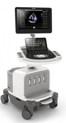
2nd International Conference on Physics and Applied Sciences (ICPAS 2021) IOP Publishing Journal of Physics: Conference Series 1963 (2021) 012107 doi:10.1088/1742-6596/1963/1/012107 The probe sends ultrasound energy into the foot, specifically the Dorslis pedis tissue, or into samples, and then receives the reflected signal echoes, which can then be converted into images on a computer monitor using an ultrasound device. Probing device selection Because of the morphological features and position (depth) of the carotid artery, a high-frequency linear array probe is commonly used for artery ultrasonography. The probe must have a center frequency of at least 7 MHz In a Doppler ultrasound unit, choose probe linear L12-4 (Model Philips 2019). 4.1Patients This research was carried out at the Ibn Al-Naphed Hospital in Baghdad, Iraq. The experimental procedure complies with the concepts of medical and biological detection. Patients were also divided into two groups, with 9 females and 21 males ranging in age from 35 to 90. By using a randomized approach, we were able to work with a total of 50 healthy and 30 Atherosclerosis patients, all of whom were clinically normal, 29 female and 21 male, and ranged in age from 20 to 69 years old. The patient is asked to have a comprehensive medical history, the length of the US technique test, and any family history of disease. Patients' biomedical factors, such as diabetes, total cholesterol, and blood pressure, are assessed as part of their diagnostic workup. While atherosclerosis can strike anyone at any age, it is more prevalent in people over 45. The exact cause of the disease is unknown. Atherosclerosis has two forms of risk factors: those that can be monitored or modified and those that cannot. The following are some of the changeable risk factors for this disease: ● Tobacco use and secondhand smoke are also harmful to your health. ● Blood pressure that is too high (hypertension) ● Cholesterol and fat levels in the blood are abnormal. ● Diabetes is a medical condition that affects ● Heart disease is a serious condition. ● Obesity is a problem that affects many people. ● Daily exercise is lacking. Other risk factors that cannot be adjusted include: ● Age is a factor. ● gender (male/female) stress 5. Results According the producer of ultrasound techniques (US) and Doppler shift Measurements in hospital. Though our study, cases were taken of 92 people, 50 cases are normal and patients were determining the blood flow average about 42 cases. As shown in table (1) the blood flow of velocity from normal range (30-118) (cm/s).the statistically was calculated in eq.(4) and (6) with variance using Doppler scattering shift in Ultrasound techniques by the value as shown in table (1) the mean of blood flow was different between the two study subgroups. The mean of blood flow of average velocity for healthy or normal was 57.596 cm/s but Atherosclerosis patients 41.49667 cm/s. 4
2nd International Conference on Physics and Applied Sciences (ICPAS 2021) IOP Publishing
Journal of Physics: Conference Series 1963 (2021) 012107 doi:10.1088/1742-6596/1963/1/012107

 Table (1) explain the clinically normal or healthy were ages from (12-69) years old and the
 disease (35-90).

 No=50
 age(years)
 subgroup
 Blood flow
 healthy average
 12-69 30-118 14-27 velocity(cm/s) fo(HZ)

 57.626
 range 50.06 17.12

 Mean 22.97926
 13.12922
 2.303389

 No.=42
 SD

 subgroup age(years)

 Blood flow
 average
 (-149) to velocity(cm/s)
 patient 35-90 (+118) 14-20 fo(HZ)

 range 61.21951 35.53415 17.60976
 11.40285 45.67128
 Mean 1.145043

 SD
 The effect of others risk on Atherosclerosis wasn’t appeared in US technique using Doppler shift
 with range source sound 14-27Hz see fig.2.

 5
2nd International Conference on Physics and Applied Sciences (ICPAS 2021) IOP Publishing
Journal of Physics: Conference Series 1963 (2021) 012107 doi:10.1088/1742-6596/1963/1/012107

 the effect of risk factor on Atherosclerosis
 patients

 MEDICAL HISTORY LPL PRESURE Diabetes
 34
 31

 14
 12

 factors of Atherosclerosis patients

 Figure 2: Explain the factors and the stronger effect is Diabetes in Atherosclerosis

 The effect of age and diabetes is stronger to Atherosclerosis patient has age 90 years at blood flow
 -149 cm/s and notes some persons were negative sign average velocity when received the signal
 from transducer prob.

 Compare the other factors such medical history ,LPL and high pressure were weak effect . The
 effect of diabetes, as shown in fig.2, was more powerful than the other effects.

 200 correlation coeff. =-0.51045
 averge velocity (cm/s)

 100

 0
 0 20 40 60 80 100
 -100

 -200
 age(year)

 Figure3: Explain the effect of age on the average blood flow for patients

 200 correlation coff.=-0.16038
 blood flow(cm/s)
 averge velocity

 100
 0
 0 100 200 300 400 500 600
 -100
 -200
 glucose

 Figure 4: The effect of diabetics on the blood flow for disease

 6
2nd International Conference on Physics and Applied Sciences (ICPAS 2021) IOP Publishing
Journal of Physics: Conference Series 1963 (2021) 012107 doi:10.1088/1742-6596/1963/1/012107

 6. Conclusions

 Ultrasound methods were used to measure average blood flow with the Doppler shift in this article.
 The benefit of the instrument is that it is non-invasive and can detect and easy by using Doppler
 shift to diagnosis Atherosclerosis early in the medical diagnosis process in dorslis pedis artery.
 Calculated Doppler shift has a benefit, and some details can be as follows:

 1) The average blood flow is used to measure the factors of for atherosclerosis
 2) The effect of factors on Atherosclerosis is distinguished for various distributions between healthy
 and sick people.
 3) Statistical data analysis was a mathematical tool for separating various variables in the early
 detection of atherosclerosis.
 4) the advantage of US technique was distinguish of the effect of age and diabetes together on
 atherosclerosis and the values of blood flow were negative indicated the backscattering accorded
 with growing age and duration of Diabetes
 5) Blood flow is inadequate in the affected arterioles. Because The walls thicken, narrowing the
 arterioles especially in the capillary of arteries is blocked. This condition is more common in
 diabetics.
 6) the technique couldn’t distinguish the influence of other biomedical factors on atherosclerosis
 such as medical history , ,LPL, and high pressure hypertension(stress/strain)
 References

 [1] Drain L (1980) The laser Doppler technique, Wiley, USA.
 [2] S. Paul Sidhu, Ultrasound of the carotid and vertebral arteries, British Medical Bulletin 2000,
 56 (No 2) 346-366.
 [3] Kenwright DA, Anderson T, Moran CM, Hoskins PR. Assessment of spectral Doppler for an
 array-based preclinical ultrasound scanner using a rotating phantom. Ultrasound Med Biol
 2015;41:2232-9.
 [4] S. A. Rajan, "Analysis of the Dynamics of Vasomtion using Laser Doppler Flow", The
 University of Texas, thesis of Master of Science in Biomedical Engineering, August 2005.
 [5] W.E Brant, Helms CA. Fundamentals of Diagnostic Radiology.University of Virginian:
 Lippincott Williams & Wilkins; 2012.
 [6] M Gerhard‑Herman, JM . M. E Gardin, Jaff, Mohler, M Roman, TZ Naqvi,. Guidelines for
 noninvasive vascular laboratory testing: A report from the American Society of
 Echocardiography and the Society for Vascular Medicine and Biology. Vasc Med 2006;11:183
 ‑200.
 [7] . X Zhou, DA Kenwright, S Wang, JA Hossack, Hoskins PR. Fabrication
 of two flow phantoms for Doppler ultrasound imaging. IEEE Trans Ultrason Ferroelectr Freq
 Control 2017;64:53‑65.
 [8]. KV Ramnarine, T Anderson, PR Hoskins. Construction and geometric
 stability of physiological flow rate wall‑less stenosis phantoms.Ultrasound Med Biol
 2001;27:245‑50.
 [9]. JY Hwang. Doppler ultrasonography of the lower extremity arteries:Anatomy and scanning
 guidelines. Ultrasonography 2017;36:111‑9.
 [10]. DA Kenwright, T .Anderson, C.M Moran, P.R Hoskins. Assessment ofspectral Doppler for
 an array‑based preclinical ultrasound scanner usinga rotating phantom. Ultrasound Med Biol
 2015;41:2232‑9.
 [11] Picot, P.A.; Rickey, D.W.; Mitchel, J.R.; Rankin, R.N.; Fenster, A. Threedimensional color
 Doppler flow imaging of the carotid artery. SPIE 1991

 7
2nd International Conference on Physics and Applied Sciences (ICPAS 2021) IOP Publishing
Journal of Physics: Conference Series 1963 (2021) 012107 doi:10.1088/1742-6596/1963/1/012107

 [12] Pellerito J, Polak JF. Introduction to Vascular Ultrasonography. Manhasset, New York:
 Elsevier Health Sciences; 2012.
 [13] Slager, C.J. et al.,“Electrical impedance of layered atherosclerotic plaques on human aortas”,
 Biomedical Engineering, IEEE Transactions, vol. 39, issue 4, 1992, pp.411 – 419.
 [14] D.A. Kenwright, T. Anderson, C.M Moran, P.R .Hoskins , Assessment of Spectral Doppler
 for an array‑based preclinical ultrasound scanner using a rotating phantom. Ultrasound Med
 Biol. 2015; 41:2232‑9.
 [15] ] Zhou X, Kenwright DA, Wang S, Hossack JA, Hoskins PR. Fabrication of two flow
 phantoms for Doppler ultrasound imaging. IEEE Trans Ultrason Ferroelectr Freq Control
 2017;64:53‑65.
 [16]Hun Myoung Park(1980). . New York: Wiley and Sons park.

 8
You can also read