H-type tracheoesophageal fistula in the neonatal period: Difficulties in diagnosis and different treatment approaches. A case series
←
→
Page content transcription
If your browser does not render page correctly, please read the page content below
56 / Arch Argent Pediatr 2020;118(1):47-60 / Brief reports

H-type tracheoesophageal fistula in the neonatal period:
Difficulties in diagnosis and different treatment approaches.
A case series
Giselle Cuestas, M.D.a, Verónica Rodríguez, M.D.a, Carolina Millán, M.D.b, Patricio Bellia Munzón,
M.D.a and Gastón Bellia Munzón, M.D.b,c

ABSTRACT                                                                  INTRODUCTION
Congenital tracheoesophageal fistula not associated with
                                                                              Tracheoesophageal fistula (TEF) is associated
esophageal atresia, known as H-type fistula, is an uncommon
anomaly. It presents with cough, choking, and cyanosis during             with esophageal atresia (EA) in 95 % of cases. Its
feeding and/or recurrent pneumonia.                                       incidence is approximately 1 in 3500 live births,
Although symptoms are usually present from birth, diagnosis               and it is slightly more frequent among males.1
is difficult. The rarity of this disease, non-specific symptoms,
                                                                              H-type TEF not associated with EA–type V
and the limitations of radiological and endoscopic confirmation
of the fistula often result in a delay between presentation               according to Ladd and Gross’ classification–
and diagnosis confirmation. Here we describe the clinical                 is a rare airway anomaly that accounts for 4 %
manifestations, assessment methods, and management of                     of all tracheoesophageal malformations. 2,3 It
3 newborn infants with H-type tracheoesophageal fistula,
                                                                          is an abnormal communication in an oblique
together with diagnosis recommendations to prevent
unnecessary delays in the management of this condition.                   course that looks like an N, located between
Key words: H-type tracheoesophageal fistula, newborn infant,              the posterior wall of the trachea (cranial orifice)
bronchoscopy, thoracoscopy.                                               and the anterior wall of the esophagus (caudal
                                                                          orifice).4 In general, it is located in the cervical
http://dx.doi.org/10.5546/aap.2020.eng.56
                                                                          region or in the thoracic inlet; the incidence of
                                                                          associated congenital malformations is lower
To cite: Cuestas G, Rodríguez V, Millán C, Bellia Munzón P,
                                                                          and it has a better prognosis among the different
Bellia Munzón G. H-type tracheoesophageal fistula in the neonatal         types of TEF.5,6
period: Difficulties in diagnosis and different treatment approaches. A       Although some articles have mentioned
case series. Arch Argent Pediatr 2020;118(1):56-60.
                                                                          cases in the neonatal period, several authors
                                                                          have described a late diagnosis, even in adult
                                                                          age.3,7,8 An early diagnosis is still a challenge for
                                                                          pediatricians because of the rarity of the condition
                                                                          and the non-specificity of symptoms. These may
                                                                          be subtle and intermittent due to the anatomical
                                                                          characteristics of the fistula (oblique course, small
                                                                          orifice, and the redundant nature of esophageal
                                                                          mucosa, which may transiently obstruct it)
                                                                          and are frequently attributed to more common
                                                                          etiologies, like gastroesophageal reflux (GER)
                                                                          and swallowing disorders or a poor feeding
a. Unit of Respiratory Endoscopy, Ear, Nose and Throat
                                                                          technique.9,10
   Department, Hospital General de Niños “Dr. Pedro de
   Elizalde,” Autonomous City of Buenos Aires, Argentina.                     The classical clinical triad of H-type TEF
b. Department of Pediatric Surgery, Fundación                             includes cough fits, cyanosis, and choking during
   Hospitalaria, Autonomous City of Buenos Aires,                         feeding, abdominal distension, and recurrent
   Argentina.
                                                                          respiratory tract infections.4-6 Diagnosis requires
c. Department of Surgery, Hospital General de Niños
   “Dr. Pedro de Elizalde,” Autonomous City of Buenos                     a high rate of clinical suspicion, and confirmation
   Aires, Argentina.                                                      is made through imaging tests, such as an upper
                                                                          gastrointestinal tract study with contrast and a
E-mail address: Giselle Cuestas: giselle_cuestas@yahoo.com.ar
                                                                          videofluoroscopy, or a tracheobronchoscopy,
Funding: None.                                                            which provides a direct view of the fistula.4
                                                                              None of these imaging tests has a sensitivity
Conflict of interest: None.                                               of 100 %. Special positioning and contrast
                                                                          administration techniques have been described to
Received: 5-9-2019
Accepted: 8-16-2019                                                       aid in diagnosis.3,9 Several studies have proposed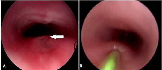
Brief reports / Arch Argent Pediatr 2020;118(1):47-60 / 57
that an endoscopy is the fundamental diagnostic in all patients (Figures 1.B and 1.C). For diagnostic
technique to confirm the presence of a fistula purposes, 2 patients had an endotracheal tube
and locate its level accurately.11 Surgery is the with balloon placed in the esophagus, then
main treatment modality, and cervical or thoracic the balloon was inflated, thus obstructing the
approach depends on the level of the fistula.5 esophageal lumen. A bronchoscope tube was
placed to ventilate the patient; the optical scope
CASE REPORTS was introduced through the tube to check that
Here we report the cases of 3 male patients the convexity of the posterior wall of the trachea
with H-type TEF seen between February 2016 and (due to the inflated balloon in the esophagus) was
February 2019. The characteristics of patients are distal to the potential fistula area. After injecting
summarized in Table 1. All patients had symptoms air, saline solution was instilled using a catheter
present at birth and had been diagnosed between placed in the esophagus above the balloon. The
9 and 30 days of life. presence of a fistula was confirmed if air and
A videofluoroscopic swallowing study (VFSS) saline solution bubbles were observed in the
showed the passage of contrast into the trachea trachea (Figure 2).
in 2 patients, but the level of the fistula was Two patients were fed with a nasogastric
accurately determined only in 1 of them (Figure tube (NGT) until the moment of the surgical
1.A). A rigid tracheobronchoscopy under general repair. The remaining patient had a Nissen
anesthesia using a 0-degree and 5-mm optical fundoplication and gastrostomy due to severe
scope confirmed the diagnosis of H-type fistula GER; an endoscopic closure of the fistula with
Table 1. Characteristics of patients with H-type fistula
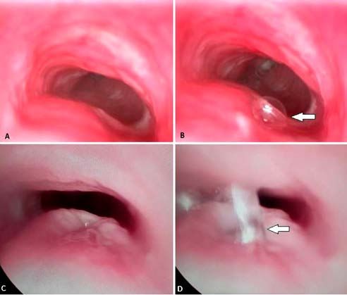
Figure 1. A. Videofluoroscopic swallowing study. Passage of contrast into the trachea (arrow) and fistula identification
(circle). B and C. Endoscopic images of the tracheal orifices of an H-type tracheoesophageal fistula (B) and a fistula associated
with esophageal atresia (C) (arrows). B. Small round orifice in the posterior wall of the cervical trachea. C. Larger orifice in
the distal (supracarinal) trachea58 / Arch Argent Pediatr 2020;118(1):47-60 / Brief reports
silver nitrate was attempted 3 times due to the during follow-up. They are fed per os and have
low morbidity of this procedure. Given the a normal height and weight growth for their age.
recurrent fistula, it was decided to perform a
surgery. Channeling of the fistula with guidewire DISCUSSION
to facilitate intraoperative identification was Congenital TEF is the result of a defective
performed in the 3 patients (Figure 3). separation of the respiratory and digestive tracts
In the post-operative period, the patients were during embryogenesis.7 The etiology of this entity
hospitalized in the intensive care unit for 7 days is multifactorial.11 There are 5 anatomical types,
and extubated on day 3. Two patients showed a with proximal EA with distal TEF being the most
favorable course. The remaining patient developed common one (90 %).1 Isolated or H-type forms
pneumothorax due to partial dehiscence of the are rare.
suture. Six tracheal rings were resected with end- The clinical presentation of H-type fistulas is
to-end anastomosis, closure of the esophageal wall, related to their caliber.12,13 Large fistulas present
and interposition of the thymus via cervicotomy with respiratory distress due to gastric distension
9 days later. The patient evolved with left and persistent airway secretions, whereas small
diaphragmatic and cord paralysis, which were ones are accompanied with recurrent cyanosis
managed through diaphragmatic plication and episodes due to saliva and milk aspiration.
liquid thickening, respectively. Symptoms improve when using a NGT for
The follow-up period ranged from 3 months feeding. A fistula presents in a delayed manner as
to 2 years. No patient had symptoms recurrence recurrent or persistent respiratory tract infections,
Figure 2. A and B. Tracheal orifice of the H-type tracheoesophageal fistula before (A) and after (B) injecting air (arrow)
through a catheter above the inflated balloon of an endotracheal tube placed in the esophagus. Mucosal congestion (tracheitis)
secondary to secretion aspiration is also seen. C and D. Tracheal orifice of the H-type tracheoesophageal fistula before (C) and
after (D) instilling saline solution (arrow) through a catheter above the inflated balloon of an endotracheal tube placed in the
esophagus.Brief reports / Arch Argent Pediatr 2020;118(1):47-60 / 59
chronic obstructive pulmonary disease, and placed in the esophagus above the inflated
bronchiectasis secondary to aspiration.6 balloon. The presence of a fistula is confirmed if
A chest X-ray may be useful to look for signs air and/or saline solution bubbles are observed
of aspiration and air in the lower esophagus.6,7 A in the trachea. Other authors had opted to instill
computed tomography is recommended to assess methylene blue to see the fluid passage.12
the lung parenchyma and sometimes helps to An endoscopy allows to channel the fistula
evidence the fistula.14 (with a ureteral or Fogarty catheter or guidewire),
Contrast tests of the upper gastrointestinal thus facilitating its identification during surgical
tract (upper gastrointestinal series or VFSS) are repair. An esophagoscopy is not recommended
part of routine care when an H-type fistula is to diagnose a TEF because the esophageal orifice
suspected.4,9 Their sensitivity ranges from 50 % to is too small and the esophageal mucosal folds
73 %.12 For a greater sensitivity, it is recommended hinder visualization.5
to perform the test with the patient in the prone Diagnosis improves significantly with
position and to insert a NGT and withdraw it tracheobronchoscopy. However, radiological
while injecting a contrast agent at different levels and endoscopic techniques complement each
of the esophagus. This technique requires video other, and using both warrants the identification
or rapid sequence films.4,5,10,12 Due to the risk for of an H-type fistula.5The differential diagnosis
aspiration, it is recommended to use a water- should include GER, swallowing disorders,
soluble contrast and have adequate resuscitation laryngeal cleft, esophageal stenosis, and extrinsic
equipment readily available.4,12 esophageal compression.12
In case of high clinical suspicion, a The treatment of choice is surgical repair. The
tracheobronchoscopy is the definite diagnostic cervical approach is indicated for fistulas above
test.5,9 It allows to identify and locate the fistula the second thoracic vertebra, whereas the thoracic
and to assess the presence of associated airway approach, for those at a more caudal level.5,12,14
anomalies (e.g., tracheomalacia, laryngeal cleft, The thoracoscopic approach has certain
stenosis, cord paralysis or a second fistula).4,5,7 advantages over the conventional thoracotomy:
The fistula is viewed as a small round opening a greater visibility of the field thanks to
in the midline of the posterior membranous wall magnification, lower post-operative pain
of the trachea, generally at the cervical level or levels, and improved aesthetic outcomes.3It is a
in the thoracic inlet.4 Secretions are commonly minimally invasive technique, but it is complex
present in the airway. and requires experienced providers.
To improve the test’s performance, it is The most common post-operative
recommended to insert an endotracheal tube complications include suture dehiscence,
with balloon in the esophagus and inject air recurrent nerve damage, and lung complications
and/or instill saline solution using a catheter secondary to GER, tracheomalacia, esophageal
Figure 3. Tracheal orifice of H-type tracheoesophageal fistula (arrow) before (A) and after (B) channeling with guidewire60 / Arch Argent Pediatr 2020;118(1):47-60 / Brief reports
stenosis or TEF recurrence.6,12 4. Ng J, Antao B, Bartram J, Raghavan A, et al. Diagnostic
difficulties in the management of H-type tracheoesopha-
In addition, 30 % of TEF are associated with
geal fistula. Acta Radiol. 2006; 47(8):801-5.
other anomalies of the digestive, urogenital 5. Jaiswal AA, Garg AK, Mohanty MK. ‘H’ type tracheo-oe-
and/or cardiovascular system, including sophageal fistula. Case reports with review of the litera-
vertebral defects, anal atresia, cardiac defects, ture. EJENTAS. 2014; 15(2):143-8.
6. Parakh H, Kapoor MS, Sharma D, Pandita A, et al. H-type
tracheoesophageal fistula, renal anomalies,
tracheo-esophageal fistula in a very low birth weight in-
and limb abnormalities (VACTERL/VATER fant: An unexpected and diagnostic challenge for neona-
association) and coloboma, heart defects, tologist. Med J DY Patil Univ. 2015; 8(3):354-7.
choanal atresia, retardation of growth, 7. Aygun D, Emre S, Nepesov S, Tekant G, et al. Presentation
of H-type tracheoesophageal fistula in two adolescents:
genital abnormalities, and ear abnormalities
Delayed diagnosis. Pediatr Neonatol. 2017; 58(2):187-8.
(CHARGE).2,5 Comorbidities determine prognosis, 8. Antabak A, Luetic T, Caleta D, Romic I. H-type tracheo-
which is otherwise very good.12 esophageal fistula in a newborn: Determining. the exact
The diagnosis of H-type fistulas implies position of fistula by intra-operative guidewire placement.
J Neonatal Surg. 2014; 3(3):36.
a high rate of clinical suspicion in newborn
9. Lee SYS, Hamouda ESM. H-type tracheoesophageal fistula
infants with cough and cyanosis during feeding diagnosed on video fluoroscopy swallowing study. BMJ
and recurrent respiratory symptoms. A rigid Case Rep. 2018; 11(1):bcr 2018227794.
tracheobronchoscopy may be considered the 10. Perry M, Eick J, Jakob K, Adolph V, et al. Clinical images
- a quarterly column: early presentation of h-type tracheo-
method of choice for diagnostic confirmation. A
esophageal fistula. Ochsner J. 2013; 13(4):483-5.
multidisciplinary approach is critical for an early 11. Donnelly P, McVea S, Flannigan C, Bali S. Incidental diag-
diagnosis and a timely management of H-type nosis of an H-type tracheo-oesophageal fistula. BMJ Case
fistulas. Rep. 2016;2016:bcr2016215419.
12. Morales Múnera OL, Valencia Chaves ML, Roya Pabón
CL, Niño Serna LF. Fístula traqueoesofágica en niños: un
REFERENCES diagnóstico para tener en cuenta. Reporte de dos casos y
1. Palacios Sánchez M, Alegría Echauri I, Alegría Echauri E, revisión de la literatura. IATREIA. 2013; 26(3):346-55.
Pérez Belmonte E, et al. Fístula en H: a propósito de un 13. Harjai MM, Holla RG, Kale R, Sharma YK. H-type tracheo-
caso. Bol Pediatr. 2013; 53(223):41-4. oesophageal fistula. Arch Dis Child Fetal Neonatal Ed. 2007;
2. Riazulhaq M, Elhassan E. Early recognition of h-type tra- 92(1):F40.
cheoesophageal fistula. APSP J Case Rep. 2012; 3(1):4. 14. Stavroulias D, Ampollini L, Carbognani P, Rusca M. Late
3. González Temprano N, Viguria Sánchez N, Ayuso González presentation of congenital H-type tracheoesophageal fis-
L, Pérez Martínez A. Fístula traqueoesofágica en H en tula in an immunocompromised patient. Eur J Cardiothorac
periodo neonatal: diferentes abordajes terapéuticos. An Surg. 2011; 40(2):e98-100
Pediatr (Barc). 2014; 81(6):e50-1.You can also read