Recurrent Blister Formation in Setting of Poorly Managed Diabetes Mellitus - Cureus
←
→
Page content transcription
If your browser does not render page correctly, please read the page content below
Open Access Case
Report DOI: 10.7759/cureus.5029
Recurrent Blister Formation in Setting of
Poorly Managed Diabetes Mellitus
Michael J. Willcox 1
1. Medicine, Tulane University School of Medicine, New Orleans, LA, New Orleans, USA
Corresponding author: Michael J. Willcox, justinwillcox@gmail.com
Disclosures can be found in Additional Information at the end of the article
Abstract
Bullous diabeticorum is a condition of unknown etiology with abrupt blister formation and
spontaneous resolution. While commonly thought as rare, it is likely underdiagnosed resulting
in mismanaged care and increased morbidity for individual patients. A shift in focus from
empiric treatment to appropriate diagnostic workup is critical for this condition in the diabetic
population.
Categories: Dermatology, Family/General Practice, Internal Medicine
Keywords: bullae, diabetes, bullous diabeticorum
Introduction
Cutaneous conditions are just some of the numerous morbidities that impact patients with
diabetes mellitus [1]. Many of these potential complications have similar initial appearances
but varying pathomechanisms. The treatments for the conditions can at times be contradictory,
thus demonstrating the importance of accurate workup [1-3]. Awareness of the various
cutaneous manifestations, the appropriate workups or treatments, and the importance of
managing the correlated poor glycemic control are important for any general practitioner caring
for this population [4].
Case Presentation
A 66-year-old Caucasian man presented to the outpatient internal medicine clinic with concern
for a non-healing ulceration on his left fourth toe and a large, previously ruptured blister on his
left heel.
The patient had previously undergone numerous detachments and amputations of digits on all
four extremities, including a below knee amputation of his right leg one year prior. The patient
Received 05/28/2019
Review began 05/31/2019 reported that all of these surgeries stemmed from spontaneous blisters on his digits. Most of
Review ended 06/15/2019 the blisters would ultimately rupture, with many becoming non-healing ulcers despite
Published 06/28/2019 aggressive antibiotic therapy and debridements.
© Copyright 2019
Willcox. This is an open access article The patient had previously undergone dermatologic workup for the frequent blistering.
distributed under the terms of the
Histological samples were inconclusive and negative on immunofluorescent staining.
Creative Commons Attribution License
CC-BY 3.0., which permits
unrestricted use, distribution, and The patient’s past medical history includes hypertension, hyperlipidemia, peripheral vascular
reproduction in any medium, provided disease, and type II diabetes mellitus. The patient’s diabetes had been poorly controlled for
the original author and source are
many years, with hemoglobin A1c values averaging over 10 for the last decade. During this
credited.
time, the patient developed sequelae of diabetic neuropathy, repeated diabetic foot ulcers with
How to cite this article
Willcox M J (June 28, 2019) Recurrent Blister Formation in Setting of Poorly Managed Diabetes Mellitus.
Cureus 11(6): e5029. DOI 10.7759/cureus.5029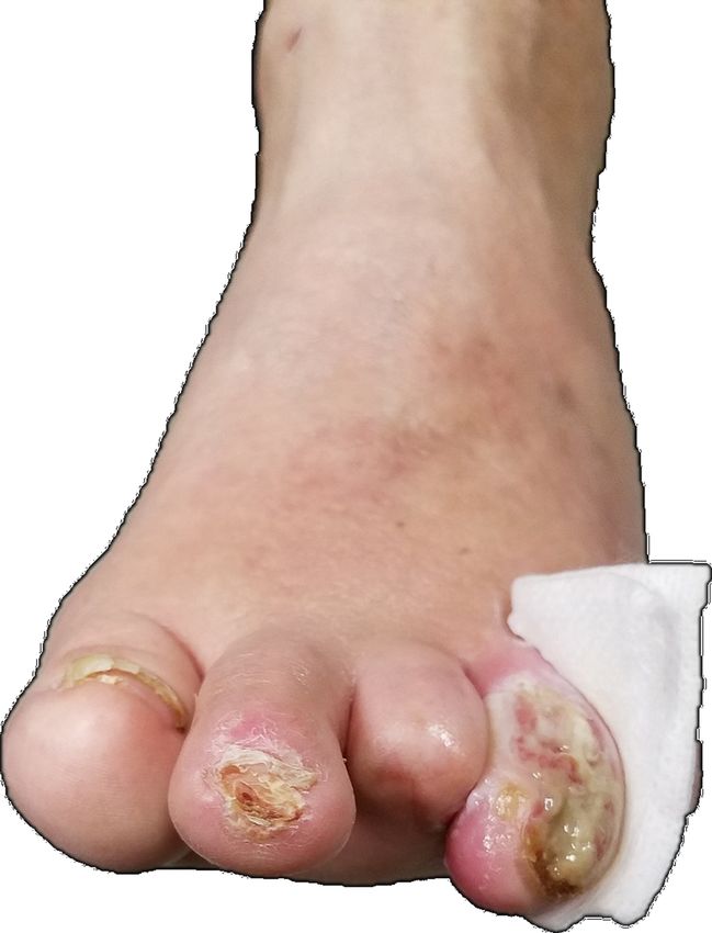
cellulitis, and end-stage renal disease with recently initiated peritoneal dialysis. Following the
initiation of peritoneal dialysis, the patient’s glucose levels have maintained a normal range.
Physical examination reveals ulceration with purulent exudate of the left fourth toe from the
entire nail bed continuing dorsally and medially to the mid-proximal phalanx (Figure 1).
FIGURE 1: Left foot with ulceration and exudate from the fourth
toe. Also visible is previously amputated third toe.
Exploration of the wound revealed tracking with bone involvement and suspected
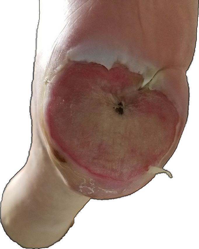
osteomyelitis. The left plantar surface has a large lesion spanning the entire width of the heel
with apparent reepithelization and a small eschar appearance in the center (Figure 2).
2019 Willcox et al. Cureus 11(6): e5029. DOI 10.7759/cureus.5029 2 of 7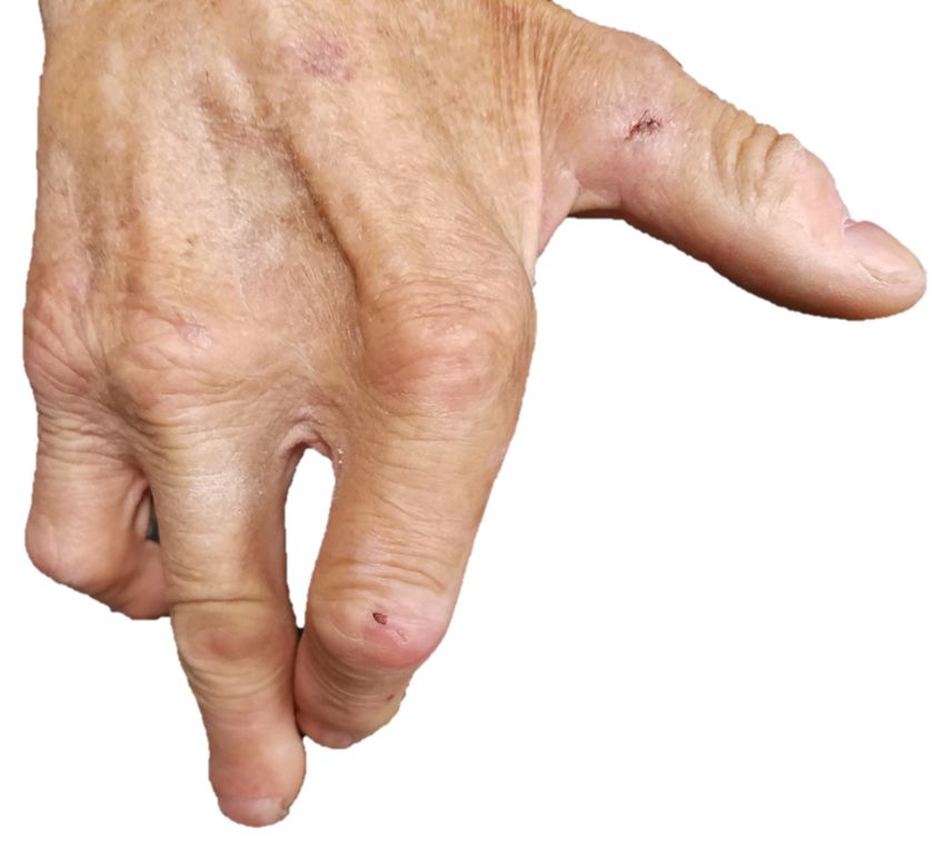
FIGURE 2: Left heel with large lesion from previously ruptured
                                      blister. Surface is dry and firm.

                                    The area is red and dry, with the appearance of a previously ruptured large blister. The rest of
                                    his foot and both hands revealed numerous previous detachments and amputations, but no
                                    present ulcerations or blisters (Figure 3-4).

2019 Willcox et al. Cureus 11(6): e5029. DOI 10.7759/cureus.5029                                                                  3 of 7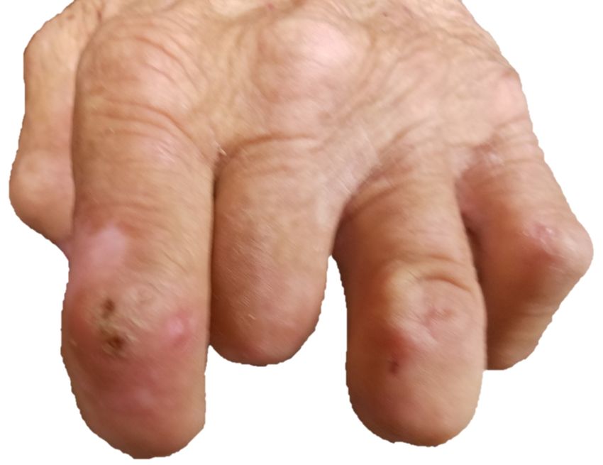
FIGURE 3: Left hand with well healed amputations of index,
                                      middle, and ring fingers.

2019 Willcox et al. Cureus 11(6): e5029. DOI 10.7759/cureus.5029                                   4 of 7
FIGURE 4: Right hand demonstrating ray amputation of the
middle finger.
The patient was referred to the wound care team for recommendations on debridement with
antibiotics vs surgical management of left fourth toe, as well as recommendations for
aggressive management of recently ruptured plantar blister.
Discussion
Cutaneous manifestations of diabetes are present in 30-70% of patients [1-4]. Bullous eruptions
are not as common as bacterial and fungal infections, but the incidence is high enough to
warrant a periodic review of the varied causes [3,5].
Bullous diabeticorum, also known as bullosis diabeticorum, presents as spontaneous eruptions
of tense, serous, and sterile fluid-filled blisters on otherwise normal skin [6-11] of patients
suffering from diabetes mellitus. They appear most frequently in the acral region: with the feet
[6,8,9,12,13], and at times hands [3,5], being the most common. The blisters appear abruptly
[12], without history of trauma [5], pain, signs of inflammation [3,11], or any further symptoms
[12]. Reported blisters have a significant variance in size, from 0.5 to 10 cm [4,10]. They tend to
have a self-limited evolution, with reepithelization of the blister floor occurring rapidly within
a few days [12] and most resolving in 2-6 weeks without scarring [2,4,5,8,9,11,13,14]. However,
most cases will reoccur [3,7,8,9,13,14]. The actual incidence of bullous diabeticorum remains
uncertain, with studies presenting an annual incidence in patients with diabetes of 0.16-0.5%
(1 in 200 to 625 patients) [4,15,16].
2019 Willcox et al. Cureus 11(6): e5029. DOI 10.7759/cureus.5029 5 of 7At the present time, the etiology remains unclear [2,3,4,8,14]. The most common presenting
comorbidities are diabetic peripheral neuropathy [3,8], followed by vasculopathy [3,5,10]. Other
less commonly presenting comorbidities include nephropathy [13,17], microangiopathy [4,18],
or disorders of metabolism of calcium, magnesium, and carbohydrates [2,5,14,19]. One
observation is that, in cases that were properly documented, the patients appear to have poor
glucose control at the time of the blisters' eruptions [4,5,11,15]. This was the case with the
presented patient, with no new blister formation noted in the year following initiation of
peritoneal dialysis and improved blood glucose regulation. Nonetheless, no presenting
condition or comorbidity has been documented as present in all cases [3,5,10,12], causing
speculation that the disease is multifactorial [11].
No specific laboratory test exists for the diagnosis of bullous diabeticorum [8,11,13], leaving it
as a clinical diagnosis of exclusion [4,8]. It is important to exclude other known bullous
conditions [8]; however, due to the increased risk of infection in diabetic patients, particularly
in the lower extremities, biopsies should only be performed in recurrent cases [4,15]. Biopsies
should be taken from the bubble region for histological analysis and from the perilesional zone
for immunofluorescence evaluation [8,13]. Additional tests should not be necessary [8].
Histopathologically bullous diabeticorum is noted for its heterogeneity [18], leaving exams
inconclusive. Findings may range from subepidermal blisters with sparse perivascular infiltrate
to intraepidermal with surrounding spongiosis [4,10]. Both direct and indirect
immunofluorescence will be negative [8,14], excluding conditions like bullous pemphigoid that
would otherwise appear similar clinically and histologically [2,8,11].
With the treatment, course, and prognosis being so different from other immune, ischemic, or
neuropathic lesions of diabetic patients [5], it is critical to perform an adequate workup. For
example, a misdiagnosis of bullous pemphigoid or pemphigus would result in treatment with
high dose corticosteroids, likely increasing risks and exacerbating the underlying diabetes
mellitus [2].
Current treatment is supportive, with primary goals to reduce discomfort and minimize the risk
of secondary infection [2,8,10,12]. Conventional therapy involves keeping the blister intact to
cover the lesion and prevent secondary infection [8,13]. Some have chosen to lance intact
bullae for controlled drainage, keeping the roof intact [4,11]. Opening the bullae would
necessitate topical antimicrobials. Authors have noted that reoccurrence at these sites is
common, but no statistics are available to compare their commonality with the recurrence of
preserved bullae. With either method, patients should be encouraged to keep their lesion clean
and protected [8,11] with cold compresses providing relief for some [2]. This would also be a
valuable time to assess and emphasize proper blood glucose regulation [4].
Conclusions
While bullous diabeticorum is generally considered to be a rare condition, the lack of a specific
diagnostic test may cause it to be significantly underdiagnosed and underreported. The
condition is likely misdiagnosed for other bullous disorders and treated inappropriately,
causing an increase in already significant morbidity and the formation of chronic ulcers. Thus,
practitioners should have an increased sensitivity for it and seek adequate workup rather than
beginning empiric treatment. In addition to ensuring proper management of individual
patients, utilization of consulting services in workup and reporting of discovered cases will
result in better assessment of incidence and pathogenesis. With more information, hopefully,
the pathogenetic mechanism underlying bullous diabeticorum can be established so that a
definitive diagnostic method can be developed and therapy can be directed at treatment instead
of simply symptom and sequela relief.
2019 Willcox et al. Cureus 11(6): e5029. DOI 10.7759/cureus.5029 6 of 7Additional Information
Disclosures
Human subjects: Consent was obtained by all participants in this study. Conflicts of interest:
In compliance with the ICMJE uniform disclosure form, all authors declare the following:
Payment/services info: All authors have declared that no financial support was received from
any organization for the submitted work. Financial relationships: All authors have declared
that they have no financial relationships at present or within the previous three years with any
organizations that might have an interest in the submitted work. Other relationships: All
authors have declared that there are no other relationships or activities that could appear to
have influenced the submitted work.
References
1. Braverman IM: Cutaneous manifestations of diabetes mellitus . Med Clin North Am. 1971,
55:1019-1029. 10.1016/S0025-7125(16)32495-6
2. Paltzik RL: Bullous eruption of diabetes mellitus. bullosis diabeticorum . Arch Dermatol. 1980,
116:474-476. 10.1001/archderm.1980.01640280111032
3. Collet JT, Toonstra J: Bullosis diabeticorum: a case with lesions restricted to the hands .
Diabetes Care. 1985, 8:177-179. 10.2337/diacare.8.2.177
4. Wilson TC, Snyder RJ, Southerland CC: Bullosis diabeticorum: is there a correlation between
hyperglycemia and this symptomatology?. Wounds. 2012, 24:350-355.
5. Allen GE, Hadden DR: Bullous lesions of the skin in diabetes (bullosis diabeticorum) . Br J
Dermatol. 1970, 82:216-220. 10.1111/j.1365-2133.1970.tb12427.x
6. Oursler JR, Goldblum OM: Blistering eruption in a diabetic bullosis diabeticorum . Arch
Dermatol. 1991, 127:247-250. 10.1001/archderm.1991.01680020115017
7. Mendes AL, Miot HA, Haddad Jr V: Diabetes mellitus and the skin . An Bras Dermatol. 2017,
92:8-20. 10.1590/abd1806-4841.20175514
8. Mota AN, Nery NS, Barcaui CB: Case for diagnosis: bullosis diabeticorum . An Bras Dermatol.
2013, 88:652-654. 10.1590/abd1806-4841.20132114
9. Kurdi AT: Bullosis diabeticorum. Lancet. 2013, 382:31. 10.1016/S0140-6736(13)60145-2
10. Hurley MY, Mattox AR: Diagnosis and management of bullous disease . Clin Geriatr Med.
2013, 29:329-359. 10.1016/j.cger.2013.01.012
11. Chatterjee D, Radotra A, Radotra BD, Handa S: Bullous diabeticorum: a rare blistering
manifestation of diabetes. Indian Dermatol Online J. 2017, 8:274-275.
10.4103/idoj.IDOJ_340_16
12. Toonstra J: Bullosis diabeticorum: report of a case with a review of the literature . J Am Acad
Dermatol. 1985, 13:799-805. 10.1016/S0190-9622(85)70226-5
13. Lipsky BA, Baker PD, Ahroni JH: Diabetic bullae: 12 cases of a purportedly rare cutaneous
disorder. Int J Dermatol. 2000, 39:196-200. 10.1046/j.1365-4362.2000.00947.x
14. Bernstein JE, Medenica M, Soltani K, Griem SF: Bullous eruption of diabetes mellitus . Arch
Dermatol. 1979, 115:324-325. 10.1001/archderm.1979.04010030032012
15. Larsen K, Jensen T, Karlsmark T, Holstein PE: Incidence of bullosis diabeticorum: a
controversial cause of chronic foot ulceration. Int Wound J. 2008, 5:591-596. 10.1111/j.1742-
481X.2008.00476.x
16. El Fekih N, Zeglaoui F, Sioud A, et al.: Bullosis diabeticorum: report of ten cases . Tunis Med.
2009, 87:747-749.
17. Cantwell AR, Jr., Martz W: Idiopathic bullae in diabetics: bullosis diabeticorum . Arch
Dermatol. 1967, 96:42-44. 10.1001/archderm.1967.01610010048005
18. Goodfield MJ, Millard LG, Harvey L, Jeffcoate WJ: Bullosis diabeticorum. J Am Acad Dermatol.
1986, 15:1292-1294.
19. Kurwa A, Roberts P, Whitehead R: Concurrence of bullous and atrophic skin lesions in
diabetes mellitus. Arch Dermatol. 1971, 103:670-675.
10.1001/archderm.1971.04000180096013
2019 Willcox et al. Cureus 11(6): e5029. DOI 10.7759/cureus.5029 7 of 7You can also read