Virtue of Patience in Surgery
←
→
Page content transcription
If your browser does not render page correctly, please read the page content below
International Journal of Otolaryngology and Head & Neck Surgery, 2014, 3, 239-247
Published Online September 2014 in SciRes. http://www.scirp.org/journal/ijohns
http://dx.doi.org/10.4236/ijohns.2014.35044

       Virtue of Patience in Surgery
       R. V. Nataraj, M. Pandare, S. Bage, M. Jagade, R. Kar, K. Parelkar
       Department of Otorhinolaryngology and Head & Neck Surgery, Grant Government Medical College and Sir J.J.
       Group of Hospitals, Mumbai, India
       Email: nataraj.rv@gmail.com

       Received 8 June 2014; revised 7 July 2014; accepted 5 August 2014

       Copyright © 2014 by authors and Scientific Research Publishing Inc.
       This work is licensed under the Creative Commons Attribution International License (CC BY).
       http://creativecommons.org/licenses/by/4.0/

       Abstract
       Managing cut throat injuries are a challenge as it may involve the three anatomical components of
       the neck namely vascular component, which involves carotid arteries, jugular veins, subclavian
       vessels and the vertebral arteries; respiratory component, which involves larynx and trachea and
       digestive component, which involves pharynx and esophagus. Injury to one or more of the com-
       ponents may lead to profuse hemorrhage from injured major blood vessels, air embolism and
       airway obstruction. Managing electrical cut throat injuries to neck are particularly challenging
       because of the damage that occurs at cellular level. We hereby report a case of electrical cut throat
       injury to neck.

       Keywords
       Electrical Cut Throat Injury, Cut Throat Injury, Collagen Dressing, Patience

       1. Introduction
       With high-voltage injuries, most of the injury appears to be thermal and most histologic studies reveal coagula-
       tion necrosis consistent with thermal injury [1]. Lee et al. [2] proposed the theory of electroporation that electri-
       cal charges, which are too small to produce thermal damage, cause protein configuration changes that threaten
       cell wall integrity and cellular function. This is a case report of a large and infected electrical cut throat injury to
       neck whose surgical outcome was excellent due to regular dressing and use of gamma sterilized collagen parti-
       cles for the wound prior to surgery.

       2. Case Report
       A 21-year-old male manual laborer, suffered from electrocution injury to neck when he came in contact with a
       live wire on 13/09/13 resulting in a large open wound in the neck. He was admitted in a nearby private hospital
       in a cyanosed and breathless state where an emergency tracheostomy was performed on the same day, a Portex

       How to cite this paper: Nataraj, R.V., Pandare, M., Bage, S., Jagade, M., Kar, R. and Parelkar, K. (2014) Virtue of Patience in
       Surgery. International Journal of Otolaryngology and Head & Neck Surgery, 3, 239-247.
       http://dx.doi.org/10.4236/ijohns.2014.35044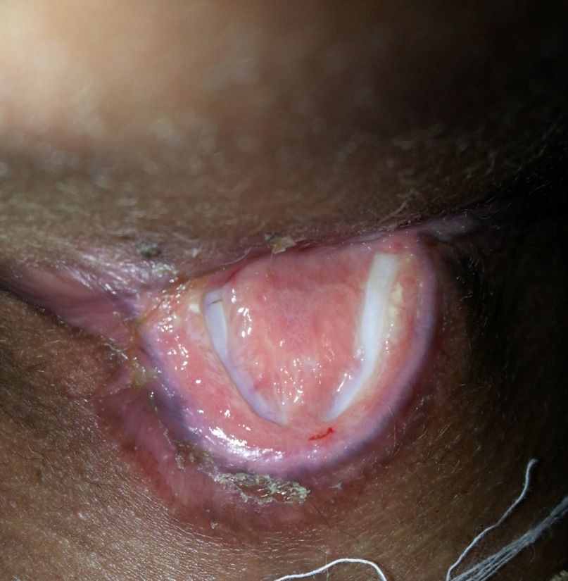
R. V. Nataraj et al.
tracheostomy tube was inserted and the neck wound was sutured.
During the post operative period the patient was not kept nil per oral and was started on liquid diet on 1st post
operative day and on semi solid diet on 2nd post operative day. Right from the 1st post operative day the patient
complained of soakage of the dressing with ingested water and food particles. On 3rd post operative day the
wound dressing was removed to reveal dehiscence of the wound with active and profuse pus discharge along
with presence of food particles on the wound & dressing. The wound was redressed and a metallic tracheostomy
tube was inserted. Following day the patient was discharged and referred to higher centre for further manage-
ment.
When the patient visited E.N.T. OPD of Sir J.J. Group of Hospitals, the above mentioned history was noted
and examination of the patient revealed following findings.
• Patient was febrile with temperature of 104˚F.
• Metallic tracheostomy tube number 32 in situ with good air blast.
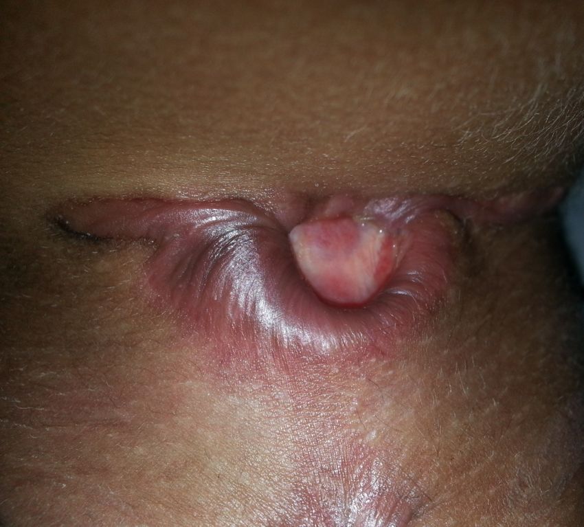
• An wound of size 6 × 2 × 2 cm with profuse slough extending superiorly up to lower body of hyoid bone,
inferiorly to the arch of cricoid cartilage, medially to 4 cm lateral to midline at the level of lower border of
body of hyoid bone and laterally to medial border of middle 1/3rd of left sternocleidomastoid muscle
(Figures 1-3).
• Both the laminae of the thyroid cartilage were exposed.
• The Cricothyroid muscle was exposed.
• Laryngeal inlet and laryngeal lumen was exposed and was covered in slough.
• Dribbling of water from the wound was noticed when the patient was asked to drink few sips of water.
An urgent CECT Neck was done and following findings were reported.
• Discontinuity of skin, subcutaneous tissue and infrahyoid muscle in anterior and lateral region of neck, more
on left than right, at the level of hypopharynx and defect in the left anterolateral wall of hypopharynx with
resultant exposure of the hypopharynx to the exterior (Figure 4 & Figure 5).
Figure 1. State of the wound upon admission.
Figure 2. A large infected wound over left side of neck.
240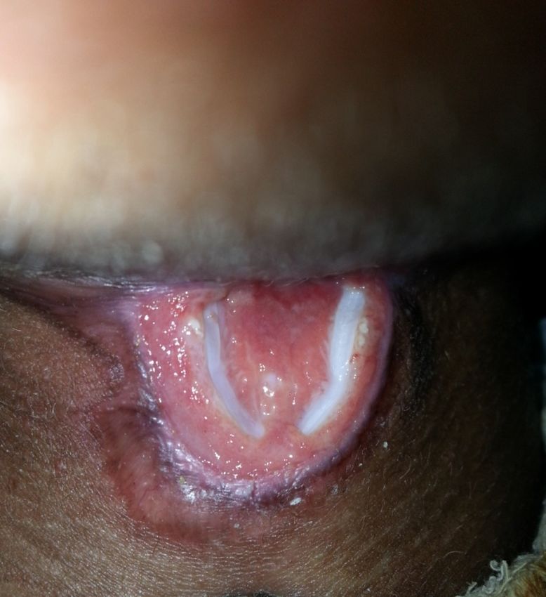
R. V. Nataraj et al.

                                     Figure 3. Hypopharynx was ex-
                                     posed to exterior.

                                     Figure 4. CT scan neck (P + C).

                                     Figure 5. CT neck (P + C).

• Deviation of epiglottis to right with disruption of left pharyngo-epiglottic fold.
• Extensive subcutaneous emphysema extending from skull base to thoracic inlet in thorax.
   Upon admission, Ryle’s tube of size 16 was inserted and high protein diet via Ryle’s tube was started. The
wound was managed by regular and twice a day dressing with Normal Saline, Eusol solution and Betadine solu-
tion under an antibiotic coverage for Gram positive & Gram negative and anaerobic bacteria. This line of ther-
apy was rigorously followed and after one week the patient showed marked improvement. The slough dried off
and granulation tissue was noticed growing on the periphery of the wound.
   But after one more week of the therapy, the rate of granulation tissue growth was noticeably slow and disap-

                                                  241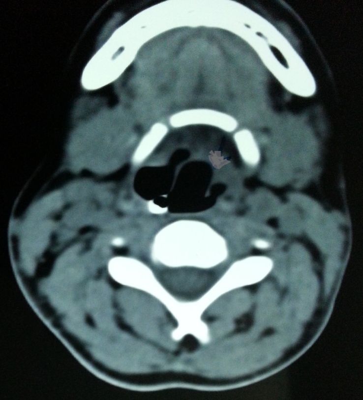
R. V. Nataraj et al.

        pointing. In order to speed up the healing process, application of gamma sterilized collagen particles was initiated.
           Collagen helps in all stages of wound healing [3]-[5].
           1) Hemostatic phase—The collagen particles are chemotactic to platelets attract the platelets towards the
        wound site. This causes aggregation of the platelets at the wound site, formation of platelet plug and thus hemo-
        stasis is achieved.
           2) Inflammatory phase—The inflammatory cells at the wound site secrete various kinds of proteolytic en-
        zymes. Collagen particles are degraded by these enzymes into protein fragments or peptides. Peptides, in turn,
        attract variety of cells like mononuclear cells, macrophages and monocytes. These cells simulate collagen syn-
        thesis and promote organized deposition of the newly formed collagen.
           3) Proliferative phase.

           4) Remodelling phase—In this phase, the collagen fibers undergo extensive cross linking to form a strong
        scar.
           Within few days of use of collagen particles, rapid growth of the granulation tissue was noticed. After three
        weeks of regular usage, the entire thyroid cartilage and the Criocothyroid muscle was covered with granulation
        tissue. The laryngeal opening was almost closed and the patient was able to swallow the saliva. Weaning of tra-
        cheostomy tube was from 24th day of admission and decannulated on 28th day without any respiratory distress.
           Picture of the wound taken at weekly intervals demonstrates the progression of the healing process (Figures
        6-11).
           The skin defect was then closed on 49th day by V-Y plasty technique (Figure 18). His post operative recovery
        was uneventful. Strict neck flexion was maintained for 10 days with help of customized neck collar. Fibreoptic
        laryngoscopy and bronchoscope was done on 7th post operative day. The larynx was found to be normal. A thin
        neat scar can be seen at the sutured site without any stenosis. Comparative CT Scans show complete healing of
        the wound without any evidence of residual defect or fistula (Figures 12-18).

        3. Discussion
        In case of cut throat or penetrating injuries of neck, one should keep in mind the possibility of following vital
        structures being injured like larynx, pharynx, trachea, oesophagus, major neck vessels, nerve roots and spinal
        cord. Clinical examination and investigations should aim towards detecting any injury to these structures. Blood

                                                            242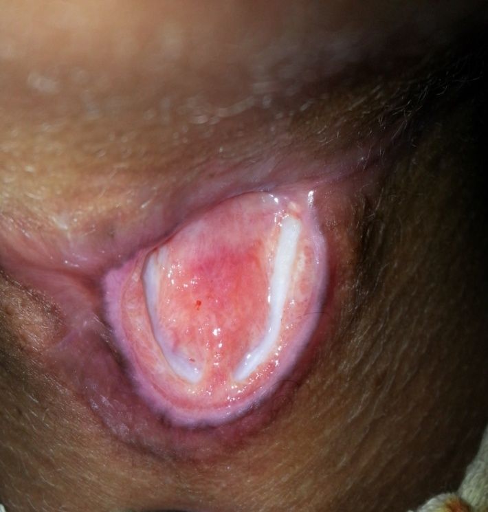
R. V. Nataraj et al.

            Figure 6. After 2 weeks.

             (a)                         (b)

Figure 7. (a) (b) After 3 weeks.

            (a)                        (b)

Figure 8. (a) (b) After 4 weeks.

            (a)                        (b)

Figure 9. (a) (b) After 5 weeks.

                              243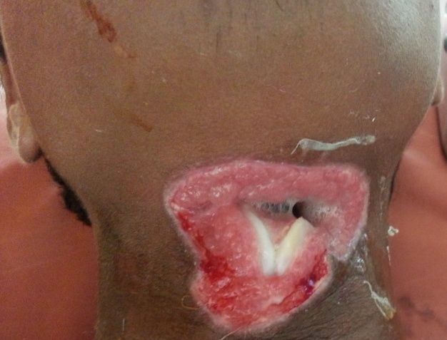
R. V. Nataraj et al.

                                  (a)                      (b)

                       Figure 10. (a) (b) After 6 weeks.

                       Figure 11. The 7th week.

                       Figure 12. CT neck before.

                                                    244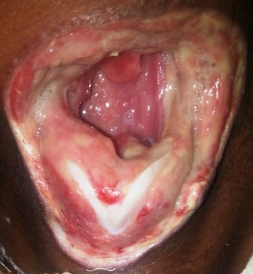
R. V. Nataraj et al.

Figure 13. CT neck after.

Figure 14. CT neck before.

Figure 15. CT neck after.

                   245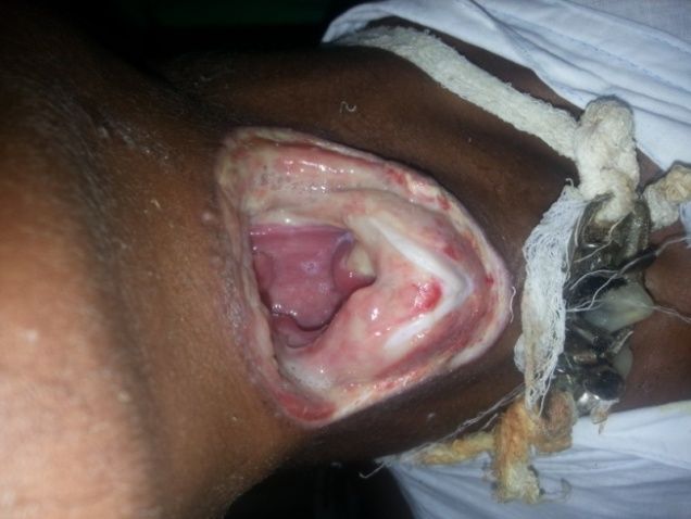
R. V. Nataraj et al.

                                         Figure 16. CT neck before.

                                         Figure 17. CT neck after.

                                         Figure 18. After primary closure.

        tests (urgent packed cell volume, urea and electrolyte levels), angiography, endoscopy (esophagoscopy, mi-
        crolaryngoscopy and bronchoscopy) and computerized tomographic (CT) scan helps to determine the extent of
        injury. Individuals are transfused with whole blood depending on the extent of blood loss. Plain radiography
        alone is not sufficient to diagnose airway trauma and the additional use of dynamic CT scan of the trachea and
        larynx and magnetic resonance imaging (MRI) can be very helpful in discovering previously undetected injuries,
        showing that some of these injuries may not even require surgical intervention.
           In the event of late presentation, such as this case, debridement of infected tissues prior to suturing is a norm.
        But debridement may also mean loss of substantial amounts of tissue to effect simple and proper closure. Con-

                                                            246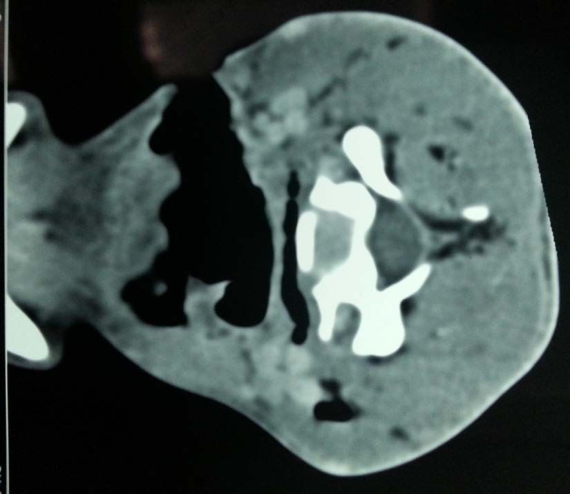
R. V. Nataraj et al.

servative debridement coupled with regular diligent dressing and [6] use of collagen particles not only helps in
normalizing the local environment but also helps in growth of new tissue. As a result, the ease and efficacy of
wound closure are increased and risk of post operative infection and wound dehiscence is also minimized.

References
[1]   Le May, S.R. (1971) Penetrating Wounds of the Larynx and Cervical Trachea. Archives of Otolaryngology, 94, 558-
      565. http://dx.doi.org/10.1001/archotol.1971.00770070858011
[2]   Lee, R.C., et al. (1988) Role of Cell Membrane Rupture in the Pathogenesis of Electrical Trauma. Journal of Surgical
      Research, 44, 709-719. http://dx.doi.org/10.1016/0022-4804(88)90105-9
[3]   Lazovic, G., Colic, M., Grubor, M. and Jovanovic, M. (2005) The Application of Collagen Sheet in Open Wound
      Healing. Archive of Annals of Burns and Fire Disasters, 18, 151-156.
[4]   Nataraj, C., Ritter, G., Dumas, S., Helfer, F.D., Brunelle, J. and Sander, T.W. (2007) Extra Cellular Wound Matrices:
      Novel Stabilization and Sterilization Method for Collagen-Based Biologic Wound Dressings. Wounds, 19, 148-156.
[5]   Motta, G., Ratto, G.B., De Barbieri, A., Corte, G., Zardi, L., Sacco, A., et al. (1983) Can Heterologous Collagen En-
      hance the Granulation Tissue Growth? An Experimental Study. The Italian Journal of Surgical Sciences, 13, 101-108.
[6]   Bhattacharya, S., Tripathi, H.N., Gupta, V., Nigam, B. and Khanna, A. (2011) Collagen Sheet Dressings for Cutaneous
      Lesions of Toxic Epidermal Necrolysis. Indian Journal of Plastic Surgery, 44, 474-477.
      http://dx.doi.org/10.4103/0970-0358.90826

                                                       247
You can also read