A Fractured Inferior Vena Cava Strut Migrating to the Right Ventricle Without Any Cardiovascular Complaint: A Case Report and Review of Literature ...
←
→
Page content transcription
If your browser does not render page correctly, please read the page content below
Open Access Case
                                          Report                                                  DOI: 10.7759/cureus.9779

                                          A Fractured Inferior Vena Cava Strut
                                          Migrating to the Right Ventricle Without
                                          Any Cardiovascular Complaint: A Case
                                          Report and Review of Literature
                                          Mohamed Elmassry 1 , Gaspar Del Rio-Pertuz 1 , Saif El-Nawaa 2 , John Abdelmalek 1 ,
                                          Mohammad M. Ansari 2

                                          1. Internal Medicine, Texas Tech University Health Sciences Center, Lubbock, USA 2. Cardiology, Texas
                                          Tech University Health Sciences Center, Lubbock, USA

                                          Corresponding author: Mohamed Elmassry, moh.elmassry88@gmail.com

                                          Abstract
                                          The use of inferior vena cava filter (IVCF) as one of the last resorts for pulmonary embolism
                                          prevention has expanded over the decades. The migration of a broken strut to the right
                                          ventricle is a very unusual complication that, when present, has been associated with life-
                                          threatening events. We report a case of a 34-year-old female with an inferior vena cava (IVC)
                                          strut that migrated and was incidentally found embedded in the right ventricle without any
                                          cardiovascular signs or symptoms. This case provides evidence that such filters probably have
                                          higher rates of complications than what has been thought because those complications might
                                          remain asymptomatic.

                                          Categories: Cardiac/Thoracic/Vascular Surgery, Cardiology, Radiology
                                          Keywords: inferior vena cava filter, strut, ivcf, right ventricle, pulmonary embolism, strut fracture,
                                          strut migration

                                          Introduction
                                          Inferior vena cava filter (IVCF) is used as one of the last resorts for pulmonary embolism
                                          prevention. There is not a consensus on when exactly IVCF is indicated, but classically
                                          speaking, IVCF is indicated in the presence of an absolute contraindication or complication to
                                          therapeutic anticoagulation, or failure of anticoagulation when there is acute proximal venous
                                          thrombosis [1-8]. One of the less common long-term complications from IVCF is migration,
                                          either the filter itself or a strut [9]. Following the venous flow, the most proximal organ for
                                          migration is the right heart. Implantation on the right ventricle can progress to life-threatening
Received 08/07/2020                       events, such as tamponade and even death [9,10]. We report a case of an IVCF strut that was
Review began 08/10/2020                   incidentally found implanted in the anterior wall of the right ventricle.
Review ended 08/11/2020
Published 08/16/2020

© Copyright 2020
                                          Case Presentation
Elmassry et al. This is an open           A 34-year-old female presented 14 years ago to an outside facility after a severe motor vehicle
access article distributed under the
                                          accident. She suffered from extensive bilateral lower extremity injuries. She underwent multiple
terms of the Creative Commons
Attribution License CC-BY 4.0., which     surgeries and hardware placement in the left hip and femur. She then developed bilateral lower
permits unrestricted use, distribution,   extremity proximal deep venous thrombosis (DVT). During hospitalization, she also developed
and reproduction in any medium,           multiple episodes of pulmonary embolism despite being on anticoagulation. The decision was
provided the original author and
                                          made to place a Greenfield inferior vena cava (IVC) filter. Since that time she has been on oral
source are credited.
                                          anticoagulation. The patient had multiple clinic visits during the following years. At one visit,

                                          How to cite this article
                                          Elmassry M, Del Rio-Pertuz G, El-Nawaa S, et al. (August 16, 2020) A Fractured Inferior Vena Cava Strut
                                          Migrating to the Right Ventricle Without Any Cardiovascular Complaint: A Case Report and Review of
                                          Literature. Cureus 12(8): e9779. DOI 10.7759/cureus.9779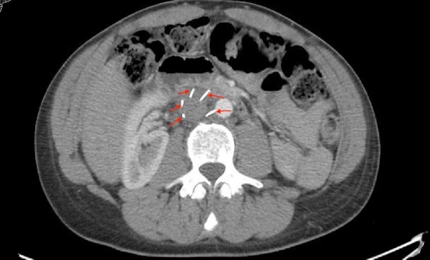
she presented with lower back pain, and as a part of her workup, an X-ray of the thoracic spine
                                   showed that the IVC filter was in place and the struts were intact (six struts) (Figure 1).

                                     FIGURE 1: X-ray of the thoracic and lumbar spine showing the
                                     inferior vena cava filter with all six struts intact.

                                   In March 2019, she presented to our facility with right flank pain. As part of her workup, she
                                   underwent a CT of the chest, abdomen, and pelvis with intravenous (IV) contrast that showed a
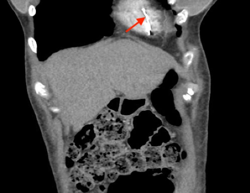
                                   high-density linear structure in the anterior wall of the right ventricle (Figures 2-4). This was
                                   likely representing a fractured IVCF strut.

2020 Elmassry et al. Cureus 12(8): e9779. DOI 10.7759/cureus.9779                                                               2 of 7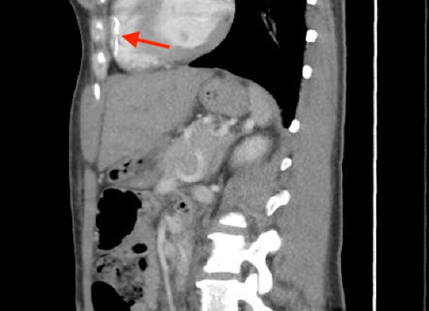
FIGURE 2: CT of the chest (axial section) showing one of the
                                     inferior vena cava filter struts embedded in the anterior wall of
                                     the right ventricle (yellow arrow).

2020 Elmassry et al. Cureus 12(8): e9779. DOI 10.7759/cureus.9779                                        3 of 7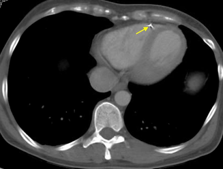
FIGURE 3: CT of the chest and abdomen (coronal section)
                                     showing the migrated inferior vena cava filter strut (red arrow).

                                     FIGURE 4: CT of the chest and abdomen (sagittal section)
                                     showing the migrated inferior vena cava filter strut (red arrow).

                                   Cardiothoracic and vascular surgery teams were consulted, and upon reviewing the CT images
                                   they agreed that the IVC filter was off-axis and missing one strut (Figure 5). They also stated
                                   that the IVC strut was likely embedded in the trabeculae of the right ventricle and
                                   endothelialized, posing minimal risk. Since the patient was asymptomatic, they decided to
                                   leave the IVC filter and the strut in place and opted for surveillance by regular follow-ups and
                                   CT scans. The patient’s right flank pain was likely musculoskeletal and responded well to the
                                   muscle relaxant. The patient was discharged home on novel oral anticoagulant with close
                                   follow-up as an outpatient.

2020 Elmassry et al. Cureus 12(8): e9779. DOI 10.7759/cureus.9779                                                                4 of 7
FIGURE 5: CT of the abdomen showing only five out of the six
struts of the inferior vena cava filter with one strut missing.
Discussion
IVCFs have been used since the 1960s for the prevention of pulmonary embolism, and since
then its use has expanded over the decades despite their complications [1]. After more than 50
years of experience, the current available societal guidelines on the use of IVCF are conflicting,
but they all agree that that the only clear indications for an IVCF placement are when a patient
had acute venous thromboembolism, in the presence of absolute contraindications to
anticoagulant. One of the reasons for this uncertainty is the lack of randomized controlled
trials evaluating the effectiveness of IVC and related complications [2-8].
Significant complications have been reported from IVCF, and those complications can be early
during placement (e.g. allergic reaction to contrast, malposition of the filter), postprocedure
(e.g. acute venous thrombosis, hematoma, arteriovenous fistula), or late after satisfactory
positioning (e.g. fracture and migration of the filter or strut, chronic thrombosis) [9]. Migration
and/or embolization of a fractured IVCF are rare but well-recognized complications of IVCF
that have been associated with life-threatening complications [9,10]. Depending on the type of
filter, the reported incidence of migration can vary from 3% to 12%, with migration to the heart
being even more unlikely, ranging from 0.1% to 0.2% [11,12].
The causes of migration and fracture are multiple: failure of engagement to the venous
wall, thrombus formation around the filter, trauma, IVC diameter increase due to physiologic
changes, increased intra-abdominal pressure, Valsalva maneuver, coughing, or extraneous
exercise [13,14]. Signs and symptoms of migration and/or embolization of the fractured IVCF
depend on the organ where the filter or strut migrated. For intracardiac migration, patients can
present with chest pain, hypotension, dyspnea, or any type of arrhythmia [14-16]. On the other
hand, it can be either indolent, patients may not be aware that a filter fractured and migrated to
an organ, like in our case where a strut from the IVCF was found as an incidental finding after
cardiothoracic imaging that was done for a noncardiovascular complaint [17].
Migration of the filter or fragments into the liver, renal vein, right atrium, left and right
2020 Elmassry et al. Cureus 12(8): e9779. DOI 10.7759/cureus.9779 5 of 7pulmonary artery, right gonadal vein, and even into the aorta has been reported [15,18-20].
Various cases of strut and filter migration to the right ventricle have been reported recently,
but all are associated with cardiovascular symptoms or outcomes ranging from a simple chest
pain complaint to cardiac tamponade symptoms [16,17]. To our knowledge, this is the first
reported case of a strut migrating from an IVCF that was found incidentally on the right
ventricle without causing any cardiovascular symptom.
In cases of suspected migration and/or embolization of a fractured IVCF to the heart, a chest CT
scan is the most suitable radiologic modality for identifying a migrated IVCF or fragments [15].
If surgery is considered, a deeper radiologic evaluation of the location and depth of penetration
of the strut or filter within the heart may assist in decision making for determining if an
endovascular or open surgical approach is more appropriate [16].
There are no available guidelines for the management approach of fractured and migrated
struts of an IVC filter, probably due to the very low incidence of such complications as
previously reported [19]. Correlation of the patient’s clinical status and location of the strut of
an IVCF filter on the CT scan is key to assist in making management decisions [14,16]. A
suggested conservative approach with close observation might be effective, especially with
asymptomatic patients like our patient.
It should be mentioned that these complications are more frequent with longer duration of
implantation [16,17,20]. Thus, we consider that the cornerstone management of this
complication is prevention, by focusing on early retrieval of the filter once protection from PE
is no longer needed. A multidisciplinary approach involving a cardiothoracic surgeon and an
interventional cardiologist or radiologist may be indicated as proposed in our case.
Conclusions
IVCFs have been widely used for decades for the prevention of pulmonary embolism despite
their complications. IVC strut fracture and migration is a rare complication that has been
reported in the literature and is usually fatal. A multimodality imaging approach is usually
required to accurately visualize the site of implantation. A conservative approach with close
observation might be effective, especially with asymptomatic patients.
Additional Information
Disclosures
Human subjects: Consent was obtained by all participants in this study. Conflicts of interest:
In compliance with the ICMJE uniform disclosure form, all authors declare the following:
Payment/services info: All authors have declared that no financial support was received from
any organization for the submitted work. Financial relationships: All authors have declared
that they have no financial relationships at present or within the previous three years with any
organizations that might have an interest in the submitted work. Other relationships: All
authors have declared that there are no other relationships or activities that could appear to
have influenced the submitted work.
References
1. Stein PD, Kayali F, Olson RE: Twenty-one-year trends in the use of inferior vena cava filters .
Arch Intern Med. 2004, 164:1541-1545. 10.1001/archinte.164.14.1541
2. Kearon C, Akl EA, Comerota AJ, et al.: Antithrombotic therapy for VTE disease:
antithrombotic therapy and prevention of thrombosis, 9th ed: American College of Chest
Physicians evidence-based clinical practice guidelines. Chest. 2012, 141:e419S-e496S.
10.1378/chest.11-2301
2020 Elmassry et al. Cureus 12(8): e9779. DOI 10.7759/cureus.9779 6 of 73. Kearon C, Akl EA, Ornelas J, et al.: Antithrombotic therapy for VTE disease: CHEST guideline
and expert panel report. Chest. 2016, 149:315-352. 10.1016/j.chest.2015.11.026
4. Kaufman JA, Kinney TB, Streiff MB, et al.: Guidelines for the use of retrievable and convertible
vena cava filters: report from the Society of Interventional Radiology multidisciplinary
consensus conference. J Vasc Interv Radiol. 2006, 17:449-459. 10.1097/01.RVI.0000203418-
39769.0D
5. Konstantinides SV, Torbicki A, Agnelli G, et al.: 2014 ESC Guidelines on the diagnosis and
management of acute pulmonary embolism: the task force for the diagnosis and management
of acute pulmonary embolism of the European Society of Cardiology (ESC). Eur Heart J. 2014,
35:3033-3080. 10.1093/eurheartj/ehu283
6. Minocha J, Smith AM, Kapoor BS, et al.: ACR Appropriateness Criteria radiologic management
of venous thromboembolism-inferior vena cava filters. J Am Coll Radiol. 2019, 16:S214-S226.
10.1016/j.jacr.2019.02.010
7. Jaff MR, McMurtry MS, Archer SL, et al.: Management of massive and submassive pulmonary
embolism, iliofemoral deep vein thrombosis, and chronic thromboembolic pulmonary
hypertension: a scientific statement from the American Heart Association. Circulation. 2011,
123:1788-1830. 10.1161/CIR.0b013e318214914f
8. Konstantinides SV, Meyer G, Becattini C, et al.: 2019 ESC Guidelines for the diagnosis and
management of acute pulmonary embolism developed in collaboration with the European
Respiratory Society (ERS): the task force for the diagnosis and management of acute
pulmonary embolism of the European Society of Cardiology (ESC). Eur Heart J. 2020, 41:543-
603. 10.1093/eurheartj/ehz405
9. Rogers NA, Nguyen L, Minniefield NE, Jessen ME, de Lemos JA: Fracture and embolization of
an inferior vena cava filter strut leading to cardiac tamponade. Circulation. 2009, 119:2535-
2536. 10.1161/CIRCULATIONAHA.108.817247
10. de Matos Soeiro A, Fernandes FL, Teixeira RP, Nicz PF, de Almeida Soeiro MCF, Serrano CV Jr,
de Oliveira MT Jr: Intracardiac embolization of inferior vena cava filter associated with right
atrium perforation and cardiac tamponade. Rev Bras Cir Cardiovasc. 2014, 29:285-288.
11. Joels CS, Sing RF, Heniford BT: Complications of inferior vena cava filters . Am Surg. 2003,
69:654-659.
12. Athanasoulis CA, Kaufman JA, Halpern EF, Waltman AC, Geller SC, Fan CM: Inferior vena
caval filters: review of a 26-year single-center clinical experience. Radiology. 2000, 216:54-66.
10.1148/radiology.216.1.r00jl1254
13. Ferris EJ, McCowan TC, Carver DK, McFarland DR: Percutaneous inferior vena caval filters:
follow-up of seven designs in 320 patients. Radiology. 1993, 188:851-856.
10.1148/radiology.188.3.8351361
14. Saeed I, Garcia M, McNicholas K: Right ventricular migration of a recovery IVC filter's
fractured wire with subsequent pericardial tamponade. Cardiovasc Intervent Radiol. 2006,
29:685-686. 10.1007/s00270-005-0136-7
15. Shennib H, Bowles B, Hickle K: Migration of a fractured inferior vena cava filter strut to the
right ventricle of the heart: a case report. J Cardiothorac Surg. 2014, 9:183. 10.1186/s13019-
014-0183-8
16. Mehta JJ, DeMarco B, Vavalle JP, Tahir KS, Rossi JS: Hemopericardium and cardiac tamponade
secondary to migrated inferior vena cava filter. Case Rep Cardiol. 2018, 2018:5919767.
10.1155/2018/5919767
17. Tam MD, Spain J, Lieber M, Geisinger M, Sands MJ, Wang W: Fracture and distant migration of
the Bard Recovery filter: a retrospective review of 363 implantations for potentially life-
threatening complications. J Vasc Interv Radiol. 2012, 23:199-205. 10.1016/j.jvir.2011.10.017
18. Nguyen NT, Barshes NR, Bechara CF, Pisimisis GT: Natural history of an intra-aortic
permanent inferior vena cava filter. J Vasc Surg. 2014, 60:784. 10.1016/j.jvs.2013.12.029
19. Bennett-Venner A, Alsumrain M, Pi J, Riar S, Vefali H, Miller R, Klukowicz A: An uncommon
complication of an inferior vena cava filter. Am J Respir Crit Care Med. 2011, 184:1209.
10.1164/rccm.201102-0309IM
20. Sharma S, Mukund A, Agarwal S, Srivastava DN: Case of a misplaced IVC filter: a lesson to
learn. Cardiovasc Intervent Radiol. 2010, 33:880-882. 10.1007/s00270-010-9825-y
2020 Elmassry et al. Cureus 12(8): e9779. DOI 10.7759/cureus.9779 7 of 7You can also read


