Ovarian endosalpingiosis mimicking hydrosalpinges. Unexpected intraoperative findings and a diagnostic rollercoaster - Oxford Academic Journals
←
→
Page content transcription
If your browser does not render page correctly, please read the page content below
Journal of Surgical Case Reports, 2021;6, 1–4

                                                                              https://doi.org/10.1093/jscr/rjab264
                                                                              Case Report

CASE REPORT

Ovarian endosalpingiosis mimicking hydrosalpinges.

                                                                                                                                                           Downloaded from https://academic.oup.com/jscr/article/2021/6/rjab264/6311008 by guest on 30 October 2021
Unexpected intraoperative findings and
a diagnostic rollercoaster
Fleur C. Muirhead1 ,*, Hong L. Lee1 and Rajeev Singh2
1
    Obstetrics and Gynaecology, Joondalup Health Campus, Joondalup, Western Australia, Australia and
2
    Obstetrics and Gynaecology, Fiona Stanley Hospital, Murdoch, Western Australia, Australia

*Correspondence address. Tel: +61 400 002 753; Fax: +61 (08) 9400 9054; E-mail: fleur-muirhead@live.com.au

Abstract
Over 80 % of ovarian cancer diagnoses are in women aged over 50 years. Post-menopausal women are at significantly increased
risk compared with other age groups. Tumour biomarkers and ultrasound assist with diagnostics. A post-menopausal
woman was referred with a possible left adnexal cyst on ultrasound. A tertiary gynaecological ultrasound suggested bilateral
hydrosalpinges without cysts. Tumour markers were negative. Unexpectedly, while undergoing a laparoscopy, the tubes were
normal in appearance; however, multiple cystic deposits suspicious for malignancy were noted on the ovaries. A laparoscopic
bilateral salpingo-oophorectomy was performed without spillage and pelvic washings obtained. On histopathology, the ovaries
contained multiple, benign epithelial cysts and pelvic washings were negative. This case demonstrates an unanticipated
peri-operative diagnostic dilemma. It highlights the surgical management decisions required to balance duty of care and
consent compliance within the scope of general gynaecology. It emphasizes the importance of histopathological examination
to confirm diagnoses.

                                                                                  Tumour biomarkers and imaging can assist with adnexal
INTRODUCTION                                                                  pathology diagnostics, particularly ultrasound [3]. Commonly
Post-menopausal women are at increased risk of malignancy                     used tumour markers include cancer antigen 125 (Ca125), carci-
compared with other age groups; however, the majority of                      noembryonic antigen (CEA) and lactate dehydrogenase (LD). An
adnexal masses are histologically benign [1]. After menopause,                increase in serum Ca125 can be caused by multiple conditions,
the reproductive tract is no longer influenced by the hormonal                many of which are benign, and however, a rise in serum Ca125 is
fluctuations of the menstrual cycle, hence the absence of                     noted in 80% of ovarian epithelial cancers [4, 5].
physiological follicles [2]. Any post-menopausal, ultrasound                      The likelihood that an ovarian mass is cancerous can be
confirmed adnexal masses should be thoroughly investigated.                   determined using the Risk of Malignancy Index I (RMI). The RMI-I
Certain sonologic ovarian cyst characteristics, such as solid                 is calculated utilizing menopausal status, a serum Ca125 result
components, complete septations, papillary projections or                     and ultrasonographic features, and is widely adopted in first
ascites, can suggest malignancy [3].                                          world countries [6, 7]. The RMI-I has a sensitivity of 78% and a

Received: April 6, 2021. Revised: May 23, 2021. Accepted: May 27, 2021
Published by Oxford University Press and JSCR Publishing Ltd. All rights reserved. © The Author(s) 2021.
This is an Open Access article distributed under the terms of the Creative Commons Attribution Non-Commercial License (http://creativecommons.org/li
censes/by-nc/4.0/), which permits non-commercial re-use, distribution, and reproduction in any medium, provided the original work is properly cited. For
commercial re-use, please contact journals.permissions@oup.com

                                                                                                                                                  1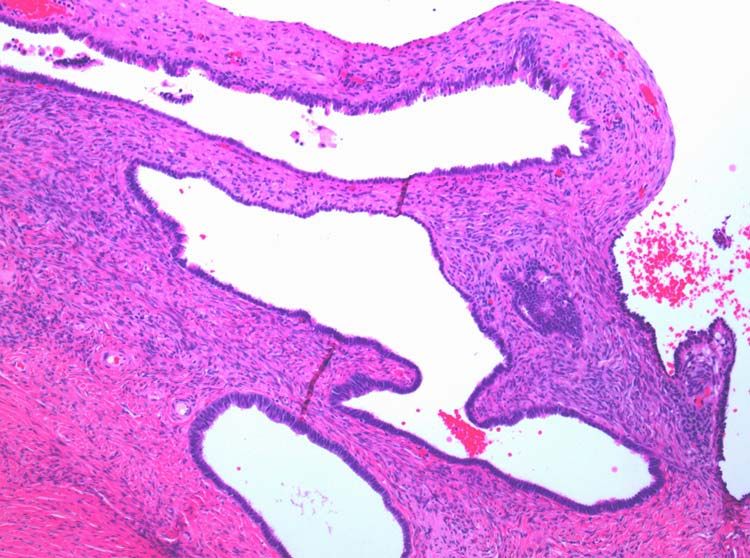
2     F. C. Muirhead et al.

                                                                                                                                                            Downloaded from https://academic.oup.com/jscr/article/2021/6/rjab264/6311008 by guest on 30 October 2021
Figure 1: The tertiary ultrasound of the right adnexa demonstrating the   Figure 2:    The tertiary ultrasound of the left adnexa demonstrating a
33 mm × 18 mm × 34 mm serpiginous cystic structure with a 13-mm lumen     30 mm × 19 mm × 27 mm cystic structure with incomplete septae in keeping
consistent with a hydrosalpinx.                                           with a possible hydrosalpinx.

specificity of 87% for ovarian malignancy when the cutoff is 200
[7].
     Evidence suggests that a referral to a multidisciplinary,
gyane-oncology team should be completed for staging and
further management if an RMI-I score is greater than 200 [6]. For a
post-menopausal woman with suspected benign pathology, the
gold standard to exclude ovarian malignancy is a laparoscopic
bilateral salpingo-oophorectomy (BSO), if appropriate. The BSO
should be performed utilizing a retrieval bag to avoid spillage           Figure 3: The intra-operative photos demonstrating the cystic appearance of the
and subsequent histopathological examination [6–8]. Surgeon               left ovary.
visualization and judgement during laparoscopy has a sensitiv-
ity of 87.5% and a specificity of 98% for discriminating malignant
from benign disease [9]. Multiple studies demonstrate that
even with a low pre-operative index of suspicion, unexpected
ovarian malignancy was noted on 0.9–8% histopathological
examinations [4, 10, 11].

CASE REPORT
A post-menopausal woman was referred to the gynaecological
outpatient clinic with an incidental finding of a possible left
adnexal cyst on a renal tract ultrasound for routine chronic              Figure 4: The intra-operative photos demonstrating multiple cystic structures
kidney disease surveillance. She was 16 years post-menopausal             covering the surface of the right ovary.
with no significant risk factors for pelvic inflammatory disease
or gynaecological cancers.
    The patient was referred for a pelvic ultrasound, which               post-menopausal status, a laparoscopic BSO was recommended
demonstrated a 2.5-cm left ovarian cyst with a thin, avascular            and the patient subsequently consented.
septum and a possible right-sided hydrosalpinx. These findings                Intraoperative findings were inconsistent with all pre-
remained stable on a repeat ultrasound 9 months later. Ca125,             operative ultrasound diagnoses. Both ovaries appeared suspi-
CEA and LD were negative.                                                 cious for malignancy with cystic lesions covering the surface of
    On review in gynaecology clinic, the patient was asymp-               the right, while the left appeared to contain two smaller cysts
tomatic and following joint discussion regarding the manage-              (Figs 3–5). There were no hydrosaplinges or free fluid within
ment options, the surgeon recommended a laparoscopic BSO.                 the pelvis. Given the abnormal features of the ovaries, the
The patient opted for surveillance management and was sub-                surgeon proceeded with the laparoscopic BSO without spillage.
sequently referred for a tertiary ultrasound and repeat tumour            Pelvic washings were performed, though there were no atypical
markers.                                                                  peritoneal or omental lesions for biopsy.
    The tertiary level gynaecological ultrasound demonstrated a               Despite the sinister appearance macroscopically, histopathol-
right adnexal serpiginous structure with a lumen and incom-               ogy was benign. The ovaries contained multiple, benign,
plete septae and a left adnexal cystic structure (Figs 1 and 2).          ciliated and epithelial cysts. Pelvic washings were negative for
The ultrasonographic features were consistent with those of               malignancy and the tubes were unremarkable.
hydrosalpinges [12]. The Ca125 remained negative. In view of                  The patient had an uneventful post-operative period and was
the ultrasound diagnosis of hydrosalpinges and the patient’s              discharged after a follow-up appointment.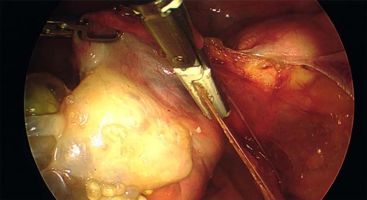
Ovarian endosalpingiosis mimicking hydrosalpinges 3
Figure 5: The intra-operative photographs of the posterior aspect of the right
ovary.
Downloaded from https://academic.oup.com/jscr/article/2021/6/rjab264/6311008 by guest on 30 October 2021
Figure 7: Normal serous epithelium with prominent delicate cilia lining the cyst
lumen; haematoxylin and eosin stain, high magnification, scale ×40.
performed. This would also have had ethical and legal issues
pertaining to consent compliance.
The case emphasizes the diagnostic importance of histopathol-
ogy, particularly in the setting of unanticipated and sinis-
ter macroscopic findings. The macroscopic appearances of
the ovaries suggested malignancy diagnosis; however, the
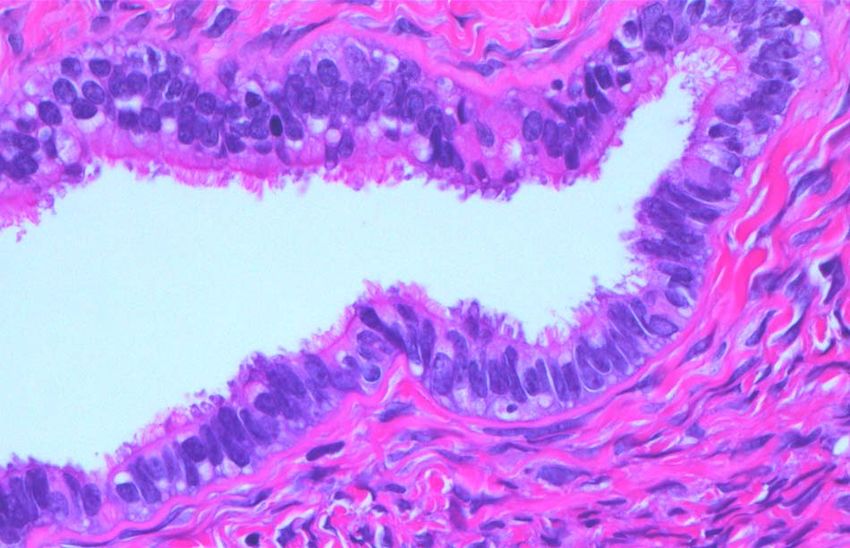
histopathology identified benign epithelial cysts [15]. The cyst
lining was comprised of a single layer of epithelial cells amid
normal ovarian stroma (Figs 6 and 7). The cysts contained
Figure 6: Multiple cysts of variable diameter lined by a single layer of epithelium, normal serous epithelium with cilia lining the cyst lumen.
separated by ovarian stroma; haematoxylin and eosin stain, medium magnifica-
These histological findings are consistent with the Müllerian
tion, scale ×10.
phenotype and are therefore endosalpingiosis, ectopic tubal cell
deposits on the surface of the ovaries, not that of hydrosalpinges
or malignant ovarian cysts as the diagnostic rollercoaster
DISCUSSION suggested [15].
This case demonstrates an unexpected, peri-operative diagnos-
tic dilemma. Despite utilizing investigations to aid diagnoses, it ACKNOWLEDGEMENTS
remained difficult to distinguish between benign and malignant
The authors acknowledge the support of Dr Matthew Bogle
pathology for this patient [9]. Pre-operatively, the surgical team
of Western Diagnostic Pathology for his expertise in anatom-
was confident that patient’s adnexal masses were tubal in origin,
ical pathology and histopathological images. The authors also
not ovarian, as demonstrated on the tertiary ultrasound. In
acknowledge the team at the JHC O&G Ultrasound Clinic for the
addition, the pre-operative RMI-I score was low risk at 30 and
use of their images for this case report.
remained so post-operatively.
The surgeon’s ethical challenge on observing the sinister
appearance of the ovaries was determining the manner in which
to proceed while balancing duty of care and consent compliance. FUNDING AND CONFLICT OF INTEREST
Management of unexpected, suspicious intra-operative findings The authors have sources of funding or no conflicts of interest
is operator dependent. There are often valid peri-operative con- to declare.
cerns regarding risks, including intra-operative spillage [7] and
seeding into post-sites [13]. Adnexectomy without intraperi-
toneal spillage of the cyst contents utilizing a specimen retrieval REFERENCES
bag is the ideal management [6, 11]. Converting a laparoscopy to 1. van Nagell J, DePriest PD. Management of adnexal masses in
laparotomy depends on the extent of abnormal pathology and post-menopausal women. Am J Obstst Gynecol 2005;193:30–5.
the operating surgeon’s skill and preference [6]. 2. Minkin MJ. Menopause: hormones, lifestyle, and optimizing
In this case, the laparoscopic BSO was performed as con- aging. Obstet Gynecol Clin North Am 2019;46:501–14.
sented without spillage using specimen retrieval bags. Despite 3. Timmerman D, Ameye L, Fischerova D, Epstein E, Melis
the intra-operative diagnosis altering from hydrosalpinges GB, Guerriero S, et al. Simple ultrasound rules to dis-
to possible ovarian malignancy, the laparoscopic BSO was tinguish between benign and malignant adnexal masses
still deemed the most appropriate management at the time. before surgery: prospective validation by IOTA group. BMJ
Obtaining pelvic washing addressed the surgeon’s suspicion of 2010;341:c6839.
non-benign pathology. In the event that the histology returned 4. Jacobs I, Oram D, Fairbanks J, Turner J, Frost C, Grudzin-
malignant, washings may have aided in staging without compro- skas JG. A risk of malignancy index incorporating CA 125,
mise consent [14]. In Australia, general gynaecological surgeons ultrasound and menopausal status for the accurate pre-
do not perform formal staging; therefore, an appendicectomy operative diagnosis of ovarian cancer. Br J Obstet Gynaecol
or further biopsies of the omentum or peritoneum were not 1990;97:922–9.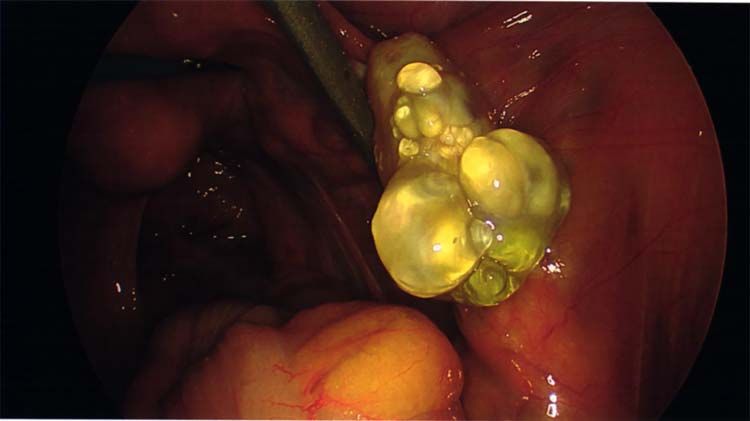
4     F. C. Muirhead et al.

 5. Milojkovic M, Hrgovic Z, Hrgovic I, Jonat W, Maass N, Buković          12-year experience with long-term follow-up. Obstet Gynecol
    D. Significance of CA 125 serum level in driscimination                 1994;83:707–12.
    between benign and malignant masses in the pelvis. Arch           11.   Muzii L, Angioli R, Zullo M, Panici PB. The unexpected ovar-
    Gynecol Obstet 2004;269:176–80.                                         ian malignancy found during operative laparoscopy: inci-
 6. Mehasseb MK, Siddiqui NA, Bryden FB. The Management of                  dence, management, and implications for prognosis. J Minim
    Ovarian Cysts in Postmenopausal Women [Document]. 2016.                 Invasive Gynecol 2005;12:81–9.
 7. Geomini P, Kruitwagen R, Bremer GL, Cnossen J, Mol BWJ.           12.   Benjaminov O, Atri M. Sonography of the abnormal fal-
    The accurate of risk scores in predicting ovarian malignancy,           lopian tube. A Merican Journal of Roentgenology 2004;183:
    a systematic review. Obstet Gynecol 2009;113:384–94.                    737–42.
 8. Lee JW, Kim CJ, Lee JE, Lee SJ, Kim BG, Lee JH, et al. Selected   13.   Kadar N. Port-site recurrences following laparoscopic oper-
    adnexal cystic masses in postmenopausal women can be                    ations for gynaecological malignancies. Br J Obstet Gynaecol
    safely managed by laparoscopy. J Korean Med Sci 2005;20:                1997;104:1308–13.
    468–72.                                                           14.   Schulte JJ, Lastra RR. Abdominopelvic washings in gyneco-

                                                                                                                                           Downloaded from https://academic.oup.com/jscr/article/2021/6/rjab264/6311008 by guest on 30 October 2021
 9. Leng JH, Lang JH, Zhang JJ, Feng FZ, Liu ZF, Sun DW, et al.             logic pathology: a comprehensive review. Diagn Cytopathol
    Role of laproscopy in the diagnosis and treatment of adnexal            2016;44:1039–57.
    masses. Chin Med J (Engl) 2006;119:202–6.                         15.   Busca A, Parra-Herran C. Pathology Outlines. 2017.
10. Canis M, Mage G, Pouly JL, Watteiz A, Manhes H, Bruhat                  https://www.pathologyoutlines.com/topic/ovarynontu
    MA. Laparoscopic diagnosis of adnexal cystic masses A                   morinclusioncyst.html (January 2021, date last accessed).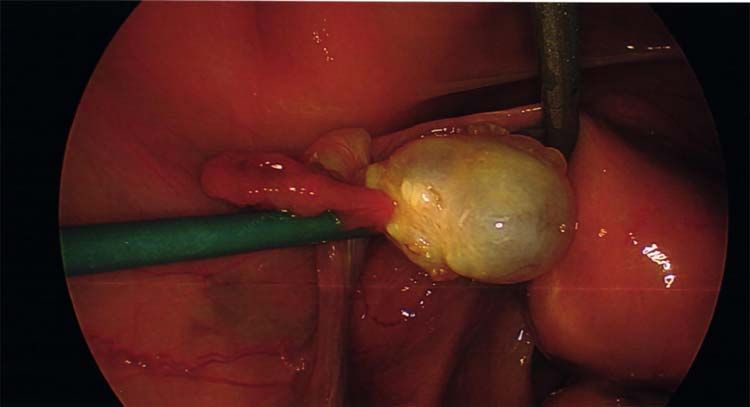
You can also read