Cutaneous and Pulmonary Sarcoidosis in Association With Tattoos
←
→
Page content transcription
If your browser does not render page correctly, please read the page content below
Cutaneous and Pulmonary Sarcoidosis in
Association With Tattoos
Maeran Chung Landers, MD, PhD; Michael Skokan, MD; Sandra Law, BS; Frances J. Storrs, MD

Sarcoidosis encompasses a heterogeneous spec-
trum of clinical presentations, including sar-
coidosis in association with tattoos. We report the
development of cutaneous and pulmonary sar-
coidosis in a patient with long-standing eyebrow
tattoos whose cutaneous sarcoidosis almost com-
pletely resolved when treated with tacrolimus
0.1% ointment. A 70-year-old woman with a                                      Figure not available online
3-year history of an erythematous eruption cir-
cumscribing her eyebrow tattoos presented with
a chronic, nonproductive cough of 8 months’
duration. Skin biopsy results demonstrated naked
tubercles consistent with sarcoidosis. Results of
radiographs and a computed tomography scan of
the chest revealed multiple pulmonary nodules
with mediastinal and hilar adenopathy. The                            Figure 1. Erythematous brown plaques with rolled
results of transbronchial biopsy were consistent                      borders and slightly atrophic centers circumscribing
with the diagnosis of pulmonary sarcoidosis.                          eyebrow tattoos.
Initial treatment with oral prednisone only
improved the pulmonary sarcoidosis. The cuta-
neous sarcoidosis almost completely resolved                          tattoo-associated cutaneous sarcoidosis has been
after the addition of tacrolimus 0.1% ointment.                       associated with the development of systemic sar-
                              Cutis. 2005;75:44-48.                   coidosis. We report the development of cutaneous
                                                                      and pulmonary sarcoidosis in a patient with long-
                                                                      standing eyebrow tattoos.

S
     arcoidosis is a disease that encompasses an
     expansive array of clinical presentations.1 One                  Case Report
     unusual clinical presentation is that of sar-                    A 70-year-old woman presented with a 3-year
coidosis in tattoos. Historically, it has been believed               history of an asymptomatic, erythematous, some-
that noncaseating granulomas may be a foreign                         what scaly eruption circumscribing eyebrow tattoos
body reaction to materials such as talc, titanium,                    that had been applied 25 years earlier. In 1999, the
and lead carbonate, which are often found in tat-                     patient had noticed a small, slowly enlarging
toos. Tattoos also can result in allergic granulomas                  erythematous plaque adjacent to her left eyebrow
caused by zirconium or beryllium. Red pigments,                       tattoo. Several months later, the patient noted a
specifically mercuric sulfide, are the most common                    similar erythematous plaque adjacent to her right
source of tattoo-related allergic reactions. Rarely,                  eyebrow tattoo. The eruptions eventually completely
                                                                      circumscribed both eyebrow tattoos (Figure 1).
Accepted for publication May 4, 2004.                                 The patient had been concealing the eruption
Drs. Landers and Storrs and Ms. Law are from the Department           with makeup for 3 years. The patient had used
of Dermatology, Oregon Health & Science University, Portland.         hydrocortisone 1% cream for several months
Dr. Skokan is from the Pulmonary Division, Oregon Clinic, Portland.
                                                                      without improvement. She had no prior history of
The authors report no conflict of interest.
Reprints: Frances J. Storrs, MD, Department of Dermatology,           skin disease.
Oregon Health & Science University, 3181 SW Sam Jackson                   The patient also reported a mild cough that had
Park Rd, OP 06, Portland, OR 97239 (e-mail: storrsf@ohsu.edu).        lasted 8 months. The dry nonproductive cough was

44 CUTIS®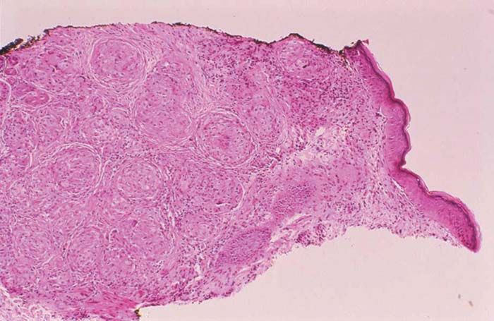
Sarcoidosis in Tattoos
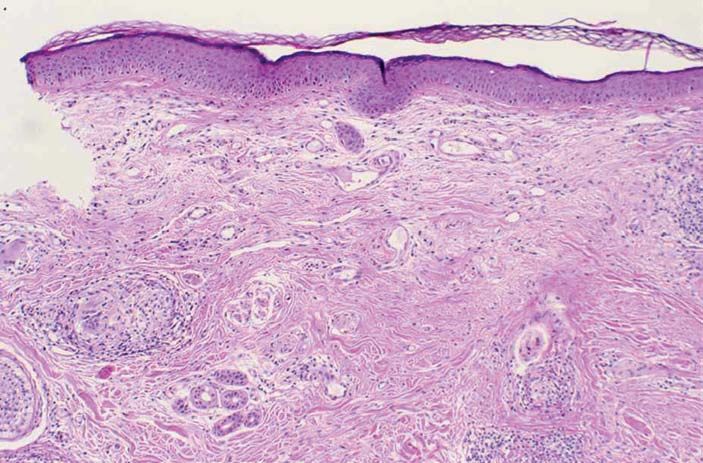
Figure 2. Skin biopsy speci-
men showing features of
sarcoidosis with noncaseat-
ing granulomas and large,
pale-staining, epithelioid
histiocytes (H&E, original
magnification 25).
Figure 3. Results of postero-
anterior (A) and lateral (B)
chest radiographs reveal
multiple pulmonary nodules,
A B bilateral hilar enlargement,
and no pleural effusion.
persistent and occurred daily without any triggering plaques were slightly atrophic. No other abnormal
or exacerbating factors. There was no significant findings were noted. Skin biopsy results revealed
medical or surgical history of dyspnea, hemoptysis, noncaseating granulomas with large, pale-staining,
postnasal drip, or gastroesophageal reflux disease. epithelioid histiocytes (Figure 2). Results of acid-
She denied smoking and occupational exposures to fast bacillus and Gomori methenamine-silver stain-
asbestos. The patient had no constitutional symp- ing procedures were negative for organisms. There
toms such as fevers, chills, weight loss, or fatigue. was no evidence of foreign body material on exam-
She reported taking no medications and having no ination under polarizing light. The histopathologic
known allergies. differential diagnosis included sarcoidosis, foreign
The findings of the visual cutaneous examina- body reaction, and allergic granulomas.
tion included nonscaly, erythematous brown Imaging studies, including posteroanterior and
plaques with rolled borders surrounding the eye- lateral chest radiographs (Figure 3) and computed
brow tattoos bilaterally. The centers of the tomography scan of the chest, were performed.
VOLUME 75, JANUARY 2005 45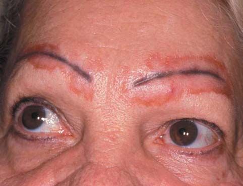
Sarcoidosis in Tattoos

Figure 4. Transbronchial
lung biopsy specimen
showing features of sar-
coidosis with noncaseating
granulomas (H&E, original
magnification 50).

The chest radiographs revealed multiple nondis-
crete nodules, 4 in the right lung field and 3 in the
left lung field. No pleural effusion was seen. The
hila were enlarged, and the radiographic appear-
ance suggested mediastinal adenopathy. The com-
puted tomography scan of the chest with contrast
revealed multiple, noncalcified, parenchymal nod-
ules. The nodules were peripherally located, with
5 in the right lung and 6 in the left lung. Hilar and
                                                                  Figure not available online
mediastinal adenopathy also was noted. Results of
a transbronchial lung biopsy demonstrated non-
caseating granulomas consistent with the diagnosis
of sarcoidosis (Figure 4). Results of special staining
procedures were negative for acid-fast bacilli and
fungi. There was no evidence of pigment or foreign
material in the granulomas. Results of laboratory
studies, including complete blood count, chemistry       Figure 5. Nearly complete clinical clearing of erythem-
panel, and liver and renal function tests, were all      atous brown plaques circumscribing the eyebrow
within reference range, as were the results of pul-      tattoos after 6 months of treatment with tacrolimus
monary function tests.                                   0.1% ointment applied twice a day.
    The patient was treated with oral prednisone
20 mg/d for pulmonary sarcoidosis. Four weeks after
starting prednisone, her nonproductive cough had         to the eyebrows.2 After 6 months of treatment with
improved. A repeat computed tomography scan of the       tacrolimus 0.1% ointment, clinical clearing was
chest performed 3 months after the initiation of         nearly complete (Figure 5). Results of a subsequent
prednisone therapy showed a reduction in the size        biopsy of the eyebrows demonstrated sparse sarcoidal
of the pulmonary nodules. At 3 months, the dose of       tubercles and scarring alopecia (Figure 6). The
prednisone was reduced to 10 mg/d for maintenance.       patient was prescribed maintenance treatment with
The patient had discontinued the hydrocortisone          tacrolimus 0.1% ointment only.
1% cream, and the oral prednisone did not signifi-
cantly improve the cutaneous manifestations.             Comment
Therefore, in addition to prednisone 10 mg/d for the     Sarcoidosis comprises a spectrum of clinical pre-
pulmonary sarcoidosis, the patient was prescribed        sentations including papular, anular, hypopig-
tacrolimus 0.1% ointment to be applied twice a day       mented, ulcerative, verrucose, and subcutaneous,

46 CUTIS®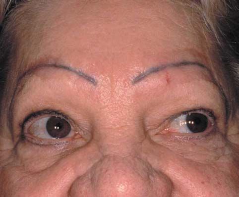
Sarcoidosis in Tattoos

                                                                                    Figure 6. Skin biopsy speci-
                                                                                    men showing features of
                                                                                    sarcoidosis with sparse non-
                                                                                    caseating granulomas and
                                                                                    scarring alopecia (H&E,
                                                                                    original magnification 25).

as well as lupus pernio and scar manifestations.1         systemic sarcoidosis, but results of a patch test were
We present this case report of pulmonary and cuta-        positive for mercury. Similar allergic reactions to
neous sarcoidosis associated with tattoos because of      green 14 and red 15 tattoo pigments have been
its unique clinical presentation.                         reported. These cases are thought to represent sar-
    The relationship of tattoo-related sarcoidosis to     coidosislike histology and allergic hypersensitivity
the development of systemic sarcoidosis is difficult      reactions to tattoo pigments. In contrast, cutaneous
to resolve. Cutaneous sarcoidosis may be seen in          sarcoidosis has been reported in the context of
relation to scarring processes, and tattoos may be        blue,4 red,5,7,11 and black6 tattoo pigments in the set-
considered to be scar-inducing conditions. Sar-           ting of systemic sarcoidosis. A case report found the
coidosis in tattoos is thought to be a variant of         presence of red tattoo pigment in both cutaneous
scar sarcoidosis due to the Köbner phenomenon.            and lung granulomas,7 suggesting that a particular
Scar sarcoidosis is most frequently seen in               tattoo pigment may have been responsible for the
patients with chronic systemic sarcoidosis. The           development of systemic sarcoidosis.
development of both scar- and tattoo-associated               The latency period between the placement of
sarcoidosis simultaneously with pulmonary sar-            the tattoo and the development of cutaneous or
coidosis in one patient supported the hypothesis          systemic sarcoidosis is variable. In this case, there
that granulomas in long-standing tattoos may be a         was a 25-year interval between the placement of
manifestation of the Köbner phenomenon in the             the eyebrow tattoos and the diagnosis of cutaneous
setting of systemic sarcoidosis.3                         and pulmonary sarcoidosis. Previous case reports
    Cutaneous and systemic sarcoidosis have been          have described latency periods of 1 year,7 10 years,4
reported in the context of tattoos in a number of         12 years,9 15 years,3,5,6 25 years,11 and 45 years.8 The
case reports.3-11 Careful evaluation to rule out sys-     wide spectrum of time from tattoo placement to
temic sarcoidosis is therefore essential and should       the diagnosis of sarcoidosis highlights the complex-
include complete history, physical examination,           ity of this association and a possible altered reac-
review of systems, blood tests, chest radiograph,         tivity of the patient to tattoos.
and, if appropriate, ophthalmologic examination.              In our patient, the eyebrow tattoos had been
Abnormal chest imaging results should be followed         placed 25 years earlier. Originally brown, brown-
up with pulmonary function testing. The most com-         black, or black, the tattoos are currently black.
mon manifestation of systemic sarcoidosis associ-         Brown tattoo pigments generally comprise inor-
ated with tattoos has been pulmonary sarcoidosis;         ganic iron oxides with combinations of red and yel-
however, there also have been reports of erythema         low oxides. Black tattoo pigments are composed of
nodosum,5 arthritis,5 and uveitis.12                      either iron oxide or carbon-based black. Tattoo pig-
    The first case of tattoo-associated sarcoidosis was   ments are suspended in hydrating fluids. Twenty-
reported in 1939.13 The patient had no evidence of        five years ago, the gold standard of hydrating fluids

                                                                                        VOLUME 75, JANUARY 2005 47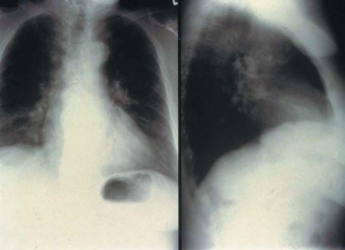
Sarcoidosis in Tattoos

was Listerine® Antiseptic. Today, hydrating fluids         performed in patients who present with tattoo-
may include ethanol, isopropyl alcohol, witch hazel,       associated cutaneous sarcoidosis. The relationship
rose water, glycerol, propylene glycol, distilled water,   between tattoos and cutaneous sarcoidosis is still
synthetic gelatins, dexpanthenol, and other healing        unclear; however, it may comprise scar with Köbner
extracts (M. J. Haake, oral communication, August 9,       phenomenon, foreign body granulomatous reactions,
2002). Because the patient was unable to locate the        or both. Treatment with prednisone and tacrolimus
tattoo artist, she could not ascertain the precise         0.1% ointment can be effective in these patients.
composition of her eyebrow tattoos.
    Foreign bodies in granulomatous cutaneous
lesions have been reported in patients with systemic       REFERENCES
sarcoidosis.16 In 22% of patients with granulomatous        1. English JC III, Patel PJ, Greer KE. Sarcoidosis. J Am Acad
cutaneous involvement, examination of the lesions              Dermatol. 2001;44:725-743.
under polarized light demonstrated the presence of          2. Katoh N, Mihara H, Yasuno H. Cutaneous sarcoidosis suc-
foreign particles. Three different clinical forms of           cessfully treated with topical tacrolimus. Br J Dermatol.
cutaneous sarcoidosis with foreign material were               2002;147:154-156.
observed, including papular sarcoidosis on the              3. Murdoch SR, Fenton DA. Sarcoidosis presenting as
knees, scar sarcoidosis, and subcutaneous sarcoido-            nodules in both tattoos and scars. Clin Exp Dermatol.
sis. The papular and scar sarcoidosis were associated          1997;22:254-255.
with Lofgren syndrome. In the tattoo process, talc          4. Collins P, Evans AT, Gray W, et al. Pulmonary sarcoido-
has often been used to enhance tattoo color. The               sis presenting as a granulomatous tattoo reaction. Br J
presence of foreign material in our patient’s tattoos          Dermatol. 1994;130:658-662.
was not seen on examination under polarized light.          5. Sowden JM, Cartwright PH, Smith AG, et al. Sarcoidosis
Energy-dispersive x-ray spectroscopy was not per-              presenting with a granulomatous reaction confined to red
formed to investigate the distinct components of the           tattoos. Clin Exp Dermatol. 1992;17:446-448.
tattoo pigment and the presence of foreign material.        6. Jones MS, Maloney ME, Helm KF. Systemic sarcoidosis pre-
    The primary treatment of cutaneous and pul-                senting in the black dye of a tattoo. Cutis. 1997;59:113-115.
monary sarcoidosis in the setting of tattoos has            7. Hanada K, Chiyoya S, Katabira Y. Systemic sarcoidal
been prednisone. In one previous case report of                reaction to tattoo. Clin Exp Dermatol. 1985;10:479-484.
tattoo-associated sarcoidosis with pulmonary                8. Weidman AI, Andrade R, Franks AG. Sarcoidosis: report of
involvement, the cutaneous eruption resolved with              a case of sarcoid lesions in a tattoo and subsequent discovery
oral steroid therapy.4,5 Our patient was treated               of pulmonary sarcoidosis. Arch Dermatol. 1966;94:320-325.
with prednisone 20 mg/d for 3 months and then               9. Kennedy C. Sarcoidosis presenting in tattoos. Clin Exp
with prednisone 10 mg/d for maintenance. Despite               Dermatol. 1976;1:395-399.
no initial improvement with prednisone 20 mg/d for         10. Dickinson JA. Sarcoidal reactions in tattoos. Arch
3 months, clinical improvement was noted after                 Dermatol. 1969;100:315-319.
6 months of therapy with prednisone 10 mg/d and            11. Blobstein SH, Weiss HD, Myskowski PL. Sarcoidal granu-
tacrolimus 0.1% ointment twice a day. Previous                 lomas in tattoos. Cutis. 1985;36:423-424.
case reports demonstrated complete resolution of           12. Rorsman H, Brehmer-Andersson E, Dahlquist I, et al.
cutaneous and pulmonary sarcoidosis at 4 months                Tattoo granuloma and uveitis. Lancet. 1969;2:27-28.
with prednisolone 15 mg/d4 and at one year with            13. Madden JF. Reactions in tattoos. Arch Dermatol.
prednisolone 40 mg/d.5                                         1939;40:256-262.
    This case illustrates the concomitant develop-         14. Lowenthal LJA. Reactions in green tattoos. Arch Dermatol.
ment of cutaneous and systemic sarcoidosis in a                1960;82:237-243.
patient with 25-year-old eyebrow tattoos. Owing            15. Goldstein N. Mercury-cadmium sensitivity in tattoos: a
to the increasing prevalence of tattoos for both               photoallergic reaction in red pigment. Ann Intern Med.
decorative and cosmetic purposes, we anticipate                1967;67:984-989.
that dermatologists will face more patients with           16. Marcoval J, Mana J, Moreno A, et al. Foreign bodies in
this unusual constellation of clinical findings. A full        granulomatous cutaneous lesions of patients with systemic
medical evaluation for systemic sarcoidosis should be          sarcoidosis. Arch Dermatol. 2001;137:427-430.

48 CUTIS®
You can also read