Case Report Malignant Degeneration of a Mature Ovarian Teratoma
←
→
Page content transcription
If your browser does not render page correctly, please read the page content below
Hindawi
Case Reports in Obstetrics and Gynecology
Volume 2021, Article ID 5527467, 4 pages
https://doi.org/10.1155/2021/5527467

Case Report
Malignant Degeneration of a Mature Ovarian Teratoma

          Mariam Mahtate ,1 Sarah Talib,2 Aziz Slaoui,2 Najia Zeraidi,1 Amina Lakhdar,1
          Brahim Rhrab,1 and Aziz Baydada1
          1
            Gynecology-Obstetrics and Endoscopy Department, Maternity Souissi, University Hospital Center IBN SINA,
            University Mohammed V, Rabat, Morocco
          2
            Gynecology-Obstetrics and Endocrinology Department, Maternity Souissi, University Hospital Center IBN SINA,
            University Mohammed V, Rabat, Morocco

          Correspondence should be addressed to Mariam Mahtate; mariammahtate28@gmail.com

          Received 14 March 2021; Accepted 9 July 2021; Published 23 July 2021

          Academic Editor: Daniel Martin

          Copyright © 2021 Mariam Mahtate et al. This is an open access article distributed under the Creative Commons Attribution
          License, which permits unrestricted use, distribution, and reproduction in any medium, provided the original work is
          properly cited.

          Mature cystic teratoma is the most common type of ovarian germ cell neoplasm, but occasionally, it can undergo malignant
          transformations, especially in postmenopausal women. These secondary malignant neoplasms are most commonly squamous
          cell carcinomas. The absence of clinical and radiological specificity of this transformation means that the diagnosis remains
          purely histological. Data is insufficient regarding the appropriate management given their rarity. However, the treatment is
          multidisciplinary and is based on surgery and a platinum-based chemotherapy regimen. We report the case of a 53-year-old
          postmenopausal female patient with malignant transformation of the ovarian teratoma who was treated surgically and whose
          outcome was favorable. The diagnosis of the teratoma was evoked on imaging, while the diagnosis of squamous cell carcinoma
          was revealed on histology. Malignant transformation is an uncommon complication of mature ovarian teratomas. No clinical,
          radiological, or biological sign is specific; therefore, resection of any ovarian mass, even asymptomatic, is required.

1. Introduction                                                           We report the case of a 53-year-old postmenopausal
                                                                      female patient with malignant transformation of the ovarian
Mature cystic teratomas (MCT) of the ovary, commonly                  teratoma who was treated surgically and whose outcome was
known as dermoid cysts, are the most common type of ovar-             favorable. The diagnosis of the teratoma was evoked on
ian germ cell neoplasms (10 to 20%). They occur mainly in             imaging, while the diagnosis of squamous cell carcinoma
young women of childbearing age [1, 2].                               was revealed on histology.
    Malignant transformation (MT) of MCT is a rare compli-
cation, with an estimated incidence of less than 2% [1, 2].           2. Case Presentation
Most often seen in the postmenopausal period, it corre-
sponds to the transformation of one of the components of              A 53-year-old female gravida 3, para 3, with no specific med-
the dermoid cyst into a cancerous tissue of a nongerminal             ical history, being in menopause for the past 6 years, pre-
nature which can be an epidermoid carcinoma, adenocarci-              sented in our department for an increase in abdominal
noma, or exceptionally a sarcoma or melanoma [2–4]. Squa-             volume and pelvic pain evolving for 4 months. The physical
mous cell carcinoma (SCC) is the most frequent malignant              and gynecological examination revealed a palpable and pain-
degeneration arising from the ectodermal component of                 ful mass on the right abdominal wall. The ultrasound images
MCT. Their clinical presentation is nonspecific and varies             revealed the presence of a pelvic mass of 68 × 55 mm which
according to the tumor stage and is similar to that of benign         had the features of a dermoid ovarian cyst (Figure 1). Pelvic
ovarian cysts. The diagnosis is established by the histological       magnetic resonance imaging (MRI) showed a round, well-
study of the surgical piece [5].                                      defined right ovarian mass of approximately 63 × 53 × 61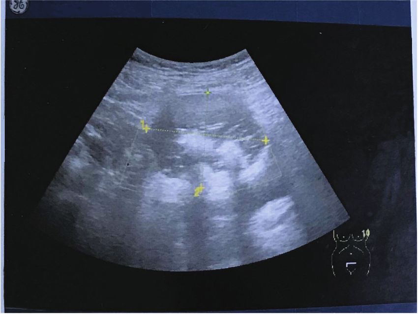
2                                                                                      Case Reports in Obstetrics and Gynecology

                                                                                       Ml 1.2   Tls 0.8 C1-5
                           03/09/18 13:27:29           ADM 15/05/81
                                                                                                        Abdomen
                                                                                                          FR    9

                                               LOGIQ                                                      CHI
                                                                                                          Frq     4.0
                                                 E9
                                                                                                       0-Gn        71
                                                                                                          D      12.0
                                                                                                        -AO%      100
                                                                                                        -
                                                                                                           CF
                      15                                                                                - Frq     2.5
                                                                                                           Gn    19.5
                                                                                                        - L/A     1/7
                                                                                                           PRF    1.0
                                                                                                        5-FO       55
                                                                                                        HS/P     3/16
                                                                                                          AO%     100
                                                                                                         -
                      −15                                                                               -
                     cm/5
                                                                                                        -

                                                                                                      10−
                                                                                                        -
                                                                                                        -

                    1 L 68.8mm
                    2 L 55.0mm
                                                                                       Ml 1.2 Tls 0.8 C1-6

Figure 1: Pelvic mass of 68 × 55 mm which had the features of a dermoid ovarian cyst with a heterogeneous echotexture and presence of
internal echoes with high echogenicity.

mm of suprauterine location, with intratumoral fat-like                   These secondary malignant neoplasms are most com-
signal intensity, which indicates a mature cystic teratoma.           monly squamous cell carcinomas. They are derived from
Tumor markers were within the normal range with cancer                the ectoderm [6]. The diagnosis is made postoperatively,
antigen 125 (CA 125) at 13.1 UI/mL and CA 19-9 at                     after surgical treatment of a supposedly benign dermoid cyst.
20 UI/mL.                                                             The clinical presentation of cysts including heaviness and
    A decision for laparotomy evaluation was made. The                pelvic pain is nonspecific, varies according to the tumor
surgical exploration found an ascites of low abundance and            stage, and is similar to that of benign ovarian cysts. In
a cystic mass adhering to the right ovary. The frozen section         advanced cancers with metastases, ascites and urinary and
diagnosis of the mass returned in favor of a squamous cell            digestive symptoms are usually the cause of the diagnosis [5].
carcinoma arising in mature cystic teratoma without infiltra-              While ultrasound is the radiological examination of
tion or rupture of the ovarian capsule (Figures 2 and 3). It          choice in the diagnosis and monitoring of mature teratomas,
was then decided to perform a total hysterectomy with bilat-          it does not detect signs of malignant transformation [7].
eral salpingo-oophorectomy and omentectomy. The patient               Indeed, ultrasound diagnosis suggests the common elements
was discharged from the hospital on the second day with an            of dermoid cysts, which are a solid organic formation, con-
uneventful postoperative course. After a multidisciplinary            taining an echogenic focus with distal acoustic attenuation
meeting, adjuvant chemotherapy was decided (bleomycin,                or shadow cone (related to the presence of the Rokitansky
etoposide, and cisplatin (BEP) regimen).                              nodule in the cystic cavity), formation containing hair, teeth,
                                                                      calcifications, and other atypical, hyperechoic, nonvascular-
                                                                      ized tissues [7]. The fatty and sebaceous contents are best
3. Discussion                                                         visualized on MRI and pelvic-abdominal CT scan [8]. Imag-
                                                                      ing diagnoses most of the time a presumably benign dermoid
Malignant transformation of mature teratomas is defined as             cyst except when there is an advanced cancer with penetra-
the development of carcinoma on one of the mature compo-              tion of the ovarian capsule and local spread [7, 8]. Some
nents of the dermoid cyst. It is an uncommon complication             authors also suggest the following as signs of malignancy:
that arises in less than 2% of patients [3]. This complication        adhesion to neighboring structures, the presence of nodules,
occurs most often during the postmenopausal period [2–4].             increased wall thickness, and the presence of necrosis and
The age of onset of this degeneration in our patient was 53           hemorrhage [7, 8]. Furthermore, the malignancy can also
years old, which is consistent with the literature with an            be suspected on intraoperative criteria such as age greater
average age of onset of 54 years old reported by several              than 40 years, large tumor size which can reach 20 cm, and
authors [2–4].                                                        the presence of hemorrhage and necrosis [1]; but it is only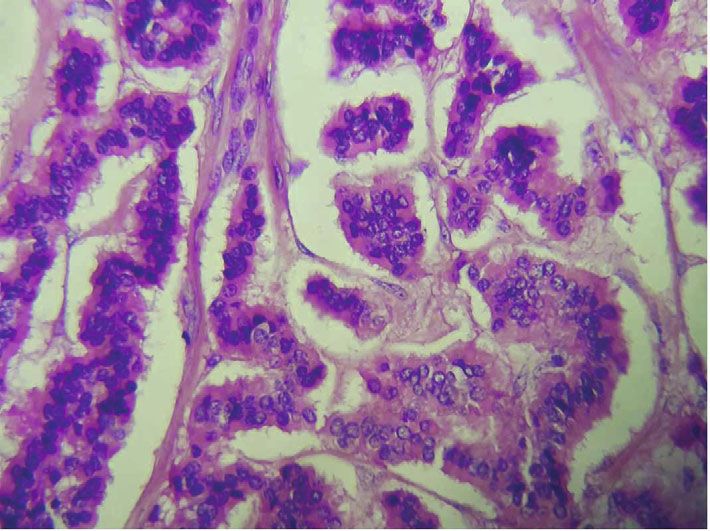
Case Reports in Obstetrics and Gynecology 3
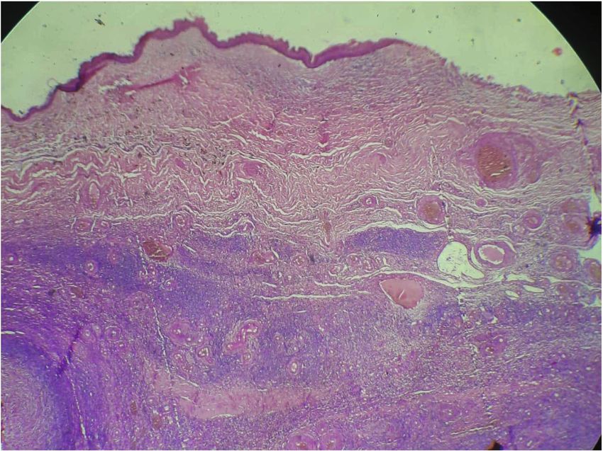
Figure 2: Cystic wall formed by a hyalinized fibrous ovarian stroma in which the covering is respiratory type with the presence of mature glial
tissue; hematoxylin and eosin staining, ×40.
ate malignant from benign ovarian tumors like in our case.
Squamous cell carcinoma (SCC) antigen may be increased
in transformed MCT associated with squamous cell carci-
noma [8, 9]. However, a low level of SCC antigen does not
formally rule out a cancerous teratoma [9]. CA 125 is a glyco-
protein secreted by the majority of serous ovarian tumors; it
is used to assess sensitivity to chemotherapy and for the diag-
nosis of recurrences. Therefore, CA 125 has a diagnostic,
prognostic, and therapeutic evaluation value [9]. Alpha-
fetoprotein (AFP) should not be interpreted as a factor of
malignancy, its production being determined by the endo-
derm tissue of the dermoid cyst. CA 19-9 may be elevated
Figure 3: High cell density tumor proliferation made up of in malignant and benign tumors [9]. In our case, tumor
anastomotic cord intercepted by monomorphic clusters with markers were within the normal range. Hackethal et al.’s
rounded nuclei or ovoid granular chromatin without mitotic meta-analysis on 277 cases of squamous cell carcinoma aris-
activity; hematoxylin and eosin staining, ×400. ing in cystic teratoma found high SCC antigen in 86.5% of
cases, high CA 125 in 71% of cases, high CA 19-9 in 77% of
cases, and ACE present in 40% of cases [9].
the histology study that confirms the degeneration of the The diagnosis of dermoid cyst is confirmed during sur-
MCT. In our case, we relied on the ultrasound and MRI gery on the macroscopic appearance with a solid ovarian cyst
results to suggest the diagnosis of mature teratoma but it is containing fat, skin, hair, and teeth. In most cases, the diag-
the frozen section that confirmed the diagnosis of a squa- nosis of malignant transformation is a surprise given by the
mous cell carcinoma arising in mature cystic teratoma. histology [8].
The tumor markers used to help characterize ovarian Surgical treatment of these malignant transformations is
lesions are not very specific and cannot be used to differenti- the same as for ovarian carcinoma: laparotomy in the majority4 Case Reports in Obstetrics and Gynecology
of cases with unilateral or bilateral adnexectomy. A second Consent
complete exploration if the frozen section was not initially
performed is necessary to respect the rules of oncology with Written informed consent was obtained from the patient for
systematic peritoneal cytology, biopsies of suspicious areas, participation and publication of this case report and any
omentectomy, total hysterectomy, and pelvic and para-aortic accompanying images. A copy of the written consent is avail-
lymphadenectomy. Chemotherapy using alkylating agents able for review by the Editor-in-Chief of this journal.
improves prognosis for advanced stages, but not the use of
radiotherapy [5–7]. In the case of young patients who wish to
maintain their fertility, having a transformed dermoid cyst Conflicts of Interest
limited to the ovarian capsule, without local or distant invasion,
conservative treatment may be possible: cystectomy or unilat- The authors declare that they have no competing interests.
eral adnexectomy with multiple peritoneal biopsies. For elderly
patients, the treatment is as for any ovarian cancer: total hyster-
ectomy with bilateral adnexectomy, omentectomy, and lymph- References
adenectomy [6–9]. Our patient underwent total hysterectomy
with bilateral salpingo-oophorectomy and omentectomy. [1] G. Ribeiro, P. Hughesdon, and E. Wiltshaw, “Squamous carci-
The prognosis depends on the stage, the presence of noma arising in dermoid cysts and associated with hypercalce-
vascular invasion, and the rupture of the ovarian capsule mia: a clinicopathologic study of six cases,” Gynecologic
[10]. To establish a prognosis, Kikkawa et al. [10] also take Oncology, vol. 29, no. 2, pp. 222–230, 1988.
into consideration the presence or absence of tumor residue; [2] P. F. Lai, S. C. Hsieh, J. C. W. Chien, C. L. Fang, W. P. Chan,
thus, the 5-year survival is 79% without tumor residue and and C. Yu, “Malignant transformation of an ovarian mature
10.1% with tumor residue. cystic teratoma: computed tomography findings,” Archives of
Gynecology and Obstetrics, vol. 271, no. 4, pp. 355–357, 2005.
[3] Y. Yamanaka, Y. Tateiwa, H. Miyamoto et al., “Preoperative
4. Conclusion diagnosis of malignant transformation in mature cystic tera-
toma of the ovary,” European Journal of Gynaecological Oncol-
Occurring preferably in the postmenopausal period, malig- ogy, vol. 26, no. 4, pp. 391-392, 2005.
nant transformation of mature cystic teratoma is a well- [4] X. Argoitia, I. Duga, E. Labeyrie, L. Toledo, C. Couteau, and
known but uncommon phenomenon. There is currently no D. Querleu, “Degeneration of dermoid cysts: a case study of
formal diagnostic criterion before the histological analysis. malignant transformation,” Gynécologie Obstétrique & Ferti-
An attempt is made to adapt the surgical aggressiveness lité, vol. 35, no. 10, pp. 1005–1008, 2007.
according to the age of the patient. Conservative surgery is [5] S. Tangjitgamol, S. Manusirivithaya, C. Sheanakul,
reserved for young women, especially nulliparous who wish S. Leelahakorn, T. Thawaramara, and S. Jesadapatarakul,
to preserve fertility. Hysterectomy and bilateral salpingo- “Squamous cell carcinoma arising from dermoid cyst: case
reports and review of literature,” International Journal of
oophorectomy are advised in postmenopausal patients.
Gynecological Cancer, vol. 13, no. 4, pp. 558–563, 2003.
[6] B. Caspi, Z. Appelman, D. Rabinerson, Y. Zalel, T. Tulandi,
and Z. Shoham, “The growth pattern of ovarian dermoid cysts:
Abbreviations a prospective study in premenopausal and postmenopausal
MCT: Mature cystic teratoma women,” Fertility and Sterility, vol. 68, no. 3, pp. 501–505,
1997.
SCC: Squamous cell carcinoma
MT: Malignant transformation [7] S. Y. Rim, S. M. Kim, and H. S. Choi, “Malignant transforma-
MRI: Magnetic resonance imaging tion of ovarian mature cystic teratoma,” International Journal
of Gynecological Cancer, vol. 16, no. 1, pp. 140–144, 2006.
CA 125: Cancer antigen 125
CA 19-9: Cancer antigen 19-9 [8] M. M. Desouki, O. Fadare, B. Chamberlain, N. Shakir, and
A. Kanbour-Shakir, “Malignancy associated with ovarian tera-
AFP: Alpha-fetoprotein
tomas: frequency, histotypes, and diagnostic accuracy of intra-
BEP: Bleomycin, etoposide, and cisplatin chemotherapy
operative consultation,” Annals of Diagnostic Pathology,
regimen. vol. 19, no. 3, pp. 103–106, 2015.
[9] A. Hackethal, D. Brueggmann, M. K. Bohlmann, F. E. Franke,
H. R. Tinneberg, and K. Münstedt, “Squamous-cell carcinoma
Data Availability in mature cystic teratoma of the ovary: systematic review and
analysis of published data,” The Lancet Oncology, vol. 9,
Supporting materials are available if further analysis is no. 12, pp. 1173–1180, 2008.
needed.
[10] F. Kikkawa, H. Ishikawa, K. Tamakoshi, A. Nawa,
N. Suganuma, and Y. Tomoda, “Squamous cell carcinoma
arising from mature cystic teratoma of the ovary: a clinico-
Ethical Approval pathologic analysis,” Obstetrics and Gynecology, vol. 89,
no. 6, pp. 1017–1022, 1997.
Ethics approval has been obtained to proceed with the
current study.You can also read