Sarcina ventriculi: A Rare Case of Life-Threatening Perforated Gastric Ulcer and Review of Literature - Journal of Gastric Surgery
←
→
Page content transcription
If your browser does not render page correctly, please read the page content below
Journal of Gastric Surgery
        Vol. 2 No. 2 (2020): Journal of Gastric Surgery, 53-56
        Case Series

Sarcina ventriculi: A Rare Case of Life-
Threatening Perforated Gastric Ulcer and
Review of Literature
  Qing Wei                             Department of Pathology, University of Alabama at
                                       Birmingham School of Medicine, Birmingham, USA
  Sixto M. Leal Jr                     Department of Pathology, University of Alabama at
                                       Birmingham School of Medicine, Birmingham, USA
  Morad Qarmali                        Department of Pathology, University of Alabama at
                                       Birmingham School of Medicine, Birmingham, USA
  Charles Mel Wilcox                   Department of Medicine, Division of Gastroenterology,
                                       University of Alabama at Birmingham School of Medicine,
                                       Birminghamn USA
  Chirag R. Patel                      Department of Pathology, University of Alabama at
                                       Birmingham School of Medicine, Birmingham, USA
  Sameer Al Diffalha                   NP 3552 North Pavilion, University of Alabama at
                                       Birmingham Hospital Birmingham, AL 35294-6823.
                                       Telephone: (205) 934-5749 Fax: (205) 975-5242

  Sarcina ventriculi is a gram-positive anaerobic bacterium reported rarely in patients with a
  history of gastrointestinal surgery and delayed gastric emptying. Sarcina has been implicated
  in the development of gastric ulcers, emphysematous gastritis, and gastric perforation. So far,
  less than 30 cases of Sarcina isolated from gastric specimens have been reported, including 3
  cases associated with life-threatening illness:
  emphysematous gastritis and gastric perforation. Herein, we report a case of a 58-yearold
  woman with history of gastric surgery who presented for evaluation of persistent gastric pain
  and incurable ulcer. She underwent total gastrectomy, and the resected stomach
  demonstrated a perforated ulcer with the presence of Sarcina microorganisms.
  We also report a second case of a 56-year-old woman with history of NSAID use who
  presented with gastric outlet obstruction. The gastric biopsy identified concurrent
  Helicobacter pylori and Sarcina. Given Sarcina's association with emphysematous gastritis
  and gastric perforation, its identification on gastric biopsies should be clearly stated in
  pathology reports and, depending on the clinical scenario, prompt clinicians to add adjunctive
  antimicrobials to anti-ulcer therapeutic regimens.

 Keywords:

Sarcina ventriculi; emphysematous gastritis; gastric perforation.

 To Cite

Wei Q, Leal SM Jr, Qarmali M, Wilcox CM, Patel CR, Al Diffalha S. Sarcina ventriculi: A Rare Case
of Life-Threatening Perforated Gastric Ulcer and Review of Literature. J Gastric Surg 2020; 2(2):
53-56

 Publication history

Received: May 10, 2020

                                                                                               1/6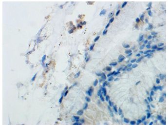
Journal of Gastric Surgery
        Vol. 2 No. 2 (2020): Journal of Gastric Surgery, 53-56
        Case Series

Accepted: May 20, 2020

Article in press: May 28, 2020

Published on line: May 30, 2020

 *Correspondence to

Sameer Al Diffalha, MD NP 3552 North Pavilion, University of Alabama at Birmingham Hospital

Birmingham, AL 35294-6823 Telephone: (205) 934-5749

Fax: (205) 975-5242

  Background:
Sarcina ventriculi is a gram-positive, non-motile, spore- forming, anaerobic bacterial coccus with a
carbohydrate fermentative metabolism.[1] Hematoxylin and Eosin (H&E) staining of gastric
biopsies reveals tetrad packets of large (3µm) basophilic cuboidal or spherical microorganisms with
a refractile wall.[1, 2] Molecular diagnosis is possible by polymerase chain reaction and sequencing
of the 16S ribosomal RNA and pyruvate decarboxylase genes.[3]

Sarcina was first identified as a human pathogen by Goodsir in 1843.[4, 5] It is a well-described
pathogen in veterinary medicine and can cause a lethal gastric bloating-like syndrome.[6] Recently
it has become increasingly implicated in human gastrointestinal disease and has been described to
be associated in patients with history of GI surgery and delayed gastric emptying.[1, 3] Most
patients exhibit GI symptoms, with some cases of severe disease, including emphysematous
gastritis and gastric perforation.[7-9] Endoscopic findings often involve retained food residue,
gastric ulcers, and inflammation or erosions.[1] Herein, we present two unique cases. One of them
is the third reported case of Sarcina associated with gastric perforation. The other presented case
is the second reported case that shows rare concurrent infections of Sarcina and Helicobacter
pylori.

  Case 1:
A 58-year-old Caucasian woman presented to the surgical oncology clinic for evaluation of a gastric
ulcer and possible total gastrectomy. She reported epigastric pain, loss of appetite, nausea, and
malnutrition. She had an open gastric bypass about 20 years prior that was taken down due to
development of an internal hernia. Afterwards, the patient suffered dysphagia, gastric reflux, and
chronic upper GI ulcers despite antacid and H. pylori treatment. Leading up to the evaluation,
esophagogastroduodenoscopy (EGD) revealed an ulcer of the posterior stomach wall distal to the
esophagogastric anastomosis, and ulcer biopsies showed benign fragments of gastric mucosa
demonstrating marked active gastritis with focal ulceration.

The patient underwent total gastrectomy, ventral hernia repair, and associated procedures.
Pathologic examination of the resected stomach showed a 2.7 cm x 2.5 cm ulcer of the proximal
lesser curvature with a 1-cm thickness perforation through the serosa (Figure 1a). H & E staining
showed multiple foci of Sarcina ventriculi tetrads (Figure 1b). Sarcina was also identified within the
gastric lumen occasionally admixed with vegetable matter, consistent with delayed gastric
emptying. Organisms were identified by the granulation tissue in the ulcer base as well as on the
serosal surface, which was consistent with perforation and the presence of organisms within the
peritoneal cavity.

The patient’s post-operative course was complicated by an anastomotic leak and polymicrobial intra-

                                                                                               2/6
Journal of Gastric Surgery
         Vol. 2 No. 2 (2020): Journal of Gastric Surgery, 53-56
         Case Series

abdominal infection that required surgical revision, antibiotics (metronidazole, vancomycin,
cefepime and piperacillin- tazobactam), and antifungal treatment. She recovered after an
approximately one-month hospitalization and was discharged.

Figure 1. Gross picture of the perforated stomach

Figure 2. The cuboid-shaped organisms were tightly packed in a tetrad formation surrounding the ulcer bed. Hematoxylin
and Eosin, 40x magnification.

  Case 2:
A 56-year-old woman presented with gastric outlet obstruction. She underwent four consecutive
outpatient upper endoscopies at a frequency of approximately once per month. The EGDs showed
an ulcer at the pre-pyloric area and a pyloric stricture, which was dilated each time. Random
biopsies of the stomach during the first EGD showed chronic active gastritis, and H. pylori was
negative by immunostaining. She started proton pump inhibitor (omeprazole 40 mg). Biopsies of
the antrum taken during her second EGD showed chronic active gastritis. Sarcina and rare H.
pylori microorganisms were identified on routine H&E stain (Figure 2a and Figure 2b). Similar to
Case 1, Sarcina species were noted in the gastric lumen admixed with vegetable matter consistent

                                                                                                               3/6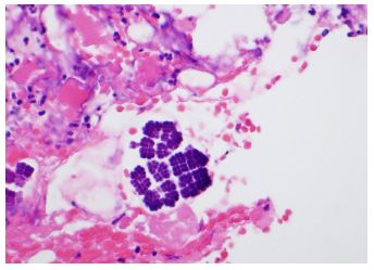
Journal of Gastric Surgery
Vol. 2 No. 2 (2020): Journal of Gastric Surgery, 53-56
Case Series
with delayed gastric emptying. By the third EGD, the ulcer had healed after the treatment. By the
fourth EGD, the patient was eating better without nausea or weight loss, and there was no further
follow-up.
Figure 3. H. pylori and Sarcina organisms co-exist in a gastric biopsy. The cuboid-shaped organisms were tightly packed in
a tetrad formation. Hematoxylin and Eosin, 40x magnification
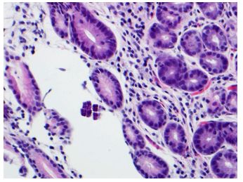
Figure 4. H. pylori and Sarcina organisms co-exist in a gastric biopsy. H. pylori were detected by anti-H pylori
immunostain (60x magnification).
Discussion:
Sarcina is a gram-positive, anaerobic, acid-tolerant coccus. It is associated with GI surgery and
exhibits a 2:1 female-to-male ratio.[1] The patients in these cases were both female; the first had a
history of GI surgery, and the second had a pyloric stricture.
The pathogenic role of Sarcina in humans remains unclear and can be an incidental finding in
gastric biopsies.[3, 10] In patients with delayed gastric emptying, retained carbohydrates and other
nutrients provide a fermentative substrate for Sarcina overgrowth.
4/6Journal of Gastric Surgery
Vol. 2 No. 2 (2020): Journal of Gastric Surgery, 53-56
Case Series
[1] In livestock, fermentative byproducts can cause deadly emphysematous gastric bloating. In
humans, fermentation byproducts may increase the risk of developing life-threatening
complications, such as emphysematous gastritis and gastric perforation.
Our first case supports the idea that Sarcina contributes to the development of gastric ulcer
perforation. The only other two reported cases of perforation were a 14-year-old male with a history
of bowel reduction due to malrotation of the small intestine [8] and a 76-year- old male with acute
abdominal pain who presented in the ER and died after surgery.[9] Our patient had a history of
gastric surgery and an ulcer resistant to H. pylori therapy. The pre-existing ulcer and delayed
gastric emptying might have provided a nidus for Sarcina overgrowth. Morphologic findings of
Sarcina microorganisms in both the gastric lumen and the serosa are consistent with clinically,
macroscopically, and microscopically confirmed perforated gastric ulcer. As Sarcina can cause
deadly emphysematous bloating in animals, it is possible that their association with human GI
pathology is not due to direct tissue invasion but rather fermentation byproducts produced by the
organism in the carbohydrate-rich gastric lumen.
Our second case demonstrates a rare example of Sarcina co-occurring with H. pylori. Only Sauter
and colleagues have previously shown co-existence of Sarcina withH. pylori in two siblings.[11]
Both H. pylori and Sarcina can survive in an acidic environment, and H. pylori infection can cause
delayed gastric emptying via smooth muscle relaxation from release of leukotrienes or nitric
oxide.[12] Although H. pylori could, in theory, be a predisposing factor in Sarcina infection, their
rare co- occurrence may be due to eradication of Sarcina by antiH. pylori treatment.
Currently, there is no consensus on treatment regimens for Sarcina.[13] Published regimens
leading to successful outcomes include anti-ulcer therapy and adjunctive use of metronidazole with
a second antibiotic (most commonly ciprofloxacin).
It is very helpful if clinicians provide clinical information about any history of delayed gastric
emptying along with the tissue that been submitted for pathology. This clinical history will alert GI
pathologists to rule out the presence of Sarcina which is usually located near the gastric mucosal
surface and has a pathognomonic tetrad morphology. To increase awareness of Sarcina’s
association with severe disease, pathologists may include the following statement in their reports:
“Sarcina has been reported to be present in association with emphysematous gastritis and gastric
perforation.”
Acknowledgements
None
Contributors
Each of the listed authors made a substantial contribution to the conception, design, and revision of
this study. All authors read and approved the final manuscript.
Funding
No funding was received for this study.
Competing interests
No benefits in any form have been received or will be received from a commercial party related
directly or indirectly to the subject of this article.
Availability of data and materials
5/6Journal of Gastric Surgery
Vol. 2 No. 2 (2020): Journal of Gastric Surgery, 53-56
Case Series
Further information is available from the corresponding author on reasonable request.
Ethics approval
Not applicable
Provenance and peer review
Not commissioned; externally peer reviewed.
Open access
This is an Open Access article distributed in accordance with the Creative Commons Attribution
Non- Commercial (CC BY-NC 4.0) license, which permits others to distribute, remix, adapt, build
upon this work noncommercially, and license their derivative works on different terms, provided
the original work is properly cited and the use is non-commercial. See: licenses/by-nc/4.0/
References
1. Al Rasheed MR, Senseng CG. Sarcina ventriculi: Review of the Literature. Arch Pathol Lab
Med. 2016;140:1441-5.
2. Behzadi J, Modi RM, Goyal K, Chen W, Pfeil S. Sarcina ventriculi as an Unknown Culprit for
Esophageal Stricturing. ACG Case Rep J. 2017;4:e118.
3. Lam-Himlin D, Tsiatis AC, Montgomery E, Pai RK, Brown JA, Razavi M, et al. Sarcina
organisms in the gastrointestinal tract: a clinicopathologic and molecular study. Am J Surg
Pathol. 2011;35:1700-5.
4. Goodsir J. XXIII: History of a case in which a fluid periodically ejected from the stomach
contained vegetable organisms of an undescribed form. J Nat Hist. 1843;11:125-6.
5. Beijerinck M. An experiment with Sarcina ventriculi. Huygens Institute-Royal Netherlands
Academy of Arts and Sciences (KNAW), Proceedings. 1911;13:1910-1.
6. DeBey BM, Blanchard PC, Durfee PT. Abomasal bloat associated with Sarcina-like bacteria
in goat kids. J Am Vet Med Assoc. 1996;209:1468-9.
7. Laass MW, Pargac N, Fischer R, Bernhardt H, Knoke M, Henker J. Emphysematous gastritis
caused by Sarcina ventriculi. Gastrointest Endosc. 2010;72:1101-3.
8. Tolentino LF, Kallichanda N, Javier B, Yoshimori R, French SW. A case report of gastric
perforation and peritonitis associated with opportunistic infection by Sarcina ventriculi.
Laboratory Medicine. 2003;34:535-7.
9. Dumitru A, Aliuş C, Nica AE, Antoniac I, Gheorghiţă D, Grădinaru S. Fatal outcome of
gastric perforation due to infection with Sarcina spp. A case report. IDCases.
2020;19:e00711.
10. Haroon Al Rasheed MR, Kim GJ, Senseng C. A Rare Case of Sarcina ventriculi of the
Stomach in an Asymptomatic Patient. Int J Surg Pathol. 2016;24:142-5.
11. Sauter JL, Nayar SK, Anders PD, D’Amico M, Butnor KJ, Wilcox RL. Co-existence of Sarcina
Organisms and Helicobacter pylori Gastritis/Duodenitis in Pediatric Siblings. J Clin Anat
Pathol (JCAP). 2013;1.
12. Zhang CL, Geng CH, Yang ZW, Li YL, Tong LQ, Gao P, et al. Changes in patients' symptoms
and gastric emptying after Helicobacter pylori treatment. World J Gastroenterol.
2016;22:4585-93.
13. Medlicott SAC. Sarcina ventricularis complicating a patient status post vertical banded
gastroplasty, a case. Journal of Gastroenterology and Hepatology Research. 2015;4:1481-4.
6/6
Powered by TCPDF (www.tcpdf.org)You can also read