INTRODUCTION TO CHILDHOOD LEUKAEMIA - B-608 Mel Greaves and Donald Pinkel
←
→
Page content transcription
If your browser does not render page correctly, please read the page content below
July 10, 2008   12:25         9in x 6in       B-608     ch01

                INTRODUCTION TO CHILDHOOD
                        LEUKAEMIA

                        Mel Greaves and Donald Pinkel

                                          1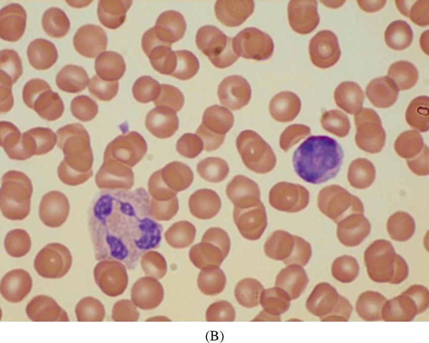
July 10, 2008   12:25        9in x 6in             B-608     ch01

                        This page intentionally left blank

                                         2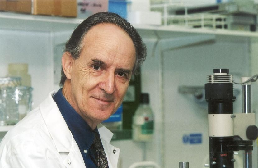
July 10, 2008   12:25           9in x 6in            B-608              ch01

                                                                               3

                 INTRODUCTION TO CHILDHOOD
                         LEUKAEMIA

                          Mel Greaves and Donald Pinkel

        Leukaemia is a cancer of blood cells, as are lymphoma and myeloma.
        In common with all cancers, they are cellular disorders driven by
        mutations or modifications of DNA, our genetic code. In an individual
        patient with leukaemia all the leukaemic cells are a clone; the progeny
        of a single, disordered blood cell.
            Leukaemia accounts for a modest fraction of adult cancers (∼ 7%)
        and, although substantially rarer (by 10 fold) in children, leukaemia
        is the major type of paediatric cancer, accounting for around one third
        of all cases. Cancer occurs throughout the animal kingdom. Several
        mammalian species, especially domesticated cats, cattle and chickens
        develop leukaemia. Medical records from antiquity in Greece and
        India backtrack the presence of cancers from more than 2000 years
        ago. In that sense, cancer is not a modern disease, as often asserted.
        But some cancers are easier to detect than others e.g. those of the
        breast and skin for example. Leukaemia posed a problem in this
        respect. It may well have existed at some level throughout human
        history but in the absence of microscopy, this “liquid” cancer would
        have escaped detection. Patients with leukaemia then would have died
        of infection or haemorrhage without a diagnosis. It was therefore not
        until the mid-19th century (with the emergence of microscopy and
        cellular pathology) that leukaemia was first observed.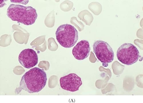
July 10, 2008   12:25                 9in x 6in                 B-608            ch01

        4                              M. Greaves and D. Pinkel

                        Fig. 1.   Rudolf Virchow, German cellular pathologist.

           In 1845, a young medical doctor, Rudolf Virchow (Fig. 1),
        observed a patient with complaints of weakness and pallor. The
        patient had a large spleen and her blood was near white because of
        very few red blood cells and excessive white blood cells. He reported
        this case in a medical journal classifying it as a case of “white blood”.
        He thought it was a new disease. His seniors doubted this idea; they
        considered this patient a victim of infectious disease. The high white
        blood cell levels were a reaction to infection. This opinion was similar
        when a French and two contemporary Scottish physicians published
        similar case reports.
           But unlike the others, young Dr Virchow embarked on an intensive
        investigation of his “new” disease. He gathered more cases of this
        fatal disorder and studied their gross and microscopic anatomical
        findings as well as clinical features. A decade later he published a
        detailed monograph including meticulous hand drawings of what he
        now called “leukaemia”, Greek for white blood. From these obser-
        vations, he postulated the cellular theory of leukaemia. The disease
        was caused by uncontrolled replication of a disordered blood cell pre-
        cursor with growth and survival advantages over normal blood cell
July 10, 2008   12:25           9in x 6in                B-608         ch01

                           Introduction to Childhood Leukaemia                5

        precursors. This unregulated proliferation eventually interfered with
        normal body function, resulting in the death of the patient. Virchow’s
        theory remains essentially correct 150 years later. It was elaborated
        by newer discoveries in cell and molecular biology and genetics as
        highlighted elsewhere in this book.
            Most of the cases of leukaemia described by Rudolph Virchow
        and other European pathologists in the mid-19th century were adults.
        From their brief descriptions and rather superficial microscopic evi-
        dence, it was probable that most of these patients were suffering
        from chronic myeloid leukaemias. A more detailed classification
        of leukaemia awaited the discoveries of Paul Ehrlich in the 1880s.
        Ehrlich discovered aniline dyes that could stain blood films which
        distinguish the morphology of different types of myeloid and lym-
        phoid blood cells. Ehrlich, and later the Swiss haematologist Naegeli,
        had the insight that clinically acute leukaemia involved “primitive”
        cells from the bone marrow that developed into distinctive myeloid
        and lymphoid lineages (Fig. 2). Thus was coined the terms acute
        lymphoblastic leukaemia (ALL) and acute myeloblastic leukaemia
        (AML). Anecdotal reports of leukaemia in children appeared in the
        late 19th century. By the turn of the century (1904), Churchill, a
        physician in Chicago, had described a series of children (15 cases)
        diagnosed with ALL. Their duration of disease before death was short
        (a few days to a few months) and the age range was newborn to
        10 years.
            The classification of acute leukaemia in children and adults are
        usually two major varieties, ALL and AML. This remained grounded
        in the simple morphology of the dye-stained cells until the 1970s,
        when immunologists introduced antibodies that could reliably distin-
        guish different types of morphologically anonymous lymphoid cells.
        Consequently, ALL was subdivided into subsets corresponding to B
        lineage precursors and T lineage precursors. In the 1980s, scientists
        carried out subcellular dissection of leukaemic cell diversity at the
        level of chromosome structure and then at the level of DNA. They
        discovered the critical and diverse underlying molecular patholology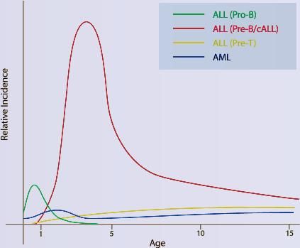
July 10, 2008 12:25 9in x 6in B-608 ch01
6 M. Greaves and D. Pinkel
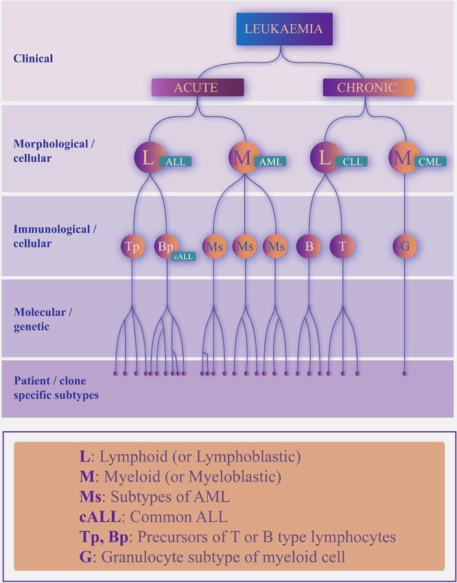
Fig. 2. Leukaemic (ALL) (A) versus normal blood (B). Courtesy of Dr B Bain. (A) The
nucleus of the leukaemic cells is stained red by special dyes. (B) Two normal white cells in
sea of smaller red cells. Large cell lower left is granulocyte. Round cell at 3.o’clock with
purple stained nucleus is normal lymphocyte.
that drives the diseases. The classification that emerges from these
sequential insights over the decades is a branching tree-like structure
(Fig. 3) used in the haematology laboratories of many hospitals.
Modern human genome scanning methods indicate that each
patient’s leukaemic cells have followed a unique evolutionary tra-
jectory as a novel subspecies of cell. The consequence is that eachJuly 10, 2008 12:25 9in x 6in B-608 ch01
Introduction to Childhood Leukaemia 7
Fig. 3. Hierarchical classification of leukaemia.
patient’s leukaemia ends up represented by an individual leaf on a
specific twig or a particular branch of the tree. The prevalence of the
major branching subtypes of childhood leukaemia vary according to
age. The peak incidence of disease at ages two to five years old reflects
mostly the “common” or B cell precursor variant of ALL (Fig. 4).
Most cancers probably harbour a similar degree of complexity
which, in part at least, may explain some of their general intransi-
gence to successful therapy — they are not one disease. The degreeJuly 10, 2008 12:25 9in x 6in B-608 ch01
8 M. Greaves and D. Pinkel
Fig. 4. Age distribution of main subtypes of childhood leukaemia.
of cellular, chromosomal and molecular diversity in leukaemia raises
some difficult but important practical issues and opportunities. At
what level of cellular and molecular characteristics is response to
therapy determined? This must surely depend upon the particular
therapy itself which has been continually modified and tailored to
cellular subtype. Currently in leukaemia, and cancer in general, it
appears that abnormal genotype, i.e. altered chromosomes and genes,
has a major impact on clinical response and outcome. This can be
rationalised as we now know that the altered or mutated genome
influences the signal pathways in cells that control life or death of
those cells in the face of therapeutic drugs or irradiation. The pat-
terns of distinctive genetic change in leukaemic cells also provide
potentially ideal therapeutic targets since they uniquely distinguish
leukaemic from normal cells. But, this in turn poses a very consid-
erable dilemma for cancer and leukaemia therapeutics. Do we designJuly 10, 2008 12:25 9in x 6in B-608 ch01
Introduction to Childhood Leukaemia 9
and tailor novel therapeutics to match each patient — with all the
technical and major financial implications this would entail — or do
we continue to strive to identify shared, generic features of leukaemia
that provide common therapeutic targets? Currently, both avenues
are being explored very actively.
Childhood acute leukaemia occurs throughout the world, though it
appears that the incidence rate of ALL is significantly higher (perhaps
by 10 fold) in more affluent or developed countries. In the latter, the
annual incidence rate is at around 30 to 40 cases per one million
children which, for paediatric populations in the UK or the USA,
translates to around 450 or 2,750 new cases each year respectively.
In absolute terms, the risk of any child developing acute leukaemia
between birth and age 15 years (in Europe, Australia or the USA) is
approximately one in 2000.
The initial, clinical diagnosis of childhood leukaemia relates to
the pathology, the common symptoms being paleness (anaemia),
bleeding tendency, fatigue, aches and pains and unexplained fevers.
Collectively, they reflect the highjack of normal bone marrow func-
tion by leukaemic cells. Treatment, as described in the chapter by
Donald Pinkel, is now relatively complex and consists of chemo-
therapy with several drugs in different combinations and various
schedules for approximately 3 years. In the case of Janine (Chapter 6),
this was effective and curative. When standard chemotherapy fails,
intensive chemotherapy using drugs and/or drug schedules not
included in the initial treatment can be curative in many children.
Very high dose chemotherapy followed by haematopoietic stem
cell transplant to replenish the child’s blood cell system with donor
graft cells (and produce an immune reaction to the leukaemia) has
been used for over three decades. This procedure can be curative
but is associated with a relatively high procedure-related early
death rate and long term disability and late death. This negative
outcome is largely due to reaction of the donor immune system
to the child’s normal organs and tissues but also to side effects of
the drugs used to suppress the reaction. This was the case, sadly,July 10, 2008 12:25 9in x 6in B-608 ch01
10 M. Greaves and D. Pinkel
for Georgie whose story is told in Chapter 5. Meaningful compar-
isons of haematopoietic stem cell transplant methods versus intent to
cure “salvage chemotherapy” for children with relapsed ALL have
been conducted in the United Kingdom and the United States. They
demonstrate no advantage in cure or quality of survival for transplant
methods.
Despite the success in treating childhood leukaemia (cure rates
are reported to be 80%) for some children with ALL, much remains
to be done. Closer attention is needed to monitoring the effects of
each element of treatment on growth and development of the children
to healthy, happy, fulfilled adults. The eventual risk/benefit ratio of
each element must be weighed and reweighed in deciding on current
treatment.
The challenges for the future lie in the development of more
specific biologically targeted treatment and in preventive measures
derived from the understanding of the causes and mechanisms of
leukaemia. Perhaps then, the many children worldwide currently
without access to curative treatment will be reached.You can also read