Squamous Cell Carcinoma Ex Pleomorphic Adenoma of the Parotid Gland: Unusual Entity and Diagnostic Pitfalls
←
→
Page content transcription
If your browser does not render page correctly, please read the page content below
CANCER DIAGNOSIS & PROGNOSIS
1: 279-283 (2021)                                                                                         doi: 10.21873/cdp.10036

Squamous Cell Carcinoma Ex Pleomorphic Adenoma of
the Parotid Gland: Unusual Entity and Diagnostic Pitfalls
XIAOQIN LIU, XIAOYAN LIAO and DONGWEI ZHANG

Department of Pathology and Laboratory Medicine, University of Rochester Medical Center, Rochester, NY, U.S.A.

Abstract. Background: Carcinoma ex pleomorphic                       salivary glands with a majority of cases noted in the parotid
adenoma (PA) of the salivary gland with squamous cell                and submandibular glands, and 20% of cases seen in minor
carcinoma (SCC) component is extremely rare and can be               salivary glands (4). CXPA can be asymptomatic and has a
easily misdiagnosed as a benign PA or SCC (primary or                similar clinical presentation as PA before it becomes widely
metastatic). Case Report: A 75-year-old male who had no              invasive. Patients frequently become aware of the cancer when
significant past medical history, presented with a rapid             they experience rapid enlargement of the mass, pain, or other
growing right parotid gland mass. A fine needle aspiration           clinical symptoms. Facial nerve involvement is present in one
revealed malignant cells. Following partial parotidectomy, a         third of cases (5). The clinical presentation may resemble a
2.4 cm ill-defined mass was grossly identified.                      multiple facial nerve schwannoma. Increased preoperative
Microscopically, it showed a keratinizing SCC with adjacent          duration of a PA increases the risk of malignant transformation
component of residual PA. Immunohistochemically, the                 into CXPA. Treatment for CXPA often involves a complete
malignant tumor cells were positive for p40, p63 and CK5.            surgical resection followed by adjuvant radiotherapy.
The residual PA was focally positive for CAM5.2, SMA, p63               The malignant components of CXPA can be divided into
and S100. The pathological features were consistent with             epithelial component only, myoepithelial component only or
SCC ex PA. The patient was well at the 7 month-follow-up             both, of which, adenocarcinoma not otherwise specified, and
post-surgery. Conclusion: SCC ex PA is a rare entity that            salivary ductal carcinoma are most common, but squamous
can be mistaken for a benign PA with squamous metaplasia,            cell carcinoma (SCC) is uncommon (1, 5, 6). We herein,
or primary or metastatic SCC. It behaves aggressively and            report an extremely rare case of CXPA of the parotid gland
has high recurrence and metastasis rate. Awareness of this           with an unusual malignant component of SCC, review the
disease and the diagnostic pitfalls are essential to avoid           literature, and discuss diagnostic pitfalls and prognosis.
misinterpretation in difficult cases.
                                                                     Case Report
Carcinoma ex pleomorphic adenoma (CXPA) is defined as a
carcinoma arising from a primary (de novo) or recurrent              A 75-year-old male with no known pre-existing PA presented
benign pleomorphic adenoma (PA) (1, 2). It accounts for 3%-          with a right cheek lump that he had noticed 3 weeks earlier. A
5% of all salivary gland neoplasms and 12% of all salivary           computerized tomographic (CT) scan revealed a 2.6 cm parotid
malignancy (2, 3). Eighty percent of CXPA occur in major             mass in the superficial lobe extending to abut the
                                                                     retromandibular vein and an enlarged lymph node nearby
                                                                     (Figure 1). A fine needle aspiration of the mass identified
This article is freely accessible online.                            malignant cells with squamous cell differentiation.
                                                                     Subsequently, the superficial lobe and portions of the deep lobe
Correspondence to: Dongwei Zhang, MD, Ph.D., Department of           of the parotid gland were surgically removed. Macroscopically,
Pathology and Laboratory Medicine, University of Rochester Medical   a 2.4 cm ill-defined mass was identified. The tumor had grey-
Center, 601 Elmwood Avenue, Rochester, New York 14642, U.S.A.        white, fibrotic cut surface. No necrosis was grossly identified.
Tel: +1 5852765653, e-mail: dongwei_zhang@urmc.rochester.edu
                                                                     Given the squamous cell differentiation noted by fine needle
Key Words: Carcinoma ex pleomorphic adenoma, squamous cell           aspiration, the entire specimen was submitted for histologic
carcinoma, parotid gland.                                            examination. Microscopic examination revealed an ill-defined
                                                                     invasive tumor composed of predominantly keratinizing
©2021 International Institute of Anticancer Resarch                  squamous cells with areas of a poorly differentiated carcinoma
www.iiar-anticancer.org                                              component. Within the tumor, a 2 mm hyalinized lesion

                                                                                                                                 279
CANCER DIAGNOSIS & PROGNOSIS 1: 279-283 (2021)
Figure 1. Radiological findings of the right parotid gland mass. Computed tomographic scan showing a 2.6 cm parotid mass (arrows) in the
superficial lobe extending to abut the retromandibular vein. (A) Axial view; (B) Coronal view.
composed of bland epithelial cells with a focal ductal structure SCC component in 4 out of 38 cases of CXPA. In 33 cases of
in a hyalinized and fibrotic stroma was identified, suggesting CXPA reported by Suzuki et al. (8) and 21 cases of CXPA
a typical PA (Figure 2A and B). The invasive SCC showed described by Lim et al. (9), only one case each was classified
perineural invasion, but no lymphovascular invasion or lymph as SCC. In contrast, Lewis et al. (6) found no cases with SCC
node metastasis was identified. Resection margin was involved component in 73 cases of CXPA, nor did Tortoledo et al. (10)
by SCC. Immunohistochemically, all malignant tumor cells among 37 cases of CXPA. SCC, as the pure malignant
were positive for p40 (Figure 2C), p63, and CK5, whereas component of CXPA, thus, seems to be rare; only a few such
negative for CK7, confirming squamous differentiation. cases are reported to our knowledge (Table I) (11-17). In our
Immunohistochemical staining performed on the tiny focus of case, a component of SCC was immediately recognized. The
hyalinized lesion showed that the epithelial (ductal) cells were differential diagnosis would be metastatic SCC (more
positive for CAM5.2. Some tumor cells were positive for common), primary parotid gland SCC and SCC ex PA. The
SMA, p63, and S100, confirming the presence of myoepithelial patient had no history of SCC, therefore metastatic SCC is
cells (Figure 2D-F). The above findings confirmed PA of this unlikely. Both primary SCC and SCC ex PA in the salivary
small hyalinized lesion. glands are extremely rare. Presence of residual PA is a key to
Overall, the histological features and immunoprofile distinguish SCC ex PA from primary SCC. In our case, with
supported the diagnosis of SCC ex PA, American Joint extensive sampling, a tiny focus of well-circumscribed nodule
Committee of Cancer (AJCC 8th edition) pathologic stage was noted within SCC, which was morphologically and
pT2N0. Postoperatively, the patient underwent radiation therapy immunohistochemically consistent with PA. The PA
for 6 weeks because of positive margin, perineural invasion, and component showed extensive hyalinization, which was a
poorly-differentiated histology. At 7 months follow-up post- significant predictor of malignant transformation in PA noted
surgery, he was well with no tumor recurrence or metastasis. in a study of atypical mixed tumors (18).
A slow growing parotid mass that has recently exhibited a
Discussion growth spurt should raise the suspicion of a malignancy (e.g.,
CXPA). CXPA can be mistaken for a benign PA. In PAs with
CXPA is defined as any epithelial malignancy arising in extensive sclerosis or hyalinization, additional sampling is
association with benign primary or recurrent PA. Carcinoma necessary to exclude a malignant component. It can also be
components of CXPA are often adenocarcinoma not otherwise misdiagnosed as another salivary gland malignancy, as
specified, salivary duct carcinoma, or myoepithelial carcinomas frequently overgrow and replace the benign area of
carcinoma. SCC ex PA is rare. Seifert et al. (7) identified an PA. Molecular testing for PLAG1 and HMGA2 rearrangement
280Liu et al: Squamous Cell Carcinoma Ex Pleomorphic Adenoma
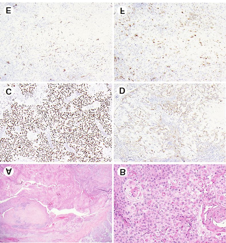
Figure 2. Histological features and ancillary studies of the right parotid gland mass. (A) Low-power view of the tumor showing hyalinized
pleomorphic adenoma (upper left) surrounded by squamous cell carcinoma and adjacent normal parotid gland tissue [hematoxylin and eosin (H&E),
×20]. (B) High-power view displaying keratinizing squamous cell carcinoma component (H&E, ×100). (C) The carcinoma component was strongly
positive for p40 by immunohistochemistry (×100). (D-F) The pleomorphic adenoma cells were positive for CAM 5.2 (D), SMA (E) and S100 (F) by
immunohistochemistry (×100).
(19, 20), known as major cytogenetic abnormality in PA, may The prognosis of CXPA is thought to be worse than that
help identify previous PA. In our case, SCC was easily of other salivary gland malignancies, with a survival rate
recognized as the malignant component, but the challenge is to varying from 25% to 65% (6, 8, 9). Tumor stage, grade,
determine whether it is primary or metastatic SCC. Primary extent of invasion, tumor size, proportion of carcinoma, high
SCC is extremely rare. Therefore, whenever SCC component proliferation index, positive margin, and perineural invasion
is identified in a salivary gland, extensive tumor sampling, are well known significant prognostic factors (6). In addition,
careful histologic assessment and scrutiny of patient’s medical some studies have revealed that histologic subtype is closely
history are required to rule out SCC ex PA vs. metastasis before related to clinical outcomes. The survival rates for patients
making a diagnosis of primary SCC. with invasive salivary duct carcinoma or adenocarcinoma not
281CANCER DIAGNOSIS & PROGNOSIS 1: 279-283 (2021)
Table I. Summary of reported squamous cell carcinoma ex pleomorphic adenomas including the current case.
Author Case, n Histology subtype Follow-up, years
SDC Adenocarcinoma NOS SCC Others
Seifert et al. (7) 38 N/A N/A 4 N/A N/A
Suzuki et al. (8) 33 8 16 1 8 Recurrence (11), metastasis (9), death (10), 3 years
Lim et al. (9) 21 10 0 1 10 Recurrence (7), metastasis (5), death (12), 68 months
Lewis et al. (6) 73 24 31 0 18 Recurrence (15), metastasis (66), death (36); 14 months-17 years
Tortoledo et al. (10) 37 13 9 0 15 Death (18)
Zbären et al. (17) 24 4 6 1 13 Recurrence (5), metastasis (6), death (5); 12 months-10 years
Ita et al. (16) 1 0 0 1 0 N/A
Mitate et al. (15) 1 0 0 1 0 No recurrence, 6 years
Current case 1 0 0 1 0 No recurrence, 7 months
SDC: Salivary duct carcinoma; NOS: not otherwise specified; SCC: squamous cell carcinoma; N/A: not available.
otherwise specified was found to be significantly poorer than 3 Antony J, Gopalan V, Smith RA and Lam AK: Carcinoma ex
other subtypes (8). Loco-regional recurrence is considered to pleomorphic adenoma: a comprehensive review of clinical,
be a major prognostic factor for patients with CXPA. Olsen pathological and molecular data. Head Neck Pathol 6(1): 1-9,
2012. PMID: 21744105. DOI: 10.1007/s12105-011-0281-z
et al. noted that the prognosis after detection of progression
4 Expertpath. 2021. Available at: https://app.expertpath.com/
or recurrence was poor, with a median survival of less than document/carcinoma-ex-pleomorphic-adenoma/21cae3e7-aa24-
1 year (5). All disease specific deaths occurred within 6 4be5-8ae7-87db5af42310?searchTerm=CXPA [Last accessed on
years after the initial operation. July 5, 2021]
In conclusion, SCC ex PA is a rare entity with significant 5 Olsen KD and Lewis JE: Carcinoma ex pleomorphic adenoma:
clinical and pathological relevance. Awareness of this entity a clinicopathologic review. Head Neck 23(9): 705-712, 2001.
and its diagnosis pitfalls are critical, as patients could PMID: 11505478. DOI: 10.1002/hed.1100
6 Lewis JE, Olsen KD and Sebo TJ: Carcinoma ex pleomorphic
potentially be inappropriately discharged without follow-up
adenoma: pathologic analysis of 73 cases. Hum Pathol 32(6): 596-
if mistakenly diagnosed with a benign PA. It generally 604, 2001. PMID: 11431714. DOI: 10.1053/hupa.2001.25000
displays aggressive behavior and has high recurrence and 7 Seifert G, Schulz J and Donath K: A pathological sub-classification
metastatic rates. Early diagnosis, adequate removal of of carcinoma of salivary gland pleomorphic adenoma. An analysis
neoplasms and careful follow-up remain the best strategy for of 38 cases (author’s transl). HNO 25(10): 337-348, 1977. PMID:
patients with CXPA. 199562.
8 Suzuki M, Matsuzuka T, Saijo S, Takahara M, Harabuchi Y, Okuni
Conflicts of Interest T, Himi T, Kakizaki T, Fukuda S, Yamada K, Nagahashi T, Abe
T, Shinkawa H, Katagiri K, Sato H, Fukui N, Ishikawa K, Suzuki
T, Kobayashi T, Saito D, Saijo S, Tateda M, Hashimoto S, Ishida
The Authors have no conflicts of interest to declare in relation to
A, Kakehata S, Suzuki O, Hashimoto Y and Omori K: Carcinoma
this study.
ex pleomorphic adenoma of the parotid gland: a multi-institutional
retrospective analysis in the Northern Japan Head and Neck
Authors’ Contributions Cancer Society. Acta Otolaryngol 136(11): 1154-1158, 2016.
PMID: 27295405. DOI: 10.1080/00016489.2016.1191671
Xiaoqin Liu, Xiaoyan Liao and Dongwei Zhang contributed to the 9 Lim CM, Hobson C, Kim S and Johnson JT: Clinical outcome
design and implementation of the study, the analysis of the results of patients with carcinoma ex pleomorphic adenoma of the
and the writing of the article. parotid gland: a comparative study from a single tertiary center.
Head Neck 37(4): 543-547, 2015. PMID: 24677516. DOI:
10.1002/hed.23638
References 10 Tortoledo ME, Luna MA and Batsakis JG: Carcinomas ex
pleomorphic adenoma and malignant mixed tumors.
1 Gnepp DR: Malignant mixed tumors of the salivary glands: a Histomorphologic indexes. Arch Otolaryngol 110(3): 172-176, 1984.
review. Pathol Annu 28 Pt 1: 279-328, 1993. PMID: 8380049. PMID: 6322732. DOI: 10.1001/archotol.1984.00800290036008
2 Nouraei SA, Hope KL, Kelly CG, McLean NR and Soames JV: 11 Kim KM, Lee A, Yoon SH, Kang JH and Shim SI: Carcinoma
Carcinoma ex benign pleomorphic adenoma of the parotid gland. ex pleomorphic adenoma of the palate – a case report. J Korean
Plast Reconstr Surg 116(5): 1206-1213, 2005. PMID: 16217459. Med Sci 12(1): 63-66, 1997. PMID: 9142663. DOI: 10.3346/
DOI: 10.1097/01.prs.0000181654.68120.0f jkms.1997.12.1.63
282Liu et al: Squamous Cell Carcinoma Ex Pleomorphic Adenoma
12 Garth RJ: Squamous liver metastases from a carcinoma arising 17 Zbären P, Zbären S, Caversaccio MD and Stauffer E: Carcinoma
within a pleomorphic adenoma of the parotid gland. J Laryngol ex pleomorphic adenoma: diagnostic difficulty and outcome.
Otol 104(2): 152-153, 1990. PMID: 2157786. DOI: 10.1017/s00 Otolaryngol Head Neck Surg 138(5): 601-605, 2008. PMID:
22215100112137 18439465. DOI: 10.1016/j.otohns.2008.01.013
13 Iino M, Yamada H, Ishikawa H, Suzuki M, Shomura E, Ide F, 18 Auclair PL and Ellis GL: Atypical features in salivary gland
Saito I and Mori Y: Carcinoma ex pleomorphic adenoma of the mixed tumors: their relationship to malignant transformation.
submandibular gland: report of a case with an unusual malignant Mod Pathol 9(6): 652-657, 1996. PMID: 8782203.
component of clear cell squamous cell carcinoma. Oral Surg 19 Martins C, Fonseca I, Roque L, Pereira T, Ribeiro C, Bullerdiek
Oral Med Oral Pathol Oral Radiol Endod 106(2): e30-e34, 2008. J and Soares J: PLAG1 gene alterations in salivary gland
PMID: 18554943. DOI: 10.1016/j.tripleo.2008.04.023 pleomorphic adenoma and carcinoma ex-pleomorphic adenoma:
14 Nakamori K, Ohuchi T, Hasegawa T and Hiratsuka H: a combined study using chromosome banding, in situ
Carcinoma ex pleomorphic adenoma of the buccal region is hybridization and immunocytochemistry. Mod Pathol 18(8): 1048-
composed of salivary duct carcinoma and squamous cell 1055, 2005. PMID: 15920557. DOI: 10.1038/modpathol.3800386
carcinoma components. Int J Oral Maxillofac Surg 38(10): 1116- 20 Röijer E, Nordkvist A, Ström AK, Ryd W, Behrendt M, Bullerdiek
1118, 2009. PMID: 19467841. DOI: 10.1016/j.ijom.2009.04.016 J, Mark J and Stenman G: Translocation, deletion/amplification,
15 Mitate E, Kawano S, Kiyoshima T, Kawazu T, Chikui T, Goto and expression of HMGIC and MDM2 in a carcinoma ex
Y, Matsubara R and Nakamura S: Carcinoma ex pleomorphic pleomorphic adenoma. Am J Pathol 160(2): 433-440, 2002.
adenoma of the upper lip: a case of an unusual malignant PMID: 11839563. DOI: 10.1016/S0002-9440(10)64862-6
component of squamous cell carcinoma. World J Surg Oncol 11:
234, 2013. PMID: 24044722. DOI: 10.1186/1477-7819-11-234
16 Ita M, Utida K, Nagatsuka H, Gondo T, Sasaki K and Ueyama
Y: A case of squamous cell carcinoma ex pleomorphic adenoma
in the palate: Immunohistochemical analysis and chromosomal Received June 18, 2021
alteration by comparative genomic hybridization. Oral Oncology Revised July 6, 2021
Extra 41(8): 170-173, 2019. DOI: 10.1016/j.ooe.2005.04.004 Accepted July 7, 2021
283You can also read