INTRAMURAL DUODENUM HAEMATOMA IN A CHILD WITH REPEATED ATTACKS OF PANCREATITIS
←
→
Page content transcription
If your browser does not render page correctly, please read the page content below
SCRIPTA MEDICA (BRNO) –78 (3): 151–160, August 2005

INTRAMURAL DUODENUM HAEMATOMA IN A CHILD WITH
       REPEATED ATTACKS OF PANCREATITIS

               KRAFKA K.1, TŮMA J.1, TEYSCHL O.1, BARTL V.1, SKOTÁKOVÁ J. 2

  1
   Department of Paediatric Surgery, Orthopaedics, and Traumatology, Faculty Hospital in Brno,
                         Faculty of Medicine, Masaryk University, Brno
      2
       Department of Paediatric Radiology, Faculty Hospital in Brno, Faculty of Medicine,
                                    Masaryk University, Brno

                                     Received after revision July

Abstract
    In this publication we present the case report of a child born in 1998, who was admitted to our
department several times with repeated attacks of pancreatitis. During the hospitalisation the patient
was checked up for ERCP, among other examinations. During further hospitalisations with the same
diagnosis a tumorous formation of duodenal haematoma appeared, which was the reason for a surgery
revision with the presurgery and histological diagnosis of intramural duodenum haematoma. This
diagnosis is rare in children and is usually associated with a blunt trauma in the abdomen, which was
not the case. In the discussion we consider the possible causes of this disease.

Key words
      Paediatric pancreatitis, ERCP, Intramural duodenal haematoma

                                         INTRODUCTION

    Pancreatitis is a relatively rare cause of pain in the abdomen in children. It is
described mainly in association with metabolic diseases (1), diseases of the biliary
tract (pigment stones in haemolytic disease, a choledochal cyst, parasites), virus
infection (cytomegalovirus, varicella, coxsackievirus B), bacterial infection (Salmo-
nella, Mycoplasmas), anomalies in the pancreatic duct (pancreas divisum), familial
chronic pancreatitis, toxins (alcohol, boric acid), and trauma (blunt abdominal trau-
ma, surgery, ERCP) (2). Metabolic causes include hypercalcaemia associated with
primary hyperparathyroidism caused by the adenoma of parathyroidoma, hyperlipi-
demia, or aminoaciduria. In 25 % of paediatric patients the cause of pancreatitis is
unknown. In these cases recurring attacks are described in as much as 28 % (3).
    One of the main clinical symptoms, similarly to adults, is nausea, vomiting, and
pain in the abdomen located in the epigastrium or the central part of the abdomen,
sometimes radiating into the back (4), muscular tension in the upper part of the

                                                                                                 151
abdomen, tachycardia, and frequently fever. Laboratory tests reveal an increased
number of leucocytes, increased haematocrit as the result of haemoconcentration
due to the loss of liquid, in 15 % of patients there is hypocalcaemia, and in up to
25 % of the patients there is hyperglycaemia during the acute attack (3).
The sensitivity of amylase in the serum is lower in paediatric patients, compared
to adults; according to some authors it can be absent in up to 40 % of cases (5),
nonetheless, its increase to triple values is considered significant. The values of se-
rum lipases are usually increased as well but the elevation has a low correlation with
the extent of pancreatitis.
If no objective cause of the illness is identified or if there are no complications
requiring surgery, the treatment is conservative, consisting of complete parenteral
nutrition with the release of gastric juices with a nasogastric probe, sufficient sub-
stitution of liquid and ions, analgetic therapy due to the Oddi’s sphincter spasm,
and coverage of the nutrition needs with parenteral supply. Antibiotics are not
a first-choice treatment; they should be limited to cases where there is a suspicion
of infection or an increased number of leucocytes and CRP, and the patient is fever-
ish. (3) The treatment of the illness is strictly individual, sometimes in paediatric
patients the condition improves quickly but there are also cases of difficult recovery
with the necessity of surgical revision. Besides the sonographic check-up, computer
tomography and magnetic resonance, endoscopic retrograde cholangiopancreatog-
raphy is used in paediatric patients to identify the possible pathology in the area of
the biliary tract and pancreatic duct. We emphasise this type of examination as one
of the possible causes of the illness listed in the casuistics. Bleeding into the retro-
peritoneum or duodenum occurs more frequently in children in relation to a blunt
abdominal trauma (7). Haematoma of the duodenum wall in this case is a relatively
rare diagnosis and there is not sufficient experience with the accurate diagnostics
and therapy (8). Spontaneous intramural duodenum haematoma caused by pan-
creatitis is rare in adults and is mostly induced by anticoagulation treatment (9).
Other sources present the rupture of a pancreatoduodenal artery aneurysm as
the cause for duodenum haematoma (10). The origin of intramural haematoma of
the duodenum wall can relate to the biopsy of the intestine walls during histological
examination (11). We did not discover that our patient suffered from any metabolic
diseases or any other aetiological factors.
The haematoma of the duodenal wall without a traumatic cause is a rare diagno-
sis in paediatric patients.
MATERIAL AND METHODS
Our casuistics debates a female child born in January 1998. The anamnesis did not identify any he-
reditary inclination; prior to the first hospitalisation the patient underwent only an adenotomy without
complications. Pre-surgery examinations identified a slightly prolonged APTT, the other coagulation
status was within physiological limits.
152Fig. 1
Endoscopic retrograde cholangiopancreatography with negative findings (June 2002)
Fig. 2
Endoscopic retrograde cholangiopancreatography with negative findings (June 2002)
153The patient was repeatedly admitted to our department with pains in the abdomen and with in-
creased values of serum amylase; the problems usually occurred after a dietary mistake, for example
after eating meatloaf or grilled sausage.
For the first time the patient was admitted from 29 March to 2 April 2002. In terms of anamnesis,
the pain in the abdomen lasted 6 hours before admission, the patient vomited twice at home and twice in
hospital, the pain was localised in the epigastrium. The ultrasound examination showed an enlarged pan-
creas: capita 16 mm, body 18 mm, cauda 25.2 mm. Laboratory tests were within limits, prolonged APTT
(1.3, 1.2). Level F XII 77 %. After one-day tea diet and subsequent gradual introduction of dietary food
the condition improved and the patient was released on 2 April with a normal level of serum amylase.
The second admission to our department was from 19 June 2002. Admitted with pain in the epi-
gastrium, vomited twice. Amylase at admission 23, CRP O, Leu 10. Ultrasound examination negative.
During hospitalisation ERCP examination carried out with a negative finding (Figs. 1–2). Following
a conservative therapy the patient was released on 27 June 2002 with a negative clinical finding and
a normalised level of serum amylase. Administration of Pancreolan 3x1.
A third admission took place from 19 August 2002, when the patient came with pain in the epigas-
trium, without vomiting. Amylase in serum at admission 14.12, at release 3.48, maximum Leu during
hospitalisation 8.8, CRP 0. Ultrasound identified a diffusion enlarged pancreas echogenicity, the thick-
ness in the body was 15–16 mm, without dilatation of ductus pancreaticus, the pancreas surrounding
without free liquid. Following a conservation treatment the patient was released on 26 August 2002,
recommended Pancreolan and pancreatitic diet.
The next admission took place from 9 May 2003 with pains in the epigastrium and mesogastrium.
Laboratory tests identified amylase values at admission 26.2, Leu 18, CRP O, with gradual improve-
ment following conservative therapy. At admission, the ultrasound examination identified that the
pancreas was difficult to view due to pneumatosis, a 24 x 27 mm formation suspected in the left adre-
nal gland area. A review ultrasound examination on 12 May was negative; due to the negative finding
the patient was released from the hospital and invited for a check-up in one month, diet, Pancreolan.
Ultrasound examinations were carried out on 23 and 27 May concentrating at pancreas and the left
adrenal gland area; both sonographic findings were negative.
On 22 January 2004 the patient was admitted again with pain in the abdomen and vomiting three times.
Condition after the antibiotics treatment of tonsillitis one week before admission. No trauma in the abdomen
was identified through questioning. Amylase in serum at admission 2.3, max. 4.9. Ultrasound examination
on 22 January identified a dilated duodenum ansa around the submerged capita of the pancreas (capita
and body 11.2 mm, cauda 18 mm). In the area between the pancreas capita and the gall bladder a solid
focus of 47 x 43 x 70 mm was identified with a central share of liquid. This diagnosis was confirmed
by a CT examination describing a globular cystoid formation with the content of higher densities
40–50 HU, size 37 x 39 x 70 mm (Figs. 3–4).
Part of the duodenum is compressed and dilated before the formation, the formation is likely
to cause a light obstruction in the papilla localisation, the pancreatic tract is more spacious. The
gland itself is not enlarged, of normal structure and density. A passage was made through the gas-
trointestinal tract: dilatation of the duodenum bulb and the proximal part of DI, a significant duode-
num antiperistaltic and duodenogastric reflux. A remarkable right-side impression about 58 mm long
in the descending part of the duodenum with stagnation of the contrast substance above the spot.
RES: stenosis D II conditioned by a pathological focus of extramural aetiology (Fig. 5).
Operation on 29 January 2004: Attempt at pre-operational endoscopy: due to stenotic process it
was impossible to penetrate into the duodenum. Laparoscopy: in the sub-hepatic area on the duode-
num a solid cylindrical formation identified, passing to the next duodenum section. Conversion: after
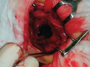
mobilisation according to Kocher a solid formation found in the duodenum (Fig. 6) wall in areas D II
and D III, thrombus vacuumed and coagulum evacuated (histology) after discission following a punc-
ture and histological sampling. A broader discission was carried out, a papilla located 40 mm from the
discission in the proximal direction was checked. Transverse suture of the duodenum. Further post-
operation condition was without complications. On 9 February 2004 the patient was released from the
hospital with a healed operation wound; the amylase values were normal.
154Fig. 3
CT scan – preoperative investigation showing solid formation of size 37x39x70 mm
Fig. 4
CT scan – preoperative investigation
155Fig. 5
x–ray passage – preoperative investigation with duodenal compression and stenosis
156Fig. 6
Intramural blood tumor of the duodenal wall – intraoperative view
Fig. 7
Ultrasonic investigation – postoperative with negative findings (May 2004)
157Conclusion of the histological examination: haemorrhage in muscularis and adventitia of the duo-
denum wall.
So far the last admission took place on 15 February 2004. The cause was a short history of pain in
the abdomen; amylase in serum at admission was 16.39, at release 3.68, CRP O, Leu 12,2. Ultrasound
examination of the abdomen was negative in the area of the pancreas capita, in the area of the gland
a small amount of liquid was suspected.
The passage through the duodenum shows a discrete narrowing of D II which is freely developing
with a free flow of the contrast substance into other sections of the small intestine. After conservative
therapy the child was released without any symptoms; diet and Pancreolan were recommended.
On 26 May 2004 the last ultrasound examination took place at the outpatient department with
a negative finding (Fig. 7). At that time the child was without clinical symptoms.
DISCUSSION
Pancreatitis in childhood and haematoma of the duodenum without a traumatic
cause are very rare diagnoses in children; in our case they occurred only in one pa-
tient. As regards pancreatitis, it is a disease without any aetiological grounds, where
the literature supposes the highest probability of relapse. In the case of the haemato-
ma of the duodenum we considered an association with the ERCP examination but
this was carried out 18 months before the diagnosis and a number of check-ups in
the meantime were negative. The parents ruled out the possibility of an accident
and the clinical finding did not identify any signs of abdominal trauma. The slightly
prolonged APTT in the haemocoagulation status did not cause any bleeding condi-
tions in the patient and is not likely to be the cause for the haematoma. The litera-
ture rarely mentions bleeding into the jejunum wall in adults induced by long-term
administration of coumarin preparations (12). In the aetiology of the haematoma
origin we incline toward the bleeding induced by the pancreatitis described in the
literature; however, this theory is rather a hypothesis. The operation revision in
our case was required due to the fact that an accurate differential diagnostic of the
possible tumour duodenum was not possible and the haematoma obstructed the
intestine passage. Repeated attacks of pancreatitis were undoubtedly also caused by
the failure to observe the recommended diet.
Acknowledgement
The publication is funded from grant ND 7648–3/ 2OO3.
158Krafka K., Tůma J., Teyschl O., Bartl V., Skotáková J.
INTRAMURÁLNÍ HEMATOM DUODENA U DÍTĚTE S OPAKOVANÝMI ATAKAMI
PANKREATITIDY
Souhrn
V naší kasuistice uvádíme případ dítěte, které bylo na naší klinice hospitalizováno opakovaně
s atakami pankreatitidy. Během těchto hospitalizací byla provedena řada vyšetření: opakované
sonografie, ERCP a CT vždy s negativními nálezy. V lednu 2004 byl bez jakékoliv traumatické anam-
nézy zjištěn intramurální hematom duodena, který jsme řešili operací. U dítěte kromě lehce vyšších
hodnot APTT nebyly rovněž během hospitalizací zjištěny poruchy koagulace. Uvedená diagnóza
bez traumatické příčiny je v dětském věku ojedinělá, u dospělých pacientů je popisována například
v souvislosti s dlouhodobým užíváním kumarinových preparátů. V závěru se zamýšlíme nad možnými
příčinami vzniku onemocnění, zda se jednalo o spontánní krvácení do stěny dvanácterníku při opako-
vaných atakách pankreatitidy, nebo zda by se mohlo jednat o pozdní následek vyšetření ERCP.
REFERENCES
1. Ashraft KW, Holder TM et al. Pediatric Surgery, 2nd edition, 1993, pp 527–529.
2. Pezzilli R, Morselli-Labate AM, Castellano E, Barbera C et al. Acute pancreatitis in children: an Ital-
ian multicentre study. Dig Liver Dis 2002; 34(5): 343–348.
3. Uretsky G, Goldschmiedt M, James K. Childhood pancreatitis. American Family Physician 1999;
59 (No 9), May 1.
4. Lerner A, Branski D, Lebenthal E. Pancreatic diseases in children. Pediatr Clin North Am l996;
43: 125–56.
5. Mehta DI. Acute and chronic pancreatitis in childhood. Ind J Pediatr 1999; 66 (Suppl): 81–86.
6. Keil R, Šnajdauf J, Štůj J, Kalousová J et al. Endoscopic retrograde cholangiopancreatography in
infants and children. Ind J Gastroenterology 2000; 19: 175–177.
7. Jewwet TC Jr, Caldarola V, Karp MP, Allen JE et al. Intramural hematoma of the duodenum. Ar-
chives of Surgery 1988; 123 (No 1, January 1988): 54.
8. Lotti R, Gaetano Perri S, Gola P, Leardi S et al. An intramural hematoma of the duodenum. Ann Ital
Chir 2000; 71: 519–523.
9. Dubois J, Guy F, Porcheron J. A pancreatic-induced intramural hematoma: a case report and litera-
ture review. Hepatogastroenterology 2003; 50: 1689–1692.
10. Kazama I, Maruyama M, Horiki N, Fujita Y. A case of intraluminal duodenum hematoma, sup-
posed to be caused by the rupture of the aneurysma. Nippon Shokakibyo Gakkai Zasschi 2002;
99: 1476–1480.
11. Camarero C, Herrera D, Corbaton J, Mingo A et al. Intramural hematoma of the duodenum fol-
lowing endoscopic biopsy: an unusual complication of non-therapeutic endoscopy in children.
Eur J Pediatr 2004; 163: 418–419.
12. Avent ML, Canaday BR, Sawyer WT. Warfarin-induced intramural hematoma of the small intestine.
Clin Pharm 1992; 11: 632–635.
159160
You can also read