The Yoga Mat Technique in Postless Hip Arthroscopy - Dr ...
←
→
Page content transcription
If your browser does not render page correctly, please read the page content below
Technical Note

             The Yoga Mat Technique in Postless Hip Arthroscopy
    Antonio Porthos Salas, M.D., Eder Mendez-Perez, Jacek Mazek, Hector Velasco-Vazquez,
                                   and Ara Castillo-Trevizo

Abstract: Hip arthroscopy for the treatment of femoroacetabular impingement syndrome with anti-sliding techniques
and without the use of a perineal post to achieve hip distraction has increased greatly in the past 5 to 10 years. To access
the hip joint, distraction is mandatory to treat intra-articular disorders such as labral tears, acetabular cartilage defects,
loose bodies, ligamentum teres tears, and avascular necrosis of the hip. In hip distraction, counter-distraction is needed,
and this is achieved with a bulky and cushioned perineal post. Most of the described techniques in hip arthroscopy
worldwide use a perineal post, but iatrogenic pudendal nerve, genital lacerations, hematomas, and groin complications
have been reported to occur. In Latin American countries, disposable hip pad devices are expensive, and not all the
arthroscopic companies provide them. Our yoga mat technique provides enough countertraction to achieve adequate hip
distraction. Labral repair, labral reconstruction, and decompression of femoroacetabular impingement syndrome have
been achieved properly, reliably, and reproducibly, and no Trendelenburg position is needed. Postless hip arthroscopy is
made simple, and positioning the patient is not difficult. Hip surgeons can adapt this technique to a fracture table, a hip
distractor, and a standard operating room table.

P    ostless hip arthroscopy (HA) is increasingly
     becoming a very common arthroscopic surgical
procedure; its diagnostic and therapeutic uses are
                                                                                  Entering the CC is the most important step when
                                                                                initiating HA. This is performed blindly with the use of
                                                                                cannulated needles and obturators; an image intensifier
numerous and commonly directed to treat labral tears,                           is also used in this important step. Sufficient force is
intra-articular cartilage lesions, ligamentum teres tears,                      needed to distract the head of the femur approximately
and femoroacetabular impingement syndrome (FAIS).                               10 mm away from the acetabulum, as measured on the
Distraction of the hip joint is very important and                              image intensifier. This takes no direct account of the
mandatory in HA when entering the central compart-                              thickness of the articular cartilage, which most often is
ment (CC), and because of this, the surgeon must apply                          not directly visible.1-3
enough force of traction. Although hip distraction is                             HA can cause some significant iatrogenic lesions, such
necessary to treat intra-articular disorders, traction-                         as scuffing of the femoral head, labral penetrations, and
related injuries such as foot palsies, foot lacerations,                        labral punctures while accessing the CC. This access is
vaginal or scrotal lacerations, and pudendal nerve                              more difficult than with other joints because of the hip
dysfunction are very common, and several investigators                          being highly constrained, deep, and strong in muscu-
have recommended limiting traction time to less than                            lature and anatomy. Surgeons typically use a hip
2 hours.                                                                        fracture table or a commercial specialized hip distractor
                                                                                to obtain proper distraction and perform a reproducible
  From Hip Arthroscopy Mexico (A.P.S.) and Hospital Ángeles Valle Oriente       and reliable surgery.3 Hip counter-distraction is needed
(E.M.-P., H.V.-V., A.C.-T.), San Pedro Garza García, México; and Ortopedika
                                                                                in HA and is achieved with the placement of a bulky
Hospital Hip Unit, Warsaw, Poland (J.M.).
  The authors report that they have no conflicts of interest in the authorship   cushioned perineal post measuring approximately 25 to
and publication of this article. Full ICMJE author disclosure forms are         30 cm; this width is necessary and very important
available for this article online, as supplementary material.                   to avoid lesions of the pudendal and perineal
  Received December 29, 2020; accepted February 13, 2021.                       nerve, erectile dysfunction, and vaginal or scrotal
  Address correspondence to Antonio Porthos Salas, M.D., Hip Arthroscopy
                                                                                lacerations.4-7
Mexico, San Pedro Garza Garcia, Nuevo Leon, Mexico. E-mail: dr.
porthossalas@gmail.com
  Ó 2021 Published by Elsevier on behalf of the Arthroscopy Association of
North America. This is an open access article under the CC BY-NC-ND license
                                                                                Surgical Technique (With Video Illustration)
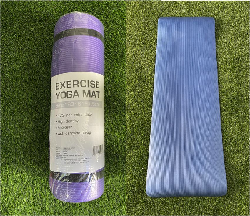
(http://creativecommons.org/licenses/by-nc-nd/4.0/).                              A yoga mat is placed on a standard operating room
  2212-6287/202079                                                              (OR) table. The yoga mat (Sunshine Yoga, Charlotte,
  https://doi.org/10.1016/j.eats.2021.02.020                                    NC) is cut in half with conventional scissors; one half

                                           Arthroscopy Techniques, Vol 10, No 6 (June), 2021: pp e1525-e1530                      e1525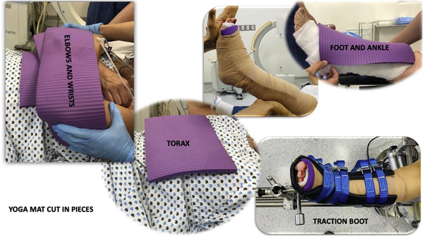
e1526                                              A. P. SALAS ET AL.

will be placed on the posterior trunk of the patient on       portal used for vision, immediately anterior to the
top of the OR table and the other half will be cut in         trochanteric tip; a paratrochanteric space portal is used
pieces to protect the bony prominences (Fig 1). HA is         as a working portal, situated 3 cm distally from the
performed with the patient in the modified supine              anterolateral portal and over the anterior trochanteric
position. The hip is distracted with a hip distractor         border; and a new modified mid-anterior portal, which
(ArthroMX; San Pedro Garza Garcia, Mexico) or a hip           is located 1.5 cm above and between the anterolateral
fracture table (MAQUET Gmbh, Rastatt, Germany),               and paratrochanteric space portals, which we call the
with the patient receiving general anesthesia, and no         trochanteric triangle portal (Fig 4). The anterior hip
muscle relaxants are used, although they may be               bursa is resected with a shaver or a radiofrequency
added. The standard OR table is covered by the yoga           wand, and a longitudinal capsulotomy is performed
mat foam placed under the upper body of the patient           posteriorly to access the headeneck junction to perform
(Fig 2). Both arms are placed over the chest in a figure       a femoro-osteochondroplasty for the cam asphericity. If
of 8 or X position, and the patient is secured on the OR      there is a pincer deformity, an acetabuloplasty is
table. Bony prominences are protected with the yoga           performed without distraction.
mat foam strips at the level of the elbows, wrists, feet,        Afterward, an arthroscopic dynamic impingement test
and ankles and are rolled with Coban (3M, Austin, TX)         is performed with the help of the surgical assistant, fellow,
(Fig 3). The patient’s hands are left free for intravenous    or nurse, and it will be useful to evaluate the site of
line and medication passage, which are double checked         impingement and the femoro-osteochondroplasty. After
by the anesthesiologist. A long blanket is placed around      finishing on the PC, a surgical assistant or nurse techni-
the patient’s upper body. Heavy-duty duct tape (3M) is        cian will place the foot and attach it to the hip fracture
placed in an X or figure of 8 fashion posteriorly,             table to distract the hip under controlled arthroscopic
securing the patient’s upper body on the OR table.            vision (Figs 5 and 6). It is very important to tell the
  The patient is prepped and draped in a standard             anesthesiologist when you are beginning your distrac-
fashion with a vertical isolation drape. Radiographic         tion, because if the yoga mat foam is not well secured, the
and anatomic landmarks are marked on the patient’s            patient can slide under the sterile field and fall from the
operative hip, and portals are established to access the      OR table. This is easily prevented by the anesthesiologist’s
peripheral compartment and CC: an anterolateral               careful monitoring (Table 1) (Video 1).7-9

                                                                                         Fig 1. The yoga mat for postless
                                                                                         hip arthroscopy.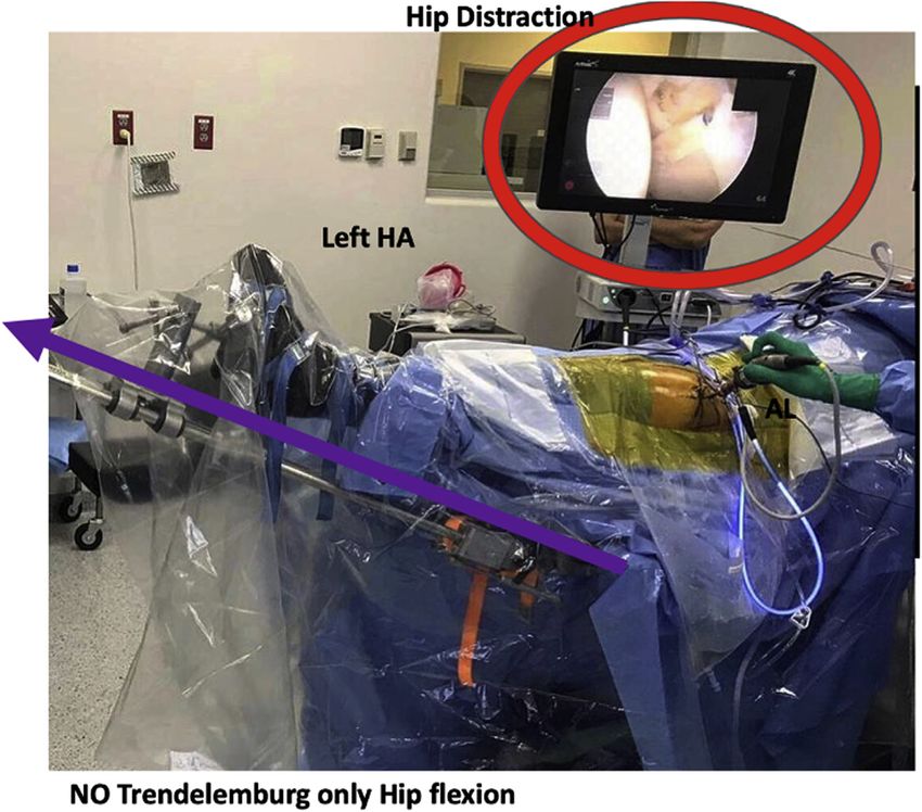
THE YOGA MAT TECHNIQUE                                                  e1527

Fig 2. The yoga mat placed in a standard operating room table.

                       Discussion                                 special disposable hip pad equipment is usually not
  Postless HA has evolved greatly in the past 5 years             available in third-world countries, and most of the time
because of the attention paid to the iatrogenic pudendal          the arthroscopic companies will not provide them;
nerve palsy and the devastating groin lesions. The                therefore, performing HA safely can become very

Fig 3. The yoga mat is cut in pieces and placed at the thorax, elbows, wrists, and operative foot to protect bony prominences and
avoid iatrogenic lesions and nerve dysfunctions.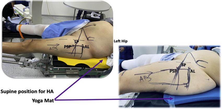
e1528                                                 A. P. SALAS ET AL.

Fig 4. Hip arthroscopy with the patient in the supine position with the yoga mat placed on the standard operating room table
under the posterior trunk of the patient. Observe the anterolateral portal (AP; vision), the peritrochanteric space portal (PSP;
working portal), and triangle portal (TP; anchor placement and working).

                                                                                             Fig 5. Hip arthroscopy (HA) in
                                                                                             the supine position with no
                                                                                             Trendelenburg, only hip flexion.
                                                                                             Observe the distraction (red cir-
                                                                                             cle) of more than 2 cm.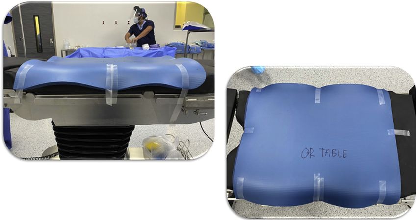
THE YOGA MAT TECHNIQUE                                                       e1529

                                                                             mention that while considering the risks and limita-
                                                                             tions of their technique to achieve safe distraction, the
                                                                             surgeon should keep in mind the following complica-
                                                                             tions: lighter patients, i.e., less than 120 pounds, may
                                                                             require greater degrees of Trendelenburg to achieve
                                                                             distraction.
                                                                                Jimenez et al.10 conclude that postless surgery has
                                                                             become a popular method of performing HA and that
                                                                             although various techniques exist for achieving traction
                                                                             without a post, the majority require special beds and
                                                                             special equipment. Their technique can be performed
                                                                             with any hip traction table, at zero additional cost, and
                                                                             with the patient leveled before procedure starts.
                                                                                Salas et al.11 started performing postless HA with the
                                                                             yoga mat technique because of the expensive pink pad
                                                                             or the disposable hip kit ($100-500 USD), which most
                                                                             of the time is unattainable for patients and insurance
                                                                             companies. Their technique does not require a Tren-
Fig 6. Left hip with an acetabular rim recession. Observe the
distraction in the intraoperative fluoroscopic image.
                                                                             delenburg position to achieve a hip distraction.
                                                                                Limitations exist with the postless HA technique, as
                                                                             with all arthroscopic techniques. We always recom-
difficult and sometimes impossible. Merrell et al.8                           mend performing a trial traction test manually before
performed HA without a perineal post, but they used                          surgery and posteriorly with the hip distractor with
a deflated taped beanbag positioned and contoured in                          fluoroscopic control, because of the risk of the patient
the lateral portion of the patient’s body, and they added                    sliding down the OR table and because of the risk of not
a blanket to the abdomen of the patient, similar to                          producing the proper countertraction (Table 2). Our
our technique, although beanbags are not always                              yoga mat technique provides enough countertraction to
available in hospitals. They conclude that this is a very                    achieve adequate hip distraction; none of our patients
simple technique, and the risk of nerve injuries is                          has ever slid from the OR table while in surgery. Labral
minimal.                                                                     repair, labral reconstruction, and decompression of
  Mei-Dan et al.9 demonstrate in their surgical tech-                        FAIS have been achieved properly, reliably, and
nique no difficulty in accessing the hip joint, distraction                   reproducibly. We believe that postless HA is made
is not an issue, and their positioning is quick and easy                     simple with the yoga mat, which is also cost-effective.
and requires no special equipment. Their theory is that                      Positioning the patient is not difficult, and the sur-
distraction without a post should eliminate the perineal                     geon can adapt this technique to a fracture table, a hip
pressure-related complications of the pudendal and                           distractor, or a standard OR table. Adequate hip
perineal nerves, as well as the delicate tissues of the                      distraction is achieved, and the surgeon can perform
perineum. They use a Trendelenburg position to                               anything from a basic FAIS decompression to a complex
use gravity as countertraction. Kollmorgen et al.1                           labrum reconstruction.

Table 1. Pearls and Pitfalls of the Yoga Mat Technique in Postless Hip Arthroscopy
                                   Pearls                                                                    Pitfalls
The posterior part of the yoga mat has to be in contact with the posterior         Patient slippage due to poor patient positioning on the OR
  trunk of the patient to prevent slippage.                                          table.
Always secure the patients upper body on the OR table to help with the             The patient’s positioning toward the OR table has to be very
  counter traction when distracting the hip.                                         smooth to prevent nerve dysfunction to any bony
                                                                                     prominences.
Protect bony prominences of the thorax, elbows, wrists, and operative foot         Blockage of the intravenous line on hands or arms due to
  with the cut pieces of the yoga mat.                                               excessive compression when positioning the patient.
Always perform a manual trial traction before surgery to observe if your           Do not place any blankets between the yoga mat and the
  patient slips off the OR table.                                                    patient.
Perform a trial traction with your hip fracture table or distractor to observe     A failed distraction of the hip and an incomplete surgery.
  whether your patient slips off the operating room table.
Ask the anesthesiologist to observe motion and slippage of the patient when
  distracting the hip.
 OR, operating room.
e1530 A. P. SALAS ET AL.
Table 2. Advantages of the Yoga Mat Technique in Postless 3. Dienst M, Seil R, Kohn DM. Safe arthroscopic access to
Hip Arthroscopy the central compartment of the hip. Arthroscopy 2005;21:
1. There is no need of the perineal post. 1510-1514.
2. There are no iatrogenic lesions to the perineal nerve and pudendal 4. Domb B, Hanypsiak B, Botser I. Labral penetrations rate
nerve by compression with the post. in a consecutive series of 300 hip arthroscopies. Am J
3. No scrotal or vaginal lacerations occur. Sports Med 2012;40:864-869.
4. The patient can be positioned on a regular operating room table. 5. Kuhns BD, Erickson BJ, Bressler LR, Sarmast SM, Nho SJ.
5. There is no need for gravity or a Trendelenburg position to help Perineal post padding technique to improve hip distrac-
with the distraction for countertraction. tion in tall patients. Arthrosc Tech 2015;4:e747-e750.
6. Hip injections are easily performed with this technique.
6. Salas AP. Radiographic and anatomic landmarks to
7. The surgeon can perform anything from a basic hip arthroscopy for
approach the anterior capsule in hip arthroscopy. J Hip
FAIS to a labrum reconstruction, as well as AVN core. decom-
pression, open FAIS surgery, and other intra-articular disorders of Preserv Surg 2015;2:431-437.
the hip. 7. Salas AP, Gonzalez-Campos M, Gonzalez-Bazaldua JLA,
8. The procedure is reliable and reproducible. Salas-Perez AD. The Mayo table technique. Arthrosc Tech
9. Low cost (the yoga mat can be re-used). 2016;5:e459-e463.
10. Body weight is not a contraindication (we have used this tech- 8. Merrell G, Medvecky M, Daigneault J, Jokl P. Hip
nique in patients who are thin, patients with normal- weight, and arthroscopy without a perineal post: A safer technique for
patients with obesity). hip distraction. Arthroscopy 2007;23:107.e1-107.e3.
AVN, avascular necrosis; FAIS, femoroacetabular impingement 9. Mei-Dan O, McConkey MO, Young DA. Hip arthroscopy
syndrome. distraction without the use of a perineal post: Prospective
study. Orthopedics 2013;36:e1-e5.
10. Jimenez M, Haneda M, Pascual-Garrido C. The Hip
References Arthroscopy Post-less Procedure Impingement (HAPPI)
1. Kollmorgen R, Ellis T, Lewis B, Harris J. Achieving post- technique: Achieving distraction with standard hip
free distraction in hip arthroscopy with a pink pad tables at zero additional cost. Arthrosc Tech 2020;9:
patient positioning device using standard hip distraction e1697-e1701.
tables. Arthrosc Tech 2019;8:e363-e368. 11. Salas AP, Mazek J, Araujo-Reyes D, Gonzalez-Campos M,
2. Eriksson E, Arvidsson I, Arvidsson H. Diagnostic and oper- Castillo-Trevizo A, Garcia JM. The Tuthankamun technique
ative arthroscopy of the hip. Orthopedics 1986;9:169-176. in hip arthroscopy. Arthrosc Tech 2018;7:e1167-e1171.You can also read


