Partial Obstruction and Intestinal Bleeding Secondary to a Congenital Duodenal Diverticulum in a Dog
←
→
Page content transcription
If your browser does not render page correctly, please read the page content below
Acta Scientiae Veterinariae, 2021. 49(Suppl 1): 597.

                                                                    CASE REPORT                                                     ISSN 1679-9216
                                                                    Pub. 597

                          Partial Obstruction and Intestinal Bleeding Secondary to a
                                  Congenital Duodenal Diverticulum in a Dog

              Carla Aparecida Batista Lorigados1, Aline Machado Zoppa2, Andre Schiller³, Iara Tiburcio³,
                                    Fabio Giusti Calderón4 & Marianna Pantano5

                                                                 ABSTRACT
Background: Intestinal diverticulum is an abnormality resulting in the formation of a blind-ended saccular pouch that can
be acquired either congenital, true (involving all intestinal layers) or false (involving the mucosa and submucosa), with
extraluminal and intraluminal type. In humans, the acquired is more frequent, the colon is the most affected segment fol-
lowed by duodenum; and majority cases of duodenal diverticula remains asymptomatic, but biliary obstruction, recurrent
acute pancreatitis, hemorrhagic ulcer, proximal intestinal obstruction and perforation may occur. The aim of this report is
to present a case of a congenital disease in dogs, prone to misdiagnosis due to non-specific clinical signs.
Case: An 8-month-old male Boxer was evaluated due to recurrent hyporexia, vomiting, melena and syncope over three
months with signs of a possible intestinal obstruction. Physical examination showed no abnormalities except for pale
mucous membranes. Complete blood count revealed anemia and leukocytosis. Platelets and biochemical profiles were
normal. Abdominal ultrasound examination indicated a dilated duodenum, measuring approximately 3.36 cm in diameter,
with heterogeneous fluid content and hyperechoic structures with acoustic shadow, peristalsis appeared decreased and
non-progressive. The gastrointestinal positive contrast study was performed to better evaluate abnormalities detected at
ultrasonography. Images after 30 min of contrast administration demonstrated a marked distension of the duodenum, filled
with contrast and a mildly filled stomach displaced to the left. Sixty min after contrast administration a marked distension
of the entire duodenum, with tortuous aspect and filled with contrast was seen. The caudal duodenal flexure was connected
to a large barium filled saccular structure that measured approximately 7 cm in diameter, consistent with a duodenal di-
verticulum. A blood transfusion was performed and surgical treatment indicated. The diverticulum and a small portion of
the caudal duodenal segment were resected, an end-side enteroanastomosis was made due to the difference in diameter
between intestinal segments. The patient was medicated with sucralfate (12.5 mg/kg), ranitidine (2 mg/kg), metronidazole
(25 mg/kg), dipyrone (25 mg/kg), tramadol (2 mg/kg) and recovered quickly from surgery. Histopathological examina-
tion characterized the diverticular tissue as a true diverticulum by the presence of all intestinal layers. Post-operative and
follow-up evaluations showed no recurrence of clinical signs.
Discussion: In veterinary practice, congenital duodenal diverticulum is a rare condition documented in dogs, curiously all
Boxers. None of the reported cases in literature had the diagnostic of duodenal diverticulum made exclusively by ultra-
sonography. Other diagnostic imaging modalities, such as gastrointestinal barium study or endoscopy, were necessary. In
one case a diagnostic was made during exploratory laparotomy. The marked dilatation of the duodenal segment impaired
ultrasound evaluation, allowing recognition of an obstructive pattern, not the diverticulum itself. At histopathological ex-
amination, the diverticular tissue was characterized by a thickened wall with a hypertrophied muscle layer, characterizing
a true duodenal diverticulum. The location, breed and age of the dogs affected with duodenal diverticulum were similar in
all veterinary cases reported. Dogs presenting signs of gastrointestinal disease and abdominal pain are common in patients
referred to ultrasound examination. However, despite the rare reports described, we must consider this affection as a differ-
ential diagnosis, whenever Boxer puppies present these clinical signs associated with gastrointestinal bleeding and syncope.
Keywords: duodenum, congenital disease, diagnostic imaging, canine.

                                                          DOI: 10.22456/1679-9216.100991
Received: 5 August 2020                                      Accepted: 20 November 2020                                   Published: 22 January 2021
1
 Department of Surgery, Faculty of Veterinary Medicine and Animal Science (FMVZ), University of São Paulo (USP), São Paulo, SP, Brazil. 2Department
of Surgery, Faculty of Veterinary Medicine, Anhembi Morumbi University (UAM), São Paulo. 3Department of Surgery, Faculty of Veterinary Medicine,
Metropolitan Colleges United (FMU), São Paulo. 4PetCare Veterinary Hospital & 5EVET Veterinary Specialties, São Paulo. CORRESPONDENCE: C.A.B.
Lorigados [clorigados@usp.br] Department of Surgery, FMVZ - USP. Av. Prof. Dr. Orlando Marques de Paiva n. 87. CEP 05508-270 São Paulo, SP, Brazil.

                                                                        1
C.A.B. Lorigados, A.M. Zoppa, A. Schiller, et al. 2021. Partial Obstruction and Intestinal Bleeding Secondary to a Congenital
Duodenal Diverticulum in a Dog. Acta Scientiae Veterinariae. 49(Suppl 1): 597.
INTRODUCTION The aim of this report is to present a rare case
Intestinal diverticulum (ID) is an abnormality of a congenital disease in dogs, prone to misdiagnosis
that results in the formation of a blind-ended saccular due to non-specific clinical signs.
pouch in the intestinal wall and can produce signs of CASE
gastrointestinal disease. The duodenal type (DD) was
primarily described in humans by Chomel in 1710, An 8-month-old intact male Boxer was presen-
and there are reports of ID in animals, including dogs, ted to the hospital with recurrent signs of hyporexia,
pigs, horses and sheep [10,13]. vomiting, melena and syncope over three months.
Duodenal diverticulum can be acquired or Physical examination showed no abnormalities except
congenital, the last contains all intestinal layers, with for pale mucous membranes. Complete blood count
an extraluminal (EDD) and intraluminal (IDD) type revealed anemia, red blood cells 2.3 x 10⁶/μL (reference
[10,12,15]. The origin of IDD seems to be caused range 5.0 - 8.0), hematocrit 16% (reference range 37-
by the presence of a luminal membrane due to reca- 54%), and leukocytosis 22,196 uL (reference range
nalization failure of the foregut lumen during fetal 6,000 - 15,000). Platelets and biochemical profiles
stages, later peristaltic and feeding pressure extends were normal. Abdominal ultrasound examination in-
it, leading to an intraluminal duodenal diverticulum dicated a dilated duodenum, measuring approximately
[1,5,6,11]. The acquired EDD is more frequent and 3.36 cm in diameter, wall thickening with preserved
characterized by herniation of the submucosa and stratification, fluid content and heterogeneous ingesta
mucosa, through a muscularis defect or a weak spot associated with small hyperechoic structures that
of the wall, such as an artery entry; it’s also named as produces acoustic shadow (Figure 1). Peristalsis appe-
pseudodiverticulum [8,10,12]. The etiopathogenesis of ared decreased and non progressive within the dilated
the intestinal congenital diverticulum remains unclear. portion. There were no sonographic changes in other
In humans it may be a remaining pouch of the cecal portions of the small intestine and colon.
appendix during embryological stages [12]. The ID is To better evaluate the abnormalities detected
a common finding in gastrointestinal imaging studies, at ultrasonography a gastrointestinal barium study
and the most usual location is in the colon, followed by was performed. Radiographs after 30 min of con-
duodenum. Most of the duodenal diverticulum remain trast administration demonstrated a distension of the
asymptomatic, but serious complications as biliary duodenum, filled with contrast and a mildly filled
obstruction, recurrent acute pancreatitis, hemorrhagic stomach displaced to the left. Sixty min after contrast
ulcer, proximal intestinal obstruction and perforation administration persisted the marked distension of the
may occur [2,5,7,14]. duodenum, with tortuous aspect. The caudal duodenal
Figure 1. Ultrasonography images of the duodenum. A- Note a marked distended segment of duodenum with fluid and heterogeneous ingesta, associ-
ated a thickened wall. B- A hyperechoic structure producing acoustic shadowing between the markers, measuring 2.4 cm in length, compatible with a
small foreign body.
2C.A.B. Lorigados, A.M. Zoppa, A. Schiller, et al. 2021. Partial Obstruction and Intestinal Bleeding Secondary to a Congenital
Duodenal Diverticulum in a Dog. Acta Scientiae Veterinariae. 49(Suppl 1): 597.
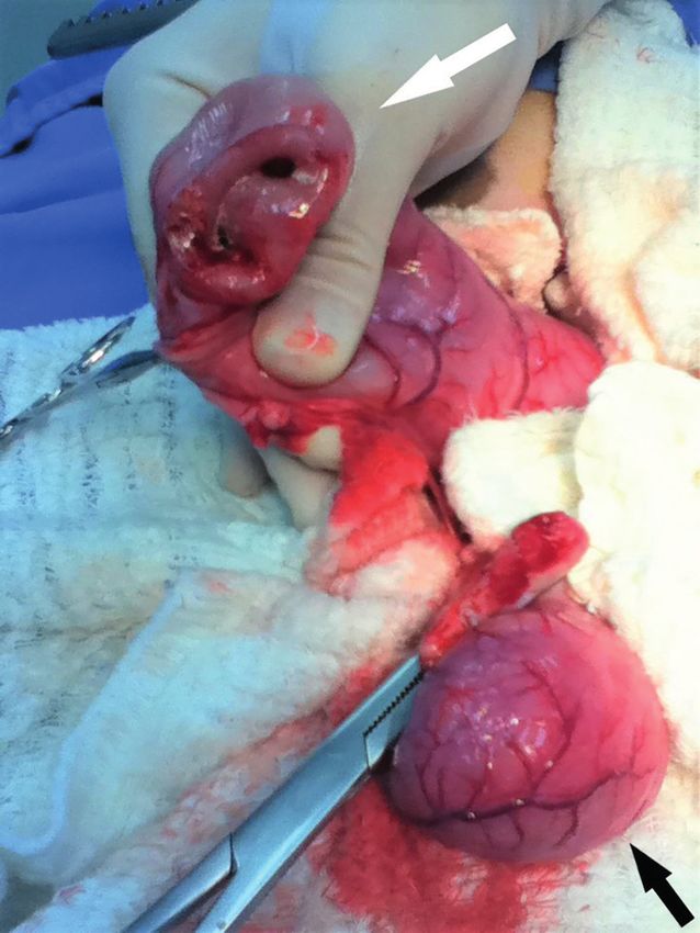
Figure 3. Intraoperative view of the enterectomy site showing duodenal
diverticulum (black arrow) and proximal duodenum (white arrow).
[2 mg/kg/PO] and metronidazole2 [25 mg/kg/PO] twice
daily, and recovered from surgery without complica-
Figure 2. Lateral (A) and ventrodorsal (B) views 60 min after barium tions. In follow-up evaluations no recurrence of clinical
administration. Note the persistence of contrast in the dilated and tortuous
segment of the duodenum (black and white arrows) and a pocket of bowel
signs was observed.
at the duodenal caudal flexure diverticulum (arrowhead). Jejunum is filled
with a contrast medium, with normal diameter and distribution. Stomach DISCUSSION
is empty (asterisk).
Congenital duodenal diverticulum is a rare
flexure was connected to a large barium filled saccular condition and it has been already reported in four
structure that measured approximately 7 cm in dia- dogs, interestingly, all Boxers [3,13,16], however, there
meter, consistent with a duodenal diverticulum. The are reports of jejunal [4] and rectal [9] diverticulum
jejunal loops were normal, with uniform diameter and involving other breeds.
distribution. The stomach was almost empty with a left The clinical signs of these four cases of duode-
displacement of pylorus (Figure 2). nal diverticulum [3,13,16] were similar to those found
A blood transfusion was performed, and a la- in this dog, including melena, anorexia, vomiting,
parotomy was indicated. The diverticulum, projected anemia and syncope. Melena and anemia were likely
from the antimesenteric border of the duodenum, and caused by the bleeding process due to inflammation,
a small portion of the caudal duodenal segment were ulcerations and rupture of superficial blood vessels
resected and an end-side enteroanastomosis was per- in consequence of food stasis in duodenum and
formed, due to the difference in diameter between duo- diverticulum.
denum and jejunum (Figure 3). Post surgical analgesia None of the reported cases in literature had
was assured by giving tramadol1 [2 mg/kg/PO] and di- the diagnosis of duodenal diverticulum made exclu-
pyrone2 [25 mg/kg/PO], twice a day. The dog was also sively by ultrasonography. Other diagnostic imaging
medicated with sucralfate3 [12.5 mg/kg/PO], ranitidine4 modalities, such as gastrointestinal barium study or
3C.A.B. Lorigados, A.M. Zoppa, A. Schiller, et al. 2021. Partial Obstruction and Intestinal Bleeding Secondary to a Congenital
Duodenal Diverticulum in a Dog. Acta Scientiae Veterinariae. 49(Suppl 1): 597.
endoscopy, were necessary to confirm the diagnosis despite the rare reports described, we must consider
[13,16]. In one case diagnostic was made during ex- this affection as a differential diagnosis, whenever
ploratory laparotomy secondary to a foreign body [3]. Boxer puppies present these clinical signs associated
The marked dilatation of the intestinal segment with gastrointestinal bleeding and syncope.
filled with ingesta impaired ultrasound evaluation, MANUFACTURERS
allowing recognition of an obstructive pattern, not 1
Cristália Produtos Químicos Farmacêuticos Ltda. Itapira, SP,
the diverticulum itself, in this case. Histopathological Brazil.
examination of the diverticular tissue showed signs of
2
Sanofi Aventis Farmacêutica Ltda. Suzano, SP, Brazil.
3
EMS S/A. Hortolândia, SP, Brazil.
chronic inflammation and a hypertrophied muscle layer 4
Glaxosmithkline Brasil Ltda. Rio de Janeiro, RJ, Brazil.
of the intestinal wall, characterizing a true duodenal
diverticulum. The location (caudal flexure of the duo- Acknowledgments. The authors would like to thank PhD Profes-
sor Stefano Hagen (University of São Paulo) for his valuable
denum), breed (Boxer) and age (immature) of the dogs and constructive comments and expertise during article revision
affected with duodenal diverticulum were similar in all and to PhD Professor Rodolfo Nurmberger Jr., in memoriam,
veterinary cases reported in literature. Dogs presenting for his knowledge in histopathology.
signs of gastrointestinal disease such as vomits, regur- Declaration of interest. The authors report no conflicts of
gitation, anorexia and abdominal pain are common in interest. The authors alone are responsible for the content and
patients referred to ultrasound examination. However, writing of the paper.
REFERENCES
1 Angus L., Larson B., Egodage T. & Raju S.G. 2013. Traumatic blow out of a duodenal diverticulum: a rare clinical
finding. Injury Extra. 44(10-12): 95-98.
2 Bittle M.M., Gunn M.L., Gross J.A. & Rohrmann C.A. 2012. Imaging of duodenal diverticula and their complica-
tions. Current Problems in Diagnostic Radiology. 41(1): 20-29.
3 Blesch M., Livet V., Cabom Q. & Cachon T. 2016. Obstruction intestinale par un corps étranger au sein d’un diverti-
cule duodénal chez un chiot Boxer. Revue de Médecine Vétérinaire. 167(3-4): 71-76.
4 Buote N.J., Kovak J.R., Fischetti A.J. & Monette S. 2007. What is your diagnosis? Mineralized-opaque-rimmed soft
tissue mass in the right caudoventral portion of the abdomen. Journal of American Veterinary Medical Association.
231(4): 527-528.
5 Chambenois E., Derhy S. & Arrive L. 2015. Intraluminal duodenal diverticulum. A rare cause of recurrent acute
pancreatitis. Clinics and Research in Hepatology and Gastroenterology. 39(3): 278-279.
6 Eusebio M., Ramos A. & Guerreiro H. 2016. Intraluminal duodenal (windsock) diverticulum: a rare cause of gas-
trointestinal bleeding. Portuguese Journal of Gastroenterology. 23(2): 113-115.
7 Glener J., Poris S., Foles B. & Harmon R. 2016. Perforated duodenal diverticulum case report. International Journal
of Surgery Case Reports. 29: 100-102.
8 Kassir R., Boueil-Bourlier A., Baccot S., Abboud K., Dubois J., Petcu C.A., Boutet C., Chevalier U., Montveneur
M., Cano M.I., Ferreira R., Debs T. & Tiffet O. 2015. Jejuno-ileal diverticulitis: etiopathogenicity, diagnosis and
management. International Journal of Surgery Case Reports. 10: 151-153.
9 Mehrjerdi H.K., Mirshahi A. & Afkhami A. 2013. Rectal diverticulum in a Terrier dog: a case report. Veterinary
Research Forum. 4(1): 63-67.
10 Penades M., Guerrero I., Peña A.B. & Corpa J.M. 2010. Duodenal gland cysts and pseudodiverticula in sheep.
Journal of Veterinary Diagnostic Investigation. 22(4): 649-651.
11 Peng H.L., Su C.T., Chang C.Y. & Lau B.H. 2014. Intraluminal duodenal diverticulum in a child concomitant with
an entrapped coin and a duodenal polyp. Formosan Journal of Surgery. 47(6): 236-239.
12 Perrot T., Poletti P.A., Becker C.D. & Platon A. 2012. The complicated duodenal diverticulum: retrospective analysis
of 11 cases. Clinical Imaging. 36(4): 287-294.
13 Polf H. & Poteet B. 2010. Imaging diagnosis - duodenal diverticulum in a dog. Veterinary Radiology & Ultrasound.
51(1): 61-64.
14 Schroeder T.C., Hartman M., Heller M., Klepchick P. & Ilkhanipour K. 2014. Duodenal diverticula: potential
complications and common imaging pitfalls. Clinical Radiology. 69(10): 1072-1076.
4C.A.B. Lorigados, A.M. Zoppa, A. Schiller, et al. 2021. Partial Obstruction and Intestinal Bleeding Secondary to a Congenital
Duodenal Diverticulum in a Dog. Acta Scientiae Veterinariae. 49(Suppl 1): 597.
15 Simões V.C., Santos B., Magalhães S., Faria G., Silva D.S. & Davide J. 2014. Perforated duodenal diverticulum:
surgical treatment and literature review. International Journal of Surgery Case Report. 5(8): 547-550.
16 Van Klaveren N.J., Grinwis G.C.M., Brocks B.A.W. & Kirpensteijn J. 2008. Collapse following gastrointestinal
bleeding secondary to a congenital duodenal diverticulum in two littermate boxer pups. Journal of Small Animal
Practice. 49(2): 103-106.
CR597
http://seer.ufrgs.br/ActaScientiaeVeterinariae
5You can also read