Foam sclerotherapy and eccentric compression - Pulsus Group
←
→
Page content transcription
If your browser does not render page correctly, please read the page content below
REVIEW ARTICLE

                               Foam sclerotherapy and eccentric compression
                                                                 Vincent Crebassa1*, CH Gardon Mollard2

Crebassa V, Mollard G. Foam sclerotherapy and eccentric compression. J                      favours varicose vein treatments during the short healing period, possibly in
Phlebol Lymphol.14(1):1-4.                                                                  addition to digressive compression.
Digressive compression of 20 mm Hg does not modify the diameter of                          Eccentric compression is often put in place by the doctor and cannot be
saphenous varicose veins in a standing position. This explains the results of               replaced by the patient. Moreover, this compression is artisanal and the
Dr Hamel-Desnos’ study, which demonstrated that there was no significant                    applied pressure is not measurable.
benefit to the use of this compression after treatment with foam sclerotherapy
of the Great Saphenous Vein. Dr. Zarca will confirm, few years later, these                 We have thus studied a new device that corresponds to a standardisation
same results with a compression of 30 mm Hg.                                                of the eccentric compression that the patient can put over his clothes and
                                                                                            reposition whenever he wants, according to his sensations. Our study shows
So how can we explain the recommendations of many authors who suggest                       that this eccentric compression allows the reduction of varicose vein diameter
that we must use an eccentric compression?                                                  and therefore the volume of the saphenous veins and superficial varicose
                                                                                            veins decrease up to 70%. Blood is the public enemy of all our treatments,
This can be explained by the Laplace’s Law which explains that the more the                 thus, as the volume of blood is reduced in the treated varicose veins we
surface is plane, the lower the transmitted pressure is. This is also explained             reduce haematomas after surgery, we limit blood carbonisation around the
by the physical law of pressure transmission according to the density of the                thermal probe and we decrease the volume of foam injected.
compressed tissues: the suppler the surface is, the lower the transmitted
pressure is.                                                                                Maintaining this compression 48 hours after these treatments would
                                                                                            enable us to have a more harmonious fibrosis, in addition to reduced side
This is obviously the case for the medial part of the thigh where we would                  effects related to excessive inflammation (redness, intravenous blood traps,
like to compress the GSV and for the posterior part of the calf for the SSV                 hematomas, localized pain, pigmentation). We could also consider treating
compression. These surfaces are flat and the tissue is supple and depressible.              larger varicose veins with this eccentric compression, without increasing the
Digressive compression loses in these cases all its efficiency. On the other                volume of foam injected.
hand, eccentric compression allows us to concentrate the pressure, with
denser material, on a specific area, to promote healing, fibrosis and control               Key Words: Varicose vein; Diameter; Volume; Eccentric compression; Foam
inflammation. Under no circumstances, it plays a role in venous return. It                  sclerotherapy; Compression and foam sclerotherapy

                                INTRODUCTION

T    he first public ennemi of endovenous treatment is the blood. The
     major risk of inefficiency and complications during sclerotherapy is the
presence of blood in the treated varicose vein. If it be during thermic endo-
venous treatment [1-3] reducing the diameter of the varicose vein during
and after the treatment is a major therapeutic objective, or more specifically,
reducing the volume of blood in the varicose vein during but also after the
treatment is a major therapeutic objective. The reduction of the varicose vein
will not only improve results by obtaining uniform fibrosis but also reduce
possible side effects and potential complications [4].

  WHY ARE WE DIVIDED ON THE UTILITY OF COMPRESSION
          AFTER ENDO VENOUS TREATMENTS?

Chemical endo venous techniques are also limited by the presence of blood.
The sclerosing product is destroyed by the blood not only in vivo [5] but also                Figure 1) Foam effect: Emptying of the vein by the foam enabling a better
in vitro [6]. The “foam” effect allows us to delay the destruction of the product             contact with the vein wall and reducing the destruction of the product by the
by the blood by totally emptying the varicose vein. The contact time between                  blood
the product and the vein wall is therefore increased (Figure 1).

  1
      Department of Phlebology, Clinique du Millenaire, Montpellier, France; 2Department of Vascular Surgery, 19 rue St Pierre 43100 Brioude, France
  Correspondence: Vincent Crebassa, Department of Phlebology, Clinique du Millenaire, Montpellier, France, E-mail: vincent.crebassa@gmail.com
  Received: December 18, 2020, Accepted: January 01, 2021, Published: January 08, 2021

                              This open-access article is distributed under the terms of the Creative Commons Attribution Non-Commercial License (CC BY-NC) (http://
                              creativecommons.org/licenses/by-nc/4.0/), which permits reuse, distribution and reproduction of the article, provided that the original work is
                              properly cited and the reuse is restricted to noncommercial purposes. For commercial reuse, contact reprints@pulsus.com

J Phlebol Lymphol Vol.14 No.1 2021                                                                                                                                     6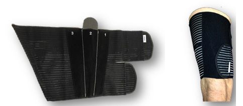
Crebassa et al.
The volume effect of the foam explains these results. However, this technique Standardised eccentric compression applies a localized, external compression,
reaches its limits when treating voluminous varicose veins. Only a limited reducing the diameter like a tumescence but without any injection, without
quantity of foam can be injected into the vein [7]. pain, without needles and consecutives risks. In addition, the diameter
reduction and immobilization effect continues as long as the patient carries
Therefore, it is highly useful to reduce the diameter of the varicose vein in this compression where a tumescence whose effects disappear in a few
order to reduce the quantity of foam to be injected. This also increases the minutes or even hours.
contact time between the product and the vein wall and allows us to treat
bigger varicose veins. WHAT IS THE IDEAL ECCENTRIC COMPRESSION SYSTEM?
Whatever the treatment, the reduction of the diameter of the treated varicose The limits of existing eccentric compression
vein after treatment and “antalgic compression“, preventing movement, is
a therapeutic objective. This favors uniform fibrosis and limits side effects Some authors, such as M. Schadek, recommend compressing the varicose
and possible complications such as: pigmentation, swelling, venous traps vein during injection with the probe, others with a weight (sandbag) but
requiring thrombectomy, localized pain, regularity defects in the fibrosis but even if this technique allows the diameter of the varicose vein to be reduced,
also hematoma, bleeding and cicatrization, complications for surgery. it does not maintain analgesic, anti-inflammatory and immobilizing pressure
after sclerosis. Its action is ephemeral and don’t limit the secondary effects
WHICH COMPRESSION, DIGRESSIVE OR ECCENTRIC [20].
COMPRESSION?
The problems that we encounter today with eccentric compression are
Certain experts defend the idea that standardized graduated compression associated with the “awkward” application after sclerosis and due to the
is not useful. C. Hamel-Desnos demonstrated this for the treatment of the fact, that after the treatment, this compression must be maintained in
GVS. She demonstrated that the use of compression stockings of less than place several days [21]. This compression is either difficult to remove or not
20 mm Hg after sclerosis was not an real advantage [8]. The use of higher removable at all (bandages, adhesive strips). Moreover, these compression
pressure does not provide significant better results [9]. systems are not standardized, the reproducibility is poor; the reliability of the
application by the patient is practically impossible (Figure 2).
Three reasons can explain these results
This can be explained by the imposed and obligatory graduation causing low
pressure on the thigh [10,11] questioning the real utility of this graduated
compression to obtain an efficient compression and that especially to the
thigh. French class II Compression stockings (15 to 20 mm Hg) will only
deliver a pressure of around 8 mm Hg on the thigh [12].
On the other hand, the Laplace law explains that the pressure transmitted
decreases on flat surfaces such as the back side of the calf muscle where the
SSV is located or on the medial side of the thigh where the GSV is situated.
Finally, the transmission of pressure decreases on a “soft” cutaneous
muscular surfaces (thigh and back side of the calf muscle) in comparison to
“hard” surfaces (tendon and bone) [13].
P=nT/r
P: the delivered pressure is depending of the curvature (r). A flat surface
is not compressed (posterior face of the calf), an angular surface is too
compressed (Tibia ridge); nT is a constant for a same device).
Some authors have demonstrated that French class II graduated compression
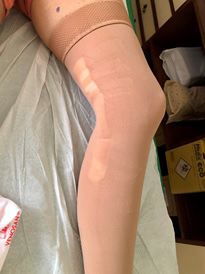
Figure 2) With accord of Dr F. Bastos: Eccentric compression under digressive
(15 to 20 mm Hg) did not modify the diameter of saphenous veins raising
compression
the question that compression, in fact, did not reduce the diameters of
saphenous and superficial veins [14,15]. In the same way, material delivering
a pressure of 20 to 30 mm Hg at the ankle does not compress the superficial To improve reproducibility we therefore try to use a standardised
veins in a standing position. compression stocking or under an overlap of two compression stocking by
interposing a localized overpressure system (silicone foam compress). The
This explains that many experts recommend using eccentric compression. use of standardised compression stockings allows for possible self-installation
by the patient [22].
Luggli uses eccentric compression on the thigh. This use significantly reduced
postoperative pain in comparison to the non-use of eccentric compression However they are difficult to put into place by the patient because the patient
[16]. must also pull on the two compression stockings to position the large foam
around the painful and that after the treatment potentially painful.
Mosti demonstrated the more efficient and better results of the use of
eccentric compression on the thigh after surgery than the use of class III It is the same for other techniques that have proved their efficiency [23]. The
graduated compression stockings [17,18]. patient cannot install it alone for most systems.
Graduated compression is used to facilitate the venous return. This is done The new device: Thanks to the experience and knowledge of our Masters
by the rebalancing of hydrostatic and oncotic pressure of the Starling law we have studied a standardized eccentric compression device reserved for
[19]. compression during and after varicose vein treatment in order to reduce
the volume of blood in the varicose vein during treatment but also after
Eccentric compression is used during treatment to reduce the diameter of treatment to improve fibrosis and to limit the adverse or exacerbated effects
the varicose vein and to reduce the blood volume of the varicose vein. It is of sclerotherapy [24].
used after treatment to control the inflammatory reaction, immobilize the
treated zone and to bring a local antalgic effect, to favor uniform fibrosis, The standardized eccentric contention-compression orthosis (Veinalgic®) is
reduce bruising and facilitate proper cicatrization. a device which is easy to use and fit alone if the patients can reach their thigh
or calf muscle themselves (Figure 3).
Graduated and eccentric compression should not be opposed but should be
considered complimentary and synergic. One facilitates the venous return
and the other facilitates cicatrization by immobilizing the treated zone.
7 J Phlebol Lymphol Vol.14 No.1 2021Foam sclerotherapy and eccentric compression
device is neither tubular nor knitted, it is adjustable, washable, sterilizable
and reusable. It is composed of a contention part which is inextensible and
a maintaining compressive part which is extensible. Patients reported no
inconvenience related to pain or specific discomfort using the compression
system. No patient reported difficulties positioning the system.
The cost is low, it is washable, sterilizable and reusable. It can be used and
conserved by the patient all along the evolution of the chronic venous disease.
The reduction of the diameter is significant with this standardized eccentric
compression with a relatively moderate pressure because the system is
evaluated at 50% extension (marker 2 for an interface pressure of 50 mm
Hg). This pressure can be increased (marker 3) giving us hope that the
Figure 3) Veinalgic®: Stiff central pocket with double density foam. High diameter can be further reduced (Table 1).
Stiffness Index (SI) and significant localized compression, extendable side parts
enable positioning and holding. Velcro fixation Some effects of this device in a recent study: 76.9% of the varicose veins
concerned the Greater Saphenous Vein (GSV) and 23.1% concerned
It is composed of an inextensible tissue pocket containing a dense foam to be the Small Saphenous Vein (SSV). The varicose veins were located in the
applied on the treated varicose vein. Thanks to the reduction of the curvature saphenous compartment in 76.6% of the cases; they were superficial
by the dense foam, the inter face pressure obtained, when the device is therefore above the superficial fascia in 24.4% of the cases. 95.4% of the
placed by patients, is high (40 to 70 mm Hg) and the Stiffness Index (SI) is varicose veins were primary and 4.6% were recurrences [25].
significant and there is no tourniquet effect. The device is maintained on the
skin or on the compression stocking by a vertical silicon strap. The pressure The reduction of the diameter is more than one third (36%) on incontinent
is adjusted by the patient using the numerical markers. This rigid part is saphenous veins and is reduced even more on superficial varicose veins (43%
maintained by two long-stretch elastic straps. This contention-compression reduction) (Table 2).
TABLE 1
Pressure and stiffness index delivered by Veinalgic®, on the thigh, according to the stretching under the elastic part and under the
stiff part
Elastic part Stiff part
Stretching Plying SI Plying SI
P-standing P-standing
30% marker 1 24.6 24.6 0 33 63.4 30.4
50% marker 2 32 36.2 4.2 40.6 73.4 32.8
100% marker 3 46.4 54 7.6 50.8 87.6 36.8
TABLE 2
Evolution of the diameter of varicose veins in a lying position with and without eccentric compression
Group Without veinalgic With veinalgic Delta Student-test (paired)
Below fascia 4.4 ± 1.3 2.8 ± 0.9 1.6 ± 1 Value T:15.4199
(n=97) IC95%:[4.1; 4.6] IC95% [2.6; 2.9] IC95%:[1.4; 1.9] p-value:Crebassa et al.
Eccentric compression allows us to imagine changes our sclerotherapy 9. Hamel-Desnos CM, Guias BJ, Desnos PR, et al. Foam sclerotherapy
indications, changes in injection volumes and an increase in contact time of the saphenous veins: randomised controlled trial with or without
allowing a reduction in the concentrations used. It makes it possible to limit compression. Eur J Vasc Endovasc Surg. 2010; 39(4):500-7.
the harmful effects even if they are not serious in sclerotherapy. 10. Zarca Ch, Bailly C, Gachet G, et al. classMousse 1: bas medicaux et
injection de mousse. Phlebologie. 2012; 65(1):11-20.
After more than 1700 Patients treated with this eccentric compression I advise
to conserve the compression 3 days and more according to the inflammatory 11. Benigni JP, Cornu Thénard A, Uhl JF. la sclérothérapie. Editions ESKA.
or the sensations. The eccentric compression is indicated specially for larger 2007; 205-7.
saphenous veins or for superficial varicose vein but further studies are 12. Benigni JP, Uhl JF, Cornu Thenard A. La thérapeutique compressive a la
however required to evaluate the consequences of eccentric compression on cuisse. Phlebologie. 2009; 62(1):77-80.
our therapeutic attitudes and their results. 13. Ricci S. La sclerotherapie. Editions ESKA. 2007:235-8.
∏ R2L=V1 14. Gardon-Mollard C. Compression médicale. Edition Masson. 219.
15. Partsch H, Menzinger G, Borst-Krafek B, et al. Does thigh compression
∏ (R × 0.57)2L=V2 improve venous hemodynamics in chronic venous insufficiency? J Vasc
Surg. 2002; 36(5):948-52.
∏ R20.572 L=V2
16. Rastel D. No reduction of diameter under the fascia with 20 mm Hg.
(∏ R2L) 0.32=V2 Phlebologie. 2014; 67(1):40-5.
17. Lord RSA, Hamilton D. Graduated compression stockings (20-30
V1 × 0.32=V2
mmHg) do not compress leg veins in the standing position. ANZ J Surg.
Demostration 1: Initial varicose volume × 0.32=Volume using Veinalgic® 2004; 74(7):581-5.
18. G Mosti. Post-treatment compression: Duration and techniques.
R=radius of the varicose vein, R × 0.57=43% reduction=new radius, Phlebology. 2013;28 Suppl 1:21-4.
L=length of the varicose vein V1=volume of the varicose vein, V2=volume 19. Mosti G, Mattaliano V, Arleo S, et al. Thigh compression after great
after compression. saphenous surgery is more effective with high pressure. Int Angiol. 2009;
28(4):274-80.
REFERENCES
20. Crebassa V. Oedème veineux chronique et compression médicale.
1. Desnos P. Mode d’action des différents Lasers endo-veineux et impact Phlebologie. 2014; 67(3):34-42.
des differentes longueurs d’onde. Phlebologie. 2013; 66(2):28-33.
21. Gachet G, Galem K. L’écho-sclérose mousse des varices sous compression
2. Anastasie B, Mordon S, Cazaubon M, et al. Lasers endo-veineux. ou tumescence externe : L’étude MOUSSECOMP. Phlebologie. 2014;
Angiologie. 2012; 64(2):50-62. 67(3):23-28.
3. Luggli M, Logo A, Guerzoni S, et al. Effects of eccentric compression 22. Benigni JP, Allaert FA, Desoutter P, et al. The efficiency of pain control
by a crossed-tape technique after endovenous laser ablation of the great using a thigh pad under the elastic stocking in patients following venous
saphenous vein: a randomized study. Phlebology. 2009; 24(4):151-6. stripping: Results of a case-control study. Perspect Vasc Surg Endovasc
4. Mosti G. Post-treatment compression: Duration and techniques. Ther. 2011; 23(4):238-43.
Phlebology. 2013; 28(1):21-4. 23. Milleret R. The sclerosis of the large saphenous veins by means of foam
5. Lurie F, Lal BK, Antignani PL, et al. Compression therapy after invasive emitted through an ultrasound-guided catheter: Alpha Technique. The
treatment of superficial veins of the lower extremities: Clinical practice conclusions of the first 1000 treatments. Phlebology. 2006;59:53-8.
guidelines of the American Venous Forum, Society for Vascular Surgery, 24. Gardon-Mollard C, Crebassa V. Évaluation de la pression interface
American College of Phlebology, Society for Vascular Medicine, and délivrée et de l’Indice deRigidité (IR) d’un nouveau dispositif combine
International Union of Phlebology. J Vasc Surg Venous Lymphat de contention-compression excentrée pour les suites des traitements
Disord. 2019; 7(1):17-28. de volumineuses varices saphènes ou superficielles. Phlebologie. 2014;
6. Parsi K, Exner T, Low J, et al. In vitro Effects of Detergent Sclerosants 67(4):32-64.
on Clot Formation and Fibrinolysis. Eur J Vasc Endovasc Surg. 2011; 25. Crébassa V, Galleze B, Gardon-Mollard C, et al. Réduire de 70% le
41(2):266-77. volume des varices pendant et après traitements. Tumescence externe
7. Watkins MR. Deactivation of Sodium Tetradecyl Sulphate Injection by par compression excentrée standardisée. Phlebologie. 2017; 70(3):20-8.
Blood Proteins. Eur J Vasc Endovasc Surg. 2011; 41(4):521-5. 26. Fegan WG, Fitzgerald DE. A histologic assessment of continuous
8. Rabe E, Breu FX, Cavezzi A, et al. European guidelines for sclerotherapy compression sclerotherapy. Angiology. 1965:433-42.
in chronic venous disorders. Phlebology. 2014; 29(6):338-54.
9 J Phlebol Lymphol Vol.14 No.1 2021You can also read