Intra-aortic Balloon Pump Induced Dynamic Left Ventricular Outflow Tract Obstruction and Cardiogenic Shock: A Case Report
←
→
Page content transcription
If your browser does not render page correctly, please read the page content below
ISSN: 2474-3674
WONG and Ng. Int J Crit Care Emerg Med 2021, 7:123
DOI: 10.23937/2474-3674/1510123
Volume 7 | Issue 3
International Journal of Open Access
Critical Care and Emergency Medicine
Case Report
Intra-aortic Balloon Pump Induced Dynamic Left Ventricular
Outflow Tract Obstruction and Cardiogenic Shock: A Case
Report
Shiun Woei WONG, MRCP1,2* and Ke Xuan Jessica Ng, MRCP1
Department of Cardiology, Tan Tock Seng Hospital, Singapore
1 Check for
updates
Lee Kong Chian School of Medicine, Singapore
2
*Corresponding author: Dr. Wong Shiun Woei, Department of Cardiology, Tan Tock Seng Hospital, 11, Jalan Tan Tock
Seng, 308433, Singapore, Tel: (65)-6357-7831, Fax: (65)-6357-3772
Abstract Association (AHA) gave a class 2A indication [1] for IABP
counterpulsation in the setting of cardiogenic shock.
We present a complex case of a 50-year-old man
who presented with inferior ST-elevation myocardial
However, long term 6-year outcome of IABP-Shock II
infarction and cardiogenic shock. Our patient presented trial [2] did not demonstrate a reduction in mortality
with out-of-hospital ventricular fibrillation cardiac arrest. in the setting of cardiogenic shock. Left ventricular
Cardiopulmonary resuscitation was promptly started and outflow tract obstruction (LVOTO) has been described
return of spontaneous circulation was achieved. The patient
in Takotsubo cardiomyopathy [3] and acute anterior
underwent urgent coronary angiography with implantation
of drug-eluting stent in his right coronary artery. Intra-aortic myocardial infarction (MI) [4]. To our knowledge, we
balloon pump was inserted. However, this was complicated describe the first report of LVOTO worsened by IABP
by worsening of left ventricular outflow tract obstruction insertion in the context of inferior myocardial infarction.
and systolic anterior motion of mitral valve leaflet. He We present the following case in accordance with the
was successfully weaned off balloon pump, inotropes and
liberated from mechanical ventilation. To our knowledge,
CARE reporting checklist.
this is the first case reported of balloon pump associated left
ventricular outflow tract obstruction in a setting of inferior
Case Description
myocardial infarction. This case illustrates the importance of A 50-year-old male with no significant history
early removal of balloon pump and precise use vasopressor
apart from smoking history presented with VF arrest.
in instituting the stepwise treatment modalities leading to a
favourable outcome. Cardiopulmonary resuscitation and immediate
defibrillation were done on-site by paramedic. His no-
flow time was 5 minutes whereas his low-flow time was
Introduction close to 25 minutes. His examination was remarkable
A 50-year-old man presented with out-of-hospital for a bilateral lung crepitations but there was no audible
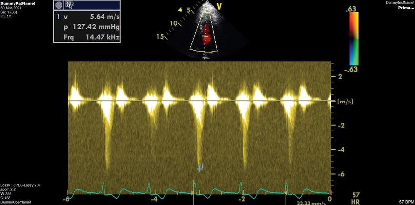
ventricular fibrillation (VF) cardiac arrest from inferior murmur. His 12-lead electrocardiogram (ECG) showed
ST-elevation myocardial infarction. The patient was sinus tachycardia with ST elevation in the inferior
intubated and started on intravenous (IV) noradrenaline. leads (Figure 1). He was given oral aspirin, ticagrelor
Primary percutaneous coronary intervention (PCI) and intravenous frusemide. However, he developed
was done to his right coronary artery (RCA) with cardiogenic shock and acute respiratory failure and he
implantation of drug-eluting stent. Intra-aortic balloon was intubated. Urgent coronary angiogram showed
pump (IABP) was inserted in view of escalating pressor triple vessel disease with acute occlusion of right
requirement and cardiogenic shock. American Heart coronary artery (Video 1 and Video 2). The left anterior
Citation: WONG SW, Ng KXJ (2021) Intra-aortic Balloon Pump Induced Dynamic Left Ventricular Out-
flow Tract Obstruction and Cardiogenic Shock: A Case Report. Int J Crit Care Emerg Med 7:123. doi.
org/10.23937/2474-3674/1510123
Accepted: July 08, 2021: Published: July 10, 2021
Copyright: © 2021 WONG SW, et al. This is an open-access article distributed under the terms of the
Creative Commons Attribution License, which permits unrestricted use, distribution, and reproduction
in any medium, provided the original author and source are credited.
WONG and Ng. Int J Crit Care Emerg Med 2021, 7:123 • Page 1 of 5 •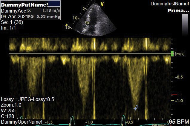
DOI: 10.23937/2474-3674/1510123                                                                                                                                                                                ISSN: 2474-3674

                  ZULKIFLI BIN OTHMAN,                                  ID:S1440219E            29-MAR-2021 22:30:22                       TAN TOCK SENG HOSPITAL PL-DEFLT           ROUTINE RETRIEVAL
                  15-MAR-1960 (61 yr)                  Vent. rate                  105   BPM      Sinus tachycardia
                  Male                                 PR interval                 166     ms     Inferoposterior infarct, acute (RCA)
                                                       QRS duration                 99     ms     Probable RV involvement, suggest recording right precordial leads
                  Room:                                QT/QTc                  355/470     ms     Baseline wander in lead(s) V1,V2,V3,V4
                  Loc:0                                P-R-T axes               77 46     131     >>> Acute MI
DOI: 10.23937/2474-3674/1510123                                                                            ISSN: 2474-3674

                    Figure 2: Transthoracic echocardiogram showed peak gradient of 127 mmHg in LVOT.

                                    Figure 3: Resolution of LVOT peak gradient to 5 mmHg.

descending artery was 100% occluded and it received             was elevated at 10.9 mmol/L, total white count 22 ×
collaterals from the right coronary artery.                     10^9/L, creatinine 300 umol/L and alkaline phosphatase
                                                                elevated at 500 IU/L.
    The patient became hypotensive after catheterization,
with systolic pressures measures between 80 and 90                 IABP was subsequently placed on 1:3 augmentation
mmHg. IABP was inserted and vasopressin was added.              and intravenous phenylephrine initiated in view of
Immediate transthoracic echocardiogram (TTE) showed             significant LVOTO. Intravenous fluid boluses were given
left ventricular ejection fraction (LVEF) of 45% with           based on dynamic parameters of fluid responsiveness.
systolic anterior motion (SAM) of the anterior mitral           Volume View (Edwards Life science) was used as
leaflet and LVOT gradient of 127 mmHg (Video 3 and              a tool for hemodynamic monitoring. The blood
Figure 2). He underwent thrombus aspiration and                 pressure improved significantly with these measures.
balloon angioplasty using a Ryurei 2.0 × 15 mm NC               Vasopressin (maximum infusion rate of 1.8 IU/kg/hr)
balloon, inflated with high pressure under intravascular        and Noradrenaline (maximum infusion rate of 0.6 mcg/
ultrasound guidance Drug-eluting stent (Ony × 3.5 × 10          kg/min) were gradually weaned off with no rebound
mm) was implanted on the right coronary artery with             hypotension. IABP was removed with vascular closure
re-establishment of TIMI 3 flow (Video 4).                      device on day 4 of ICU stay. His heart rate remained well
                                                                controlled with IV Esmolol and Remifentanil infusion.
    His maximum troponin was 22,000 ng/L. He was                He was promptly liberated from mechanical ventilation
treated with targeted temperature management at                 on Day 6 of hospitalization. His kidney and liver function
33 C, IV Piperacillin-Tazobactam, hydrocortisone 50             improved gradually with no requirement for renal
mg 6 hourly and Atorvastatin 40 mg daily. The patient           replacement therapy. A repeat TTE demonstrated no
subsequently suffered downstream complication of                LVOTO with a peak gradient of 5.53 mmHg (Figure 3).
cardiogenic shock, i.e., ischemic hepatitis, acute kidney       There was concentric hypertrophy, mild chordal SAM
injury and ventilator associated pneumonia. His lactate         with marked improvement of LVEF to 55% (Video 5).

WONG and Ng. Int J Crit Care Emerg Med 2021, 7:123                                                         • Page 3 of 5 •
DOI: 10.23937/2474-3674/1510123 ISSN: 2474-3674
All procedures performed in studies involving would significantly improve the outcome in this critically
human participants were in accordance with the ethical ill patient. In our patient, we showed that withdrawing
standards of the institutional and/or national research inotropes and IABP timely improved his hypotension
committee(s) and with the Helsinki Declaration (as markedly.
revised in 2013). Written informed consent was
obtained from the patient.
Authors Declarations
Reporting checklist
Conclusion
The authors have completed the CARE reposting
We present a case of refractory cardiogenic shock due
checklist.
to dynamic LVOTO worsened by IABP counterpulsation.
The major tool for assessment of LVOTO, however, is Conflicts of interest
echocardiography. The spectral Doppler waveform in
our patient fits with the typical late peaking or dagger All authors report no conflict of interest.
shape pattern found in dynamic LVOTO [5]. Ethical statement
Numerous reports have highlighted the occurrence This report was in accordance with institutional
of dynamic LVOTO as a complication of ST-Elevation ethical standards and in accordance with Helsinki
myocardial infarction (STEMI) [6,7]. The actual incidence Declaration. This case report has non-identifiable clinical
of these findings is unclear, but it may be significantly data of our patient.
underdiagnosed and can indeed mimic cardiogenic
shock in an acute-care setting. Failure of the anterior Source of Support
mitral valve leaflet to coapt with the posterior leaflet in None declared.
systole results in MR. The degree and duration of mitral
SAM determine the severity of the dynamic LVOTO References
gradients and MR [8-10]. Although classically described 1. O'Gara PT, Kushner FG, Ascheim DD, Casey DE Jr,
in hypertrophic cardiomyopathy, SAM and LVOTO can Chung MK, et al. (2013) 2013 ACCF/AHA guideline for
result from various clinical settings. We speculated the management of ST-elevation myocardial infarction: A
report of the American College of Cardiology Foundation/
that compensatory basal LV hypercontractility based
American Heart Association Task Force on Practice
upon mild LV narrowing led to SAM, and initiation of Guidelines J Am Coll Cardiol 61: e78-e140.
IABP further deteriorated dynamic LVOTO, which
2. Holdger T, Ume Z, Nathalie T, Franz-Josef N, Jorg H, et
result in intractable cardiogenic shock. Loss of normal al. (2019) Intraaortic balloon pump in cardiogenic shock
ventricular geometry may also affect the tension and complicating acute myocardial infarction. Circulation 139:
position of the mitral valve apparatus making SAM 395-403.
more apparent. In addition, the right coronary artery 3. Tsuchihashi K, Ueshima K, Uchida T, Oh-mura N, Kimura
was supplying collaterals to the chronically occluded left K, et al. (2001) Transient left ventricular apical ballooning
anterior descending artery. Loss of flow in most of the without coronary artery stenosis: A novel heart syndrome
LV myocardium precipitated the ventricular arrhythmia mimicking acute myocardial infarction: Angina Pectoris-
Myocardial Infarction Investigations in Japan. J Am Coll
as well as cardiogenic shock.
Cardiol 38: 11-18.
The inotropic effects of most vasopressors and 4. Hrovatin E, Piazza R, Pavan D, Mimo R, Macor F, et al.
the stress of critical illness in the setting of a low (2002) Dynamic left ventricular outflow tract obstruction in
intravascular volume state may provoke LVOTO in the setting of acute anterior myocardial infarction: A serious
susceptible patients [11,12]. Inotropes also promote and potentially fatal complication? Echocardiography 19:
449-455.
hypercontractility [13,14], accelerate blood across the
LVOT and worsen SAM of the anterior mitral valve 5. Chockalingam A, Dorairajan S, Bhalla M, Dellsperger K
leaflet. Unrecognized LVOTO may lead to a spiral of (2009) Unexplained hypotension: The spectrum of dynamic
left ventricular outflow tract obstruction in critical care
inappropriate vasopressor/inotrope use resulting in settings. Crit Care Med 37: 729-734.
severe hypotension, collapse, and death. In many
6. Takeshi TM, Kaoru D, Akitoshi S, Kazuki M, Keishi M, et
patients the onset of LVOTO is unexpected and
al. (2012) Intra-aortic balloon pump induced dynamic left
gradients can vary significantly over a short period of ventricular outflow tract obstruction and cardiogenic shock
time [15]. Given its unpredictable nature, the presence after very later stent thrombosis in left anterior descending
of dynamic LVOTO in a hypotensive critically ill patient coronary artery. J Cardiol Cases 6: e137-e140.
should always be considered. Correct therapy involves 7. Dahhan A, Mohammad A, Kapoor D, Sharma GK (2011)
avoidance of hypovolemia, using beta blockers to Hypotension due to dynamic left ventricular outflow tract
reduce the hypercontractile state and alpha agonist obstruction after percutaneous coronary intervention. Tex
(Phenylephrine) to increase the systemic vascular Heart Inst J 38: 723-726.
resistance and reduce the LVOT gradient. 8. Pellikka PA, Oh JK, Bailey KR, Nichols BA, Monahan KH,
et al. (1992) Dynamic intraventricular obstruction during
Clinical suspicion, early recognition, and appropriate dobutamine stress echocardiography: A new observation.
management of LVOTO, along with addressing of STEMI, Circulation 86: 1429-1432.
WONG and Ng. Int J Crit Care Emerg Med 2021, 7:123 • Page 4 of 5 •DOI: 10.23937/2474-3674/1510123 ISSN: 2474-3674
9. Luria D, Klutstein MW, Rosenmann D, Shaheen J, Sergey S, obstruction is associated with hypovolemia and high
et al. (1999) Prevalence and significance of left ventricular mortality in septic shock patients. Crit Care 19: 262.
outflow gradient during dobutamine echocardiography. Eur
13. Caselli S, Martino A, Genuini I, Santhini D, Carbone I,
Heart J 20: 386-392.
Agati L, et al. (2010) Pathophysiology of dynamic left
10. Hymel BJ, Townsley MM (2014) Echocardiographic ventricular outflow tract obstruction in a critically ill patient.
assessment of systolic anterior motion of the mitral valve. Echocardiography 27: E122-E124.
Anesth Analg 118: 1197-1201.
14. Slama M, Tribouilloy C, Maizel J (2016) Left ventricular
11. Yang JH, Park SW, Yang JH, Cho SW, Kim HW, et al. (2008) outflow tract obstruction in ICU patients. Curr Opin Crit
Dynamic left ventricular outflow tract obstruction without Care 22: 260-266.
basal septal hypertrophy, caused by cathecholamine
15. Cohen R, Rivagorda J, Elhadad S (2006) Asymmetric
therapy and volume depletion. Korean J Intern Med 23:
septal hypertrophy complicated by dynamic left ventricular
106-109.
obstruction after intra-aortic balloon pump counterpulsation
12. Chauvet J-L, El-Dash S, Delastre O, Bouffandeau B, placement in the setting of anterior myocardial infarction. J
Jusserand D, et al. (2015) Early dynamic left intraventricular Invasive Cardiol 18: E207-E208.
WONG and Ng. Int J Crit Care Emerg Med 2021, 7:123 • Page 5 of 5 •You can also read