Contrast imaging ultrasound for the detection and characterization of carotid vulnerable plaque
←
→
Page content transcription
If your browser does not render page correctly, please read the page content below
Review Article on Advanced Imaging in The Diagnosis of Cardiovascular Diseases

Contrast imaging ultrasound for the detection and characterization
of carotid vulnerable plaque
Vasileios Rafailidis1#, Xin Li2#, Paul S. Sidhu1, Sasan Partovi3, Daniel Staub4
1
Department of Radiology, King’s College Hospital, London, UK; 2Department of Internal Medicine, University Hospital Cleveland Medical Center,
Case Western Reserve University, Cleveland, Ohio, USA; 3Interventional Radiology Section, Imaging Institute, Cleveland Clinic Foundation,
Cleveland, Ohio, USA; 4Department of Angiology, University Hospital Basel, University of Basel, Basel, Switzerland
Contributions: (I) Conception and design: All authors; (II) Administrative support: D Staub, S Partovi, V Rafailidis, X Li; (III) Provision of study
materials or patients: D Staub, V Rafailidis, X Li; (IV) Collection and assembly of data: None; (V) Data analysis and interpretation: None; (VI)
Manuscript writing: All authors; (VII) Final approval of manuscript: All authors.
#
These authors contributed equally to this work.
Correspondence to: Prof. Daniel Staub. Department of Angiology, University Hospital Basel, University of Basel, Basel, Switzerland. Email: daniel.staub@usb.ch.

                  Abstract: Not only the degree of luminal narrowing but also the plaque morphology and composition play
                  an important role in risk stratification of carotid atherosclerotic lesions. During the last few years, carotid
                  contrast-enhanced ultrasound (CEUS) has emerged as a valuable imaging tool to assess such vulnerable carotid
                  plaques. This review article discussed the use of CEUS for the detection of carotid plaque irregularities and
                  ulcerations as well as the quantification of intraplaque neovascularization and its correlation with histology
                  and inflammatory biomarkers. Apart from evaluating for markers of vulnerable carotid plaques, CEUS
                  enhancement is directly associated with past cerebrovascular events. More importantly, preliminary evidence
                  has shown that CEUS could be used to predict future cerebrovascular and cardiovascular events. Despite the
                  progress in CEUS imaging for carotid atherosclerotic disease, past studies still suffer from the retrospective
                  nature, small sample size, and a lack of matched, well controlled prospective studies. In the future, large
                  multi-center prospective studies addressing the relationship between CEUS findings and patient clinical
                  outcomes in carotid atherosclerotic disease are warranted.

                  Keywords: Carotid plaque; ultrasound; contrast-enhanced ultrasound (CEUS); ulceration; neovascularization

                  Submitted Dec 23, 2019. Accepted for publication Jan 07, 2020.
                  doi: 10.21037/cdt.2020.01.08
                  View this article at: http://dx.doi.org/10.21037/cdt.2020.01.08

Introduction                                                                       the degree of carotid stenosis. However, there is clear
                                                                                   evidence, that not only the degree of luminal narrowing but
Atherosclerotic disease is a leading cause of morbidity
                                                                                   also plaque morphology and plaque composition assessed
and mortality. In carotid atherosclerotic disease, high-
                                                                                   by contrast-enhanced ultrasound (CEUS) play an important
risk plaques may rupture and this can lead to neurological                         role to characterize such vulnerable carotid plaques (3). This
sequelae, such as transient ischemic attacks (TIA) and                             review article discusses the use of CEUS for the detection
cerebral ischemic infarction (1). Further, when treating                           and characterization of carotid vulnerable plaques.
symptomatic carotid stenosis, there is an inherent risk of
downstream microembolization from manipulating at-
                                                                                   CEUS—basic principles and application
risk carotid plaques. High-risk plaques are characterized as
“vulnerable” and predispose patients to an elevated risk of                        As with every imaging modality, physicians performing
cardiovascular events (2).                                                         CEUS need to familiarize themselves with the technique’s
   Duplex ultrasound imaging is well established to assess                         basic principles, in order to be able to accurately interpret

© Cardiovascular Diagnosis and Therapy. All rights reserved.            Cardiovasc Diagn Ther 2020;10(4):965-981 | http://dx.doi.org/10.21037/cdt.2020.01.08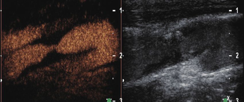
966                                                                              Rafailidis et al. Carotid contrast ultrasound and vulnerable plaque

findings, to promptly detect artifacts and to successfully                          metabolized by the liver and the internal gas is exhaled
address those, for the benefit of the patient. Being an                             by the respiratory tract. This feature is valuable as UCAs
advanced ultrasonographic technique, CEUS makes use                                 can be safely used in patients with renal failure, a patient
of the basic principles of ultrasonography (US) including                           group commonly affected by vascular pathology requiring
the emission and reception of an ultrasound beam after                              imaging. The excellent safety profile of UCAs is also
its interaction with human tissue. In addition, CEUS is                             complemented by the lack of interaction with the thyroid
characterized by some additional features, including the                            gland and the very low percentage of allergic reactions (6-8).
ultrasonographic contrast agent (UCA), the hardware
needed for the emission of specialized ultrasound beam
                                                                                    CEUS: hardware aspects
sequences and the software necessary for post-processing
and analysis. In this section, these aspects will be briefly                        The Mechanical Index, commonly known as MI, is one
discussed with regards to the carotid application of CEUS.                          of the most important parameters in US, indicating the
                                                                                    insonation power of the ultrasound beam used at any
                                                                                    moment during scanning and the “pressure” applied to the
The UCA
                                                                                    tissues scanned. In terms of mathematics, it is defined as the
Many UCAs have been made commercially available                                     ratio of peak negative pressure with the square root of US
over the last years, each one with slight differences in                            frequency. For conventional ultrasonographic techniques
composition, interaction with US beam, and variations                               such as gray-scale or colour Doppler technique, the MI is
in the approved indications in different countries.                                 typically over 1.6. For CEUS though, a much lower MI
Nevertheless, the structure of all UCAs is ubiquitous                               should be used for two reasons. First, microbubbles tend to
and includes a microbubble consisting of an internal                                react in a different way to increasing values of MI. Namely,
gas encapsulated by an external shell made of either                                when exposed to a very low MI US beam, microbubbles
phospholipids or albumin. Nowadays, SonoVue® (Bracco)                               will oscillate in a linear pattern, meaning that they will
is the UCA most frequently used for vascular applications,                          expand at the same rate that they will contract. This pattern
being approved for macro-vascular applications including                            of response will reflect the same exact frequency with the
cerebral arteries, extracranial carotid or other peripheral                         one initially emitted by the probe. If higher but still low
arteries and the portal vein in Europe (4).                                         values of MI (ranging from 0.08 to 0.1 for vascular clinical
    S o n o Vu e ® ( B r a c c o ) i s a m i c r o b u b b l e o f s u l f u r      applications) are applied, microbubbles will expand to a
hexafluoride (SF6) contained inside a monolayer shell of                            higher degree than they contract (a pattern termed non-
phospholipids. Given that this shell is hydrophilic on the                          linear oscillation), thus not only reflecting the baseline
outer surface and hydrophobic on the inner, it is stable and                        frequency but also generating harmonic frequencies.
keeps the gas successfully contained within the microbubble.                        Of course, this reaction cannot last for more than a few
On the other hand, the phospholipid shell is flexible enough                        minutes, by which time the microbubbles rupture. The same
to allow changes in size and shape; a phenomenon termed                             will happen if high MI values (those used for conventional
“oscillation” which is crucial for the generation of CEUS                           techniques) are applied to microbubbles. The second
signal (5). SonoVue® microbubbles have a mean diameter                              reason for which a low MI should be used for CEUS is that
of 2.5 μm, which is a crucial feature for clinical practice not                     static tissues (not microbubbles) will produce linear signals
only because it allows the microbubbles to travel through                           when exposed to a low-MI beam but will also produce
the entire vascular bed of the human body, reaching the                             harmonic frequencies similar to those of microbubbles if a
smallest capillary and traversing the pulmonary circulation,                        high-MI pulse is emitted. As a consequence, the harmonic
but also because it prevents the microbubbles from crossing                         frequencies emitted by static tissues will be confounded
the endothelium and hence exiting the vascular lumen. As                            with those originating from the UCA, making it difficult to
a result, microbubbles are strictly intravascular contrast                          exclusively visualize UCA (9-11).
agents, which is a property of paramount importance,                                    Historically, the first generation of CEUS techniques
particularly in vascular applications of CEUS (5). Similar                          was based on colour or power Doppler technique and
to CT and MRI contrast agents, UCAs are administered                                made use of a high-MI intermittent US beam which caused
intravenously but unlike the former, these are never                                disruption of the microbubbles and hence production of
excreted by the kidneys, as the phospholipid capsule is                             strong signal intensity and improvement of signal-to-noise

© Cardiovascular Diagnosis and Therapy. All rights reserved.              Cardiovasc Diagn Ther 2020;10(4):965-981 | http://dx.doi.org/10.21037/cdt.2020.01.08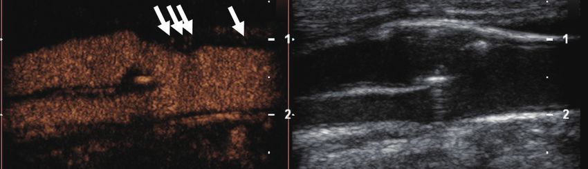
Cardiovascular Diagnosis and Therapy, Vol 10, No 4 August 2020                                                                                967

ratio. Nevertheless, this form of CEUS suffered from all                 time, leading to the time-intensity curves (TIC) (4,13).
the inherent Doppler artifacts such as overwriting artifact                  Temporal maximum intensity projection (MIP) is a
and lacked the real-time nature of currently available                   useful tool for vascular imaging, available in most US
techniques (12). Having previously described the principles              scanners. In this mode, a high-MI pulse is initially used to
governing the microbubbles—US interaction, it becomes                    disrupt microbubbles and completely erase the signal in the
evident that currently available US machines can use the                 field-of-view. Subsequently, low-MI continuous scanning is
different frequencies generated by UCA and static tissue                 performed, and every frame is aggregated to the previous
to differentiate them. In the pulse-inversion technique,                 one, thus creating a complex image containing signal from
which is currently the most widely used mode of CEUS,                    every microbubble imaged over a period of time. In essence,
the US probe emits two pulses identical in amplitude                     the scanner acts as an “open shutter camera” and creates
and frequency but with a difference of 180° in phase. As                 detailed images of macro- or micro-vascular anatomy (14).
a result, when those pulses are linearly reflected by static
tissues, the second pulse cancels the first, being its inverted
                                                                         CEUS: protocol
copy. However, when these pulses hit microbubbles,
harmonic frequencies are produced and reflected towards                  Ideally, CEUS should be performed after the completion of
the transducer. Therefore, hardware is able to selectively               the conventional US study and the area of interest has been
visualize microbubbles by suppressing the signal from static             identified. In this way, the scanning location for the CEUS
tissues at the same time (6).                                            examination can be determined and a focused scan can be
    Two of CEUS’ advantages over CT and MRI are the                      performed. Of course, both carotid arteries can be scanned
ability for prolonged scanning of the contrast agent and                 using a single dose of UCA, thanks to the prolonged
the real-time scanning pattern characterized by high                     enhancement time offered by current UCAs. A typical
spatial and temporal resolution. The enhancement pattern                 CEUS protocol for carotid CEUS examination is outlined
of structures with microbubbles is typically recorded in                 in Table 1.
cine loops in everyday clinical practice. Nevertheless,
the reperfusion technique is available in the setting of
                                                                         CEUS for the detection of plaque surface
low-MI CEUS. In this technique, a high MI pulse is
                                                                         irregularities and ulceration
instantaneously emitted, disrupting every microbubble in
the imaging field. In this way, the physician can re-observe             CEUS applications in the carotid system provides a
the arrival of microbubbles or even quantify them using                  significant amount of information both on a macro-
specialized software, making this technique a useful tool in             and micro-vascular level. Namely, CEUS can accurately
CEUS such as, identifying or quantifying carotid plaque                  delineate carotid plaque irregularities (macro-vascular/
neovascularization or confirming an area of extravasation (4).           luminal level) and can detect intraplaque neovascularization
                                                                         (micro-vascular/intraplaque level) (16). Carotid plaque
                                                                         surface irregularities and ulceration represent an issue of
CEUS: software aspects
                                                                         great clinical significance, as many studies have shown
Once acquired by the transducer, the signal produced by                  significant clinical correlation with the occurrence of
the microbubbles can be visualized on the US machine                     neurologic symptoms, embolic signals on transcranial
screen, usually using the dual-screen technique, where the               Doppler and stroke (17-19).
contrast specific image is shown next to a low-MI grayscale                  Describing carotid plaque surface morphology should be
image, helping the physicians orient themselves through the              an integral part of every imaging modality used for carotid
scanning field. Currently available devices allow for real-              atherosclerosis evaluation, as also recommended by the
time visualization of microbubbles for more than 4 minutes,              American Society of Neuroradiology (ASNR) (20). When
thus enabling qualitative evaluation of enhancement.                     it comes to US, this can be done in a straightforward way
Nevertheless, quantitative analysis of CEUS signal is now                by assessing the plaque surface morphology. Nonetheless,
feasible using both software integrated in US scanners and               it should be kept in mind that conventional US techniques
commercially available software packages. In this type of                such as colour Doppler or power Doppler may occasionally
analysis, regions of interest can be drawn over parts of the             be limited by lower sensitivity to slow blood flow (as in
CEUS image and the signal intensity can be plotted against               the case of a severely stenotic atherosclerotic lesion or

© Cardiovascular Diagnosis and Therapy. All rights reserved.   Cardiovasc Diagn Ther 2020;10(4):965-981 | http://dx.doi.org/10.21037/cdt.2020.01.08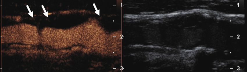
968                                                                         Rafailidis et al. Carotid contrast ultrasound and vulnerable plaque

 Table 1 A typical carotid CEUS protocol
 Parameter                                                                       Reference value/setting

 MI
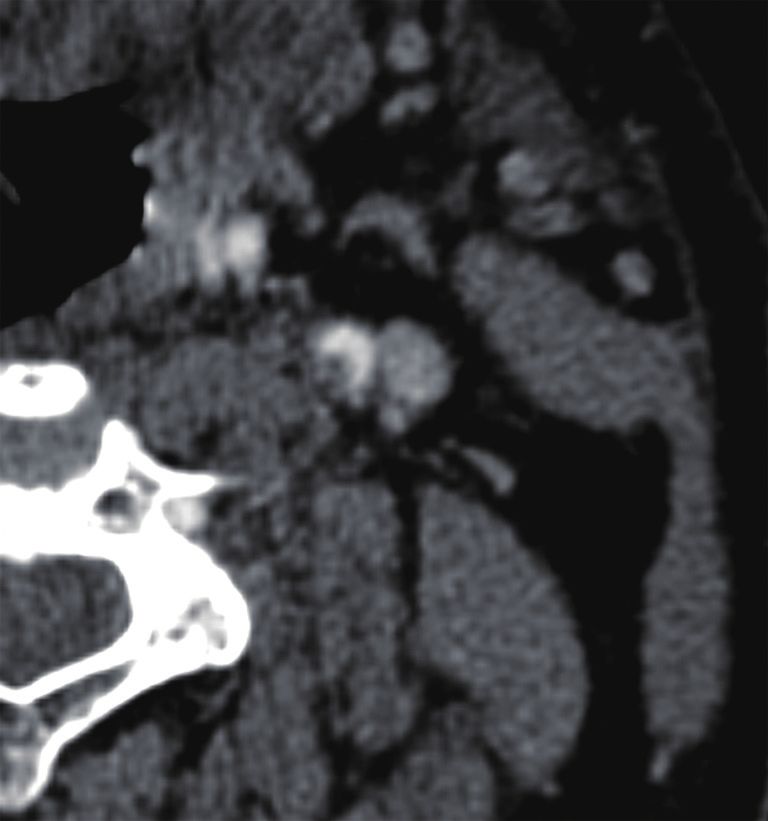
Cardiovascular Diagnosis and Therapy, Vol 10, No 4 August 2020                                                                                 969

A                                                  B                                                        C

Figure 1 An asymptomatic 63-year-old patient with ulcerated severely stenotic internal carotid artery plaque. Colour Doppler image (A)
shows the presence of echogenic plaque in the origin of internal carotid artery. Due to overwriting artifact, the plaque’s border cannot
be accurately detected, while the plaque appears irregular. On CEUS (B), an ulceration is detected (arrowhead), and the severely stenotic
lumen can be followed. Note the presence of calcification projected in this contrast-specific image (arrow), which should not be mistaken for
intraplaque neovessels or ulceration. MDCTA (C) confirming CEUS findings and the presence of ulceration (arrowhead). CEUS, contrast-
enhanced ultrasound; MDCTA, multi-detector computed tomography angiography.

conflicting results, with sensitivity values ranging from                 limitation could be overcome by the introduction of 3D
23% to 85% when the newer criteria were used (17,29).                     CEUS techniques which proved feasible in the evaluation
CEUS has been evaluated and has been directly compared                    of even complicated cases of atherosclerosis, including
to conventional colour Doppler techniques for the diagnosis               ulcerated or heavily calcified plaques. Novel 3D CEUS
of ulceration, demonstrating improved diagnostic accuracy                 achieved better agreement with angiography than colour
(Figure 1). In a study with symptomatic patients only,                    Doppler technique, in quantifying atherosclerosis (34).
colour Doppler technique was only 29% sensitive whereas                   Albeit of these promising results, it is expected that 3D
CEUS was 88% sensitive, with multi-detector computed                      CEUS might not be widely available for clinical practice
tomography angiography (MDCTA) as the reference                           in the near future. When interpreting CEUS images, it is
method (8). In a different study with both symptomatic                    important not to misinterpret echogenic parts of the plaque
and asymptomatic patients, colour Doppler technique was                   (calcifications) projecting to the contrast-specific part of a
41.2% sensitive and CEUS was 94.1% sensitive, while both                  dual-display image as ulceration. Comparison with the low-
techniques were 97.95% specific. CEUS also showed better                  MI grayscale image will readily address this issue (35).
concordance with MDCTA reference imaging (27). In a                          Quantification analysis is one of the current and most
different research setting, CEUS has been used to assess                  essential trends in many aspects of radiology, in an attempt
subclinical atherosclerosis and ulceration in patients with               to reduce subjectivity and thus to improve inter-observer
diabetes mellitus, detecting pathological plaque in 8% of                 agreement. Given the prevalence and clinical significance of
carotid segments examined (30). Thanks to its excellent                   carotid plaque irregularities, there have been some attempts
spatial and temporal resolution, CEUS can visualize a                     to quantitatively analyze and correlate plaque irregularities
swirling movement of microbubbles within ulcerations,                     with neurologic symptomatology. Although the entity
a movement pattern indicative of the arterio-arterial                     of “Bending Energy” failed to discriminate symptomatic
embolization (31). This phenomenon can be observed                        from asymptomatic plaques (36), a more recent approach
in 18% of ulcerations (27). This swirling movement                        suggested by Kanber et al. provided promising results.
had previously been shown in an experimental setting                      Using an intuitive approach, this team used a quantitative
using models of vessels (32) and can also be observed                     index deriving from the summation of angular deviation of
on colour Doppler technique in the form of the “yin-                      the plaque surface from a straight line, divided by the length
yang sign” (33). One limitation of CEUS is that it is a                   of the plaque surface. The resulting index was termed the
two-dimensional technique which may occasionally be                       surface irregularity index (SII) and was calculated using a
limited in the evaluation of complex three-dimensional                    semi-automatic software based on B-mode images. The SII
(3D) structures such as atherosclerotic blood vessels. This               was found to be an independent risk factor in predicting

© Cardiovascular Diagnosis and Therapy. All rights reserved.    Cardiovasc Diagn Ther 2020;10(4):965-981 | http://dx.doi.org/10.21037/cdt.2020.01.08
970 Rafailidis et al. Carotid contrast ultrasound and vulnerable plaque
A B
350
400
400
450 450
500
500
550
550
600
600 650
150 200 250 300 350 400 450 500 550 600 800 900 1000 1100 1200 1300
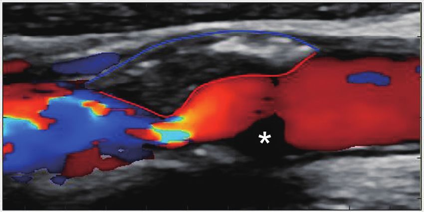
Figure 2 A male patient with a mixed echogenicity atherosclerotic plaque of the internal carotid artery. An example of a surface irregularity
quantification software using colour Doppler image (A). Note the delineation of plaque surface with a red line and the outer vascular wall
with a blue line. Parts of the lumen are not filled with blood flow signals (asterisk), while parts of the plaque surface may be obscured by
overwriting artifact. Same software using a CEUS image (B), where the lumen is fully filled with microbubbles and the plaque surface can be
better appreciated and delineated. CEUS, contrast-enhanced ultrasound.
ipsilateral hemispheric symptoms, since it was significantly depth and width measurement respectively (41). Based on
higher in symptomatic plaques, while it was not associated these findings, quantification of carotid plaque irregularities
with the degree of stenosis (37). In a later study, SII was using both Doppler techniques and CEUS appears feasible
combined with plaque grayscale median (GSM) and and a promising technique for the detection of vulnerable
stenosis to produce a multi-parametric vulnerability index carotid plaques. Further studies are needed using different
which was significantly higher in symptomatic plaques, approaches or even 3D ultrasonographic techniques.
and more importantly outperformed stenosis alone for Given its ability to better delineate plaque surface, CEUS
the prediction of symptomatic plaques (38). These studies is also expected to better detect intraluminal thrombus as a
showed that quantification of carotid plaque irregularities is complication of carotid plaque rupture (Figure 3). This has
feasible and is capable of discriminating symptomatic from been previously shown for the detection of intra-cardiac
asymptomatic plaques, using B-mode and colour Doppler thrombus and intraluminal thrombus in abdominal aortic
techniques for the delineation of plaque surface. aneurysm (42). Intraluminal thrombus in the carotid artery
The concept of SII was further confirmed using is expected to be circumferentially outlined by microbubbles
different manual software based on colour Doppler and or blood flow signals on Doppler techniques on an axial
CEUS images (Figure 2). In a study directly comparing plane, producing the previously reported “donut sign” for
subjective characterization of plaque morphology with computed tomography angiography (CTA). In long-axis
SII, the former did not significantly correlate with the images, the thrombus will be seen partially attached to a
occurrence of stroke, whereas the quantitative index was plaque (43).
again significantly higher in symptomatic plaques using
both colour Doppler and CEUS (39). In a study assessing
CEUS for the detection of intraplaque
the previously suggested multi-parametric index, CEUS
neovascularization
allowed for a slightly higher diagnostic accuracy in the
detection of symptomatic plaques, although no statistical Multiple risk factors contribute to vulnerable plaque
significance was achieved (40). In a study comparing colour formation, including a large lipid core, thin fibrous cap as
Doppler and CEUS with the histology, Hamada et al. well as inflammatory cell infiltration of the plaque (44).
concluded that CEUS was significantly superior to the In particular, aberrant vasa vasorum and intraplaque
former for the detection of histologic plaque rupture after neovascularization are implicated in the pathogenesis of
performing a quantitative analysis. Moreover, ROC analysis vulnerable plaques. The underlying atherosclerotic process
showed that CEUS was 91.3% sensitive for this diagnosis, can lead to local hypoxia and vessel wall injury, which
using the cut-off value of 1.4, 1.3 and 1.88 mm for orifice, induce inward vasa vasorum formation further leading
© Cardiovascular Diagnosis and Therapy. All rights reserved. Cardiovasc Diagn Ther 2020;10(4):965-981 | http://dx.doi.org/10.21037/cdt.2020.01.08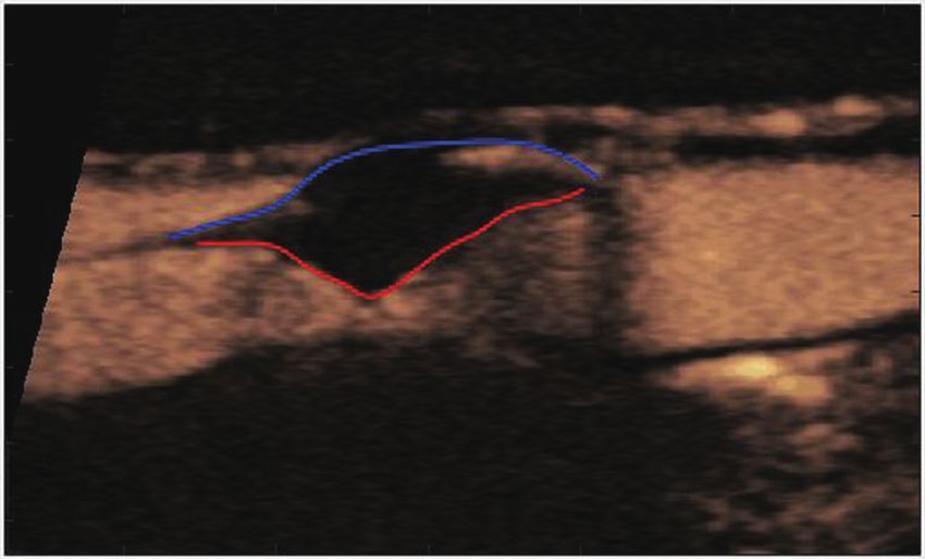
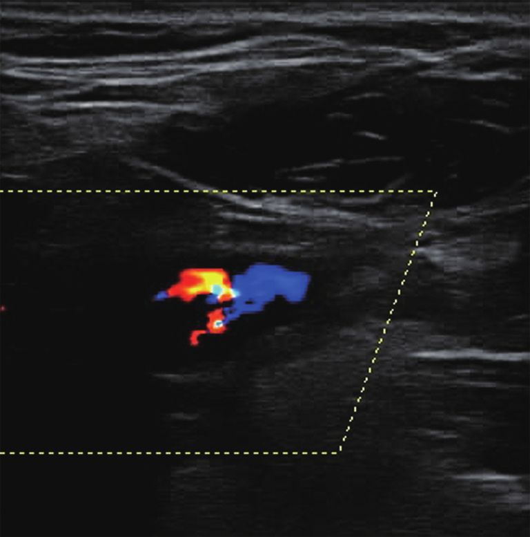
Cardiovascular Diagnosis and Therapy, Vol 10, No 4 August 2020                                                                                 971

A                                                   B                                                C

Figure 3 A 58-year-old male patient with acute stroke and free-floating thrombus in an internal carotid artery plaque. Colour Doppler
technique (A) fails to fully visualize blood flow, raising suspicion of a filling defect within the lumen. CEUS (B) accurately demonstrates an
intraluminal filling defect (asterisk), circumferentially delineated by microbubbles (arrowheads). A “donut-like” appearance is thus created
suggesting intraluminal thrombus. MDCTA (C) confirms CEUS findings, providing a similar appearance. CEUS, contrast-enhanced
ultrasound.

to intraplaque neovascularization. When functional,                       imaging of intraplaque neovascularization. The contrast
vasa vasorum are essentially a collection of small vessels                microbubbles behave similarly to red blood cells and remain
originating from the adventitia which supplies the vascular               strictly intravascular (48). Therefore, intraplaque signals
wall. However, in atherosclerotic disease, aberrant vasa                  are almost exclusively reflective of the microvasculature.
vasorum neovascularization, along with local hypoxia,                     In addition, intraplaque enhancement can represent
can lead to immature intraplaque neovascularization (45).                 intraplaque hemorrhage, a downstream effect of immature,
Such vessels are characterized by increased vascular                      leaky vasa vasorum and neovessels in vulnerable plaques.
density. These vessels oftentimes lack the critical pericytes                The intraplaque enhancement phenomenon was
to provide vascular integrity. The result is a collection                 first described by Professor Feinstein in delineating
of abundant, leaky and fragile vessels that are prone                     the plaque border in carotid stenosis (49). The authors
to intraplaque hemorrhage, which further destabilizes                     described visualizing discrete mobile microbubbles as spot
atherosclerotic plaques and may eventually lead to rupture                enhancement through the intraplaque vasculature. A pattern
of the plaque with distal embolization.                                   of adventitial vasa vasorum extending towards the core of
   In the early 2000s, a number of histological studies have              the plaque was observed on CEUS. The authors speculated
confirmed that the presence of intraplaque neovascularization             that the signal intensity might correlate with the degree
was a consistent feature in clinically significant vascular               of neovessel density. Further, it was thought that CEUS
disease. McCarthy et al. have shown that patients with                    enhancement was perhaps indicative of plaque vulnerability.
symptomatic carotid disease had significantly more                        Staub et al. have shown that a higher degree of post-contrast
intraplaque neovessels (P
972                                                                                    Rafailidis et al. Carotid contrast ultrasound and vulnerable plaque

                       A

                       B

                       C

Figure 4 Semi-quantitative, visual-based analysis of intraplaque neovascularization using a 3-point grading system. Plaques at the origin of
the internal carotid artery on B-mode ultrasound (right side) and CEUS imaging (left side) in three different patients. Grade 1 (A): carotid
plaque with no intraplaque neovascularization defined as no appearance of moving microbubbles in the plaque or confined only to the
adjacent adventitial layer. Grade 2 (B): carotid plaque with limited or moderate intraplaque neovascularization defined as moderate visible
appearance of moving bubbles in the plaque at the adventitial side or plaque shoulder (arrows). Grade 3 (C): carotid plaque with extensive
intraplaque neovascularization defined as clear visible appearance of bubbles moving to the plaque core (arrows).

with different scoring systems to grade intraplaque                                      appearance of microbubbles within the plaque) (Figure 4).
n e o v a s c u l a r i z a t i o n ( 5 1 ) . C o m m o n l y, i n t r a p l a q u e        For the assessment of intraplaque neovascularization
neovascularization was scored by a visual based 3-point                                  other studies used a quantitative analysis method by
grading system. Grade 1 was defined as no appearance of                                  measuring maximal intraplaque video-intensity after bolus
moving bubbles in the plaque or microbubbles confined only                               application of the contrast agent within the selected region
to the adjacent adventitial layer (no visible microbubbles) (50).                        of interest (TIC) (52). Furthermore, quantitative software
Grade 2 were defined as moderate visible appearance of                                   analysis of intraplaque neovascularization on CEUS used
moving bubbles in the plaque at the adventitial side or plaque                           MIP to quantify intraplaque neovascularization (51). More
shoulder (limited to moderate microbubbles), and grade 3 as                              sophisticated software with specific quantification algorithm
extensive intraplaque neovascularization, with clear visible                             have been used for automated quantification of intraplaque
appearance of bubbles moving to the plaque core (extensive                               microvessels (53-55) (Figure 5).

© Cardiovascular Diagnosis and Therapy. All rights reserved.                   Cardiovasc Diagn Ther 2020;10(4):965-981 | http://dx.doi.org/10.21037/cdt.2020.01.08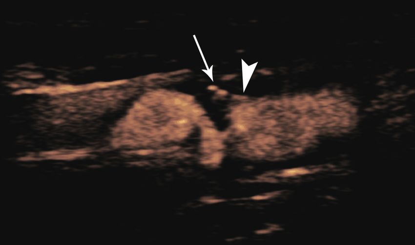
Cardiovascular Diagnosis and Therapy, Vol 10, No 4 August 2020 973
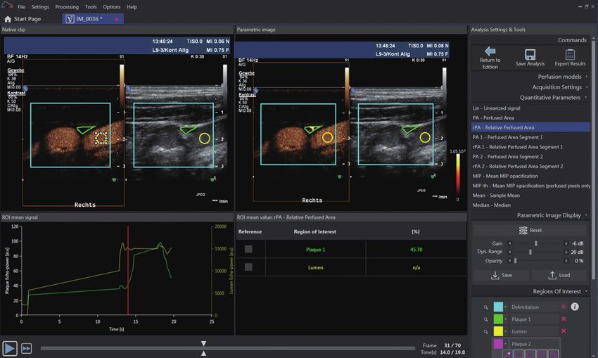
Figure 5 Quantitative analysis of intraplaque neovascularization using the VueBox® (Bracco SA) plaque package software. After bolus
injection of Sonovue® the enhancement within a region of interest of the plaque (green line) compared to the lumen (yellow line) on a time
intensity curve is used for quantitative analysis of intraplaque neovascularization. Intraplaque perfusion is visualized by a parametric color
imaging (right side). Furthermore, different perfusion parameters including the relative perfused area (rPA) of the plaque can be analysed. In
the presented analysis calculated a rPA of 45% within the plaque corresponding to moderate (grade 2) intraplaque neovascularization.
Intraplaque enhancement on CEUS compared with grade 1 was defined as no intraplaque bubbles or bubbles
histology confined to adventitia; and grade 2 was defined as bubbles
reaching plaque core or enhancement throughout the
Shah et al. provided the first qualitative measure of
plaque (57). Histological analysis was done by counting
CEUS enhancement and its histologic correlation (56).
the number of vessels in the magnified image. The study
The degree of neovascularization was visually graded
on four levels. Grade 0 was defined as no appearance showed a significantly higher number of vasa vasorum
of intraplaque enhancement. Grade 3 was defined as in grade 2 plaques in comparison to grade 1 plaques
those with pulsating arterial vessel. Grade 1 was then (3.24 vs. 1.82 mm2, P=0.005). Then in 2011, Hoogi et al.
defined as limited enhancement and grade 2 as moderate conducted the first quantitative study comparing CEUS
enhancement laying between grade 1 and 3. Histologic imaging to histologic analysis (53). Imaging quantification
analysis was carried out by measuring the level of vascular was done by segmenting all intraplaque enhancement on
markers such as CD31, CD34 and hemosiderin. It was CEUS throughout one cardiac cycle. The accumulated
found that there was a significant association between the area enhancement was then divided by the plaque volume.
degree of CEUS neovascularization and histologic vascular Histologic analysis was performed by measuring the ratio
density (r2=0.68, P=0.002). Coli et al. conducted the first of neovessel area to the total area of the plaque. The
semi-quantitative study on CEUS histology correlation. authors found that the ratio of histological vessel-to-
Enhancement patterns were graded into two categories: plaque ratio was well correlated with the degree of contrast
© Cardiovascular Diagnosis and Therapy. All rights reserved. Cardiovasc Diagn Ther 2020;10(4):965-981 | http://dx.doi.org/10.21037/cdt.2020.01.08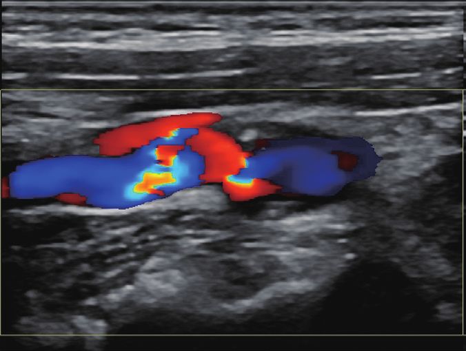
974                                                                   Rafailidis et al. Carotid contrast ultrasound and vulnerable plaque

enhancement on CEUS (r2=0.79, P
Cardiovascular Diagnosis and Therapy, Vol 10, No 4 August 2020                                                                                975

given the synergistic relationship between inflammation and              the late-phase CEUS enhancement in the assessment of
neovascularization in vulnerable plaques, it is worthwhile               plaque stability. In standard CEUS, the immediate post-
to examine the correlation between serum inflammatory                    contrast enhancement of intraplaque neovascularization
biomarkers and CEUS enhancement, a surrogate imaging                     is assessed. In comparison, late-phase CEUS utilizes the
biomarker of intraplaque neovascularization. Studies have                phenomenon where contrast microbubbles are ingested
offered evidence of a strong association between CEUS and                by monocytes. Intracellular microbubbles can remain
serum inflammatory markers.                                              acoustically stable for up to 30 minutes (74). In this study,
    In this context one of the first studied biomarkers is               late-phase CEUS signal intensity was used to compare
C-reactive protein (CRP) or high-sensitivity CRP (hs-                    with vasa vasorum density and MMP levels. The result
CRP). CRP is a non-specific inflammatory marker which                    showed significantly increased late-phase CEUS intensity
can be elevated in a variety of inflammatory states. With                in those with higher MMP-1 and MMP-3 level (P=0.043
regard to carotid atherosclerotic disease CRP is an acute                and 0.024, respectively) (75). Further, Kim et al. examined
phase reactant that accumulates in the macrophage-rich                   the relationship between the degree of neovascularization
area of the plaque (67). Therefore, a high CRP or hs-CRP                 and levels of serum biomarkers. They measured serum
level indicates a higher level of intraplaque inflammation and           levels of hs-CRP, MMP-2 and MMP-9. The degree of
potentially predicts plaque vulnerability. Alvarez Garcia et al.         neovascularization on CEUS was categorized into two
have found that unstable plaques were associated with a                  general categories per Coli et al.’s definition. It was found
higher median hs-CRP value compared to stable plaques (27.1              that both MMP-2 and MMP-9 were significantly elevated
vs. 4.1 mg/L. P
976 Rafailidis et al. Carotid contrast ultrasound and vulnerable plaque microbubbles to target tissue-bound, endovascular 2 was defined as clear intraplaque enhancement. On both inflammatory biomarkers. In pre-clinical experimental univariate and multivariate analysis, higher grade intraplaque settings, studies have shown that VCAM-1 expression level neovascularization was associated with an increased number was 24-fold higher in those with symptomatic plaques than of cardiovascular events (P=0.034 and 0.017). those without (P
Cardiovascular Diagnosis and Therapy, Vol 10, No 4 August 2020                                                                                     977

 Table 2 Sonographic markers of carotid plaque vulnerability on               biomarker. Lastly, apart from evaluating for markers
 CEUS                                                                         of vulnerable carotid plaques, CEUS enhancement is
 Marker                                   Classification                      directly associated with past cerebrovascular events.
 Carotid plaque           Smooth plaque surface                               More importantly, preliminary evidence has shown that
 surface                                                                      CEUS could be used to predict future cerebrovascular
                          Plaque surface irregularities (1–2 mm)
                                                                              imaging for carotid atherosclerotic disease, past studies still
 Carotid intraplaque      Semi-quantitative measurement:                      suffer from the retrospective nature, small sample size, and
 neovascularization                                                           a lack of matched well controlled prospective studies. In the
                            Grade 1: no vascularization
                                                                              future, large multi-center prospective studies addressing
                            Grade 2: limited or moderate
                            vascularization                                   the relationship of CEUS findings with patient outcomes in
                                                                              carotid atherosclerotic disease are warranted.
                            Grade 3: extensive vascularization

                          Semiautomatic quantitative measurement:
                          e.g., relative perfused area, plaque mean           Acknowledgments
                          intensity
                                                                              Funding: D Staub was supported by a research grant from
 CEUS, contrast-enhanced ultrasound.
                                                                              the Swiss National Science Foundation (PZ00P3_142419).
                                                                              D Staub has also received an unrestricted research grant
                                                                              from Bracco SA.
Although there were significant intergroup differences in
basic patient characteristics, it was found that the higher
grade CEUS enhancement was associated with recurrent                          Footnote
TIAs and strokes (P=0.01). However, the difference
in patient characteristics may indicate the presence of                       Provenance and Peer Review: This article was commissioned
confounding variables that can potentially interfere with                     by the Guest Editor (Luca Saba) for the series “Advanced
the results. Other recent prospective study revealed that                     Imaging in The Diagnosis of Cardiovascular Diseases”
CEUS-assessed carotid intraplaque neovascularization was                      published in Cardiovascular Diagnosis and Therapy. The
predictive of significant and complex coronary artery disease                 article was sent for external peer review organized by the
and future cardiovascular events (87).                                        Guest Editor and the editorial office.

                                                                              Conflicts of Interest: All authors have completed the ICMJE
Conclusions                                                                   uniform disclosure form (available at http://dx.doi.
In conclusion, CEUS is a valuable tool to evaluate                            org/10.21037/cdt.2020.01.08). The series “Advanced
sonographic markers of carotid plaque vulnerability by                        Imaging in The Diagnosis of Cardiovascular Diseases” was
depicting plaque surface irregularities and ulceration as                     commissioned by the editorial office without any funding
well as intraplaque neovascularization and adventitial vasa                   or sponsorship. SP and DS serve as the unpaid editorial
vasorum development (Table 2). The degree of intraplaque                      board members of Cardiovascular Diagnosis and Therapy from
neovascularization on CEUS imaging closely correlates                         July 2019 to June 2021. DS reports grants from Bracco SA,
with histological microvessel density, a marker for plaque                    outside the submitted work. PSS reports personal fees from
vulnerability. Preliminary evidence has also shown that                       Bracco SA, outside the submitted work. The authors have
CEUS could directly predict histologic grade of high-risk                     no other conflicts of interest to declare.
plaques. In addition, given the intricate interaction between
intraplaque neovascularization and inflammation, CEUS                         Ethical Statement: The authors are accountable for all
can offer valuable insights into intraplaque inflammation.                    aspects of the work in ensuring that questions related
Indeed, past studies have shown a close association between                   to the accuracy or integrity of any part of the work are
CEUS enhancement and multiple serum inflammatory                              appropriately investigated and resolved.
biomarkers. The use of tagged microbubbles can potentially
enable CEUS examination of endothelial inflammatory                           Open Access Statement: This is an Open Access article

© Cardiovascular Diagnosis and Therapy. All rights reserved.        Cardiovasc Diagn Ther 2020;10(4):965-981 | http://dx.doi.org/10.21037/cdt.2020.01.08
978 Rafailidis et al. Carotid contrast ultrasound and vulnerable plaque
distributed in accordance with the Creative Commons 11. Greis C. Technical aspects of contrast-enhanced
Attribution-NonCommercial-NoDerivs 4.0 International ultrasound (CEUS) examinations: tips and tricks. Clin
License (CC BY-NC-ND 4.0), which permits the non- Hemorheol Microcirc 2014;58:89-95.
commercial replication and distribution of the article with 12. Marshall MM, Beese RC, Muiesan P, et al. Assessment
the strict proviso that no changes or edits are made and the of portal venous system patency in the liver transplant
original work is properly cited (including links to both the candidate: a prospective study comparing ultrasound,
formal publication through the relevant DOI and the license). microbubble-enhanced colour Doppler ultrasound, with
See: https://creativecommons.org/licenses/by-nc-nd/4.0/. arteriography and surgery. Clin Radiol 2002;57:377-83.
13. Nakamura J, Nakamura T, Deyama J, et al. Assessment
of carotid plaque neovascularization using quantitative
References
analysis of contrast-enhanced ultrasound imaging is useful
1. Verhoeven B, Hellings WE, Moll FL, et al. Carotid for risk stratification in patients with coronary artery
atherosclerotic plaques in patients with transient ischemic disease. Int J Cardiol 2015;195:113-9.
attacks and stroke have unstable characteristics compared 14. Wilson SR, Jang HJ, Kim TK, et al. Real-time temporal
with plaques in asymptomatic and amaurosis fugax maximum-intensity-projection imaging of hepatic lesions
patients. J Vasc Surg 2005;42:1075-81. with contrast-enhanced sonography. AJR Am J Roentgenol
2. Naghavi M, Falk E, Hecht H, et al. From vulnerable 2008;190:691-5.
plaque to vulnerable patient--Part III: Executive summary 15. Eisenbrey JR, Daecher A, Kramer MR, et al. Effects of
of the Screening for Heart Attack Prevention and Needle and Catheter Size on Commercially Available
Education (SHAPE) Task Force report. Am J Cardiol Ultrasound Contrast Agents. J Ultrasound Med
2006;98:2H-15H. 2015;34:1961-8.
3. Staub D, Schinkel A, Coll B, et al. Contrast-enhanced 16. Rafailidis V, Charitanti A, Tegos T, et al. Contrast-
ultrasound imaging of the vasa vasorum: from early enhanced ultrasound of the carotid system: a review of the
atherosclerosis to the identification of unstable plaques. current literature. J Ultrasound 2017;20:97-109.
JACC Cardiovasc Imaging 2010;3:761-71. 17. Rafailidis V, Chryssogonidis I, Tegos T, et al. Imaging of
4. Rafailidis V, Huang DY, Yusuf GT, et al. General the ulcerated carotid atherosclerotic plaque: a review of
principles and overview of vascular contrast-enhanced the literature. Insights Imaging 2017;8:213-25.
ultrasonography. Ultrasonography 2020;39:22-42. 18. Eliasziw M, Streifler JY, Fox AJ, et al. Significance of
5. Greis C. Technology overview: SonoVue (Bracco, Milan). plaque ulceration in symptomatic patients with high-grade
Eur Radiol 2004;14 Suppl 8:P11-5. carotid stenosis. North American Symptomatic Carotid
6. Sidhu PS, Cantisani V, Dietrich CF, et al. The EFSUMB Endarterectomy Trial. Stroke 1994;25:304-8.
Guidelines and Recommendations for the Clinical 19. Rothwell PM, Gibson R, Warlow CP. Interrelation
Practice of Contrast-Enhanced Ultrasound (CEUS) in between plaque surface morphology and degree of stenosis
Non-Hepatic Applications: Update 2017 (Short Version). on carotid angiograms and the risk of ischemic stroke in
Ultraschall Med 2018;39:154-80. patients with symptomatic carotid stenosis. On behalf of
7. Piscaglia F, Bolondi L. The safety of Sonovue in abdominal the European Carotid Surgery Trialists' Collaborative
applications: retrospective analysis of 23188 investigations. Group. Stroke 2000;31:615-21.
Ultrasound Med Biol 2006;32:1369-75. 20. Saba L, Yuan C, Hatsukami TS, et al. Carotid Artery
8. ten Kate GL, van Dijk AC, van den Oord SC, et al. Wall Imaging: Perspective and Guidelines from the
Usefulness of contrast-enhanced ultrasound for detection ASNR Vessel Wall Imaging Study Group and Expert
of carotid plaque ulceration in patients with symptomatic Consensus Recommendations of the American Society of
carotid atherosclerosis. Am J Cardiol 2013;112:292-8. Neuroradiology. AJNR Am J Neuroradiol 2018;39:E9-31.
9. Chung YE, Kim KW. Contrast-enhanced ultrasonography: 21. Piscaglia F, Nolsoe C, Dietrich CF, et al. The EFSUMB
advance and current status in abdominal imaging. Guidelines and Recommendations on the Clinical Practice
Ultrasonography 2015;34:3-18. of Contrast Enhanced Ultrasound (CEUS): update 2011 on
10. Dietrich CF, Averkiou M, Nielsen MB, et al. How non-hepatic applications. Ultraschall Med 2012;33:33-59.
to perform Contrast-Enhanced Ultrasound (CEUS). 22. Saba L, Anzidei M, Marincola BC, et al. Imaging of the
Ultrasound Int Open 2018;4:E2-15. carotid artery vulnerable plaque. Cardiovasc Intervent
© Cardiovascular Diagnosis and Therapy. All rights reserved. Cardiovasc Diagn Ther 2020;10(4):965-981 | http://dx.doi.org/10.21037/cdt.2020.01.08Cardiovascular Diagnosis and Therapy, Vol 10, No 4 August 2020 979
Radiol 2014;37:572-85. 3D Contrast-Enhanced Ultrasound to Assess Internal
23. Saba L, Caddeo G, Sanfilippo R, et al. CT and ultrasound Carotid Artery Stenosis: A Pilot Study. J Neuroimaging
in the study of ulcerated carotid plaque compared 2020;30:82-9.
with surgical results: potentialities and advantages 35. Fetzer DT, Rafailidis V, Peterson C, et al. Artifacts in
of multidetector row CT angiography. AJNR Am J contrast-enhanced ultrasound: a pictorial essay. Abdom
Neuroradiol 2007;28:1061-6. Radiol (NY) 2018;43:977-97.
24. Sitzer M, Muller W, Siebler M, et al. Plaque ulceration 36. Tegos TJ, Kalomiris KJ, Sabetai MM, et al. Significance
and lumen thrombus are the main sources of cerebral of sonographic tissue and surface characteristics of carotid
microemboli in high-grade internal carotid artery stenosis. plaques. AJNR Am J Neuroradiol 2001;22:1605-12.
Stroke 1995;26:1231-3. 37. Kanber B, Hartshorne TC, Horsfield MA, et al.
25. Brinjikji W, Rabinstein AA, Lanzino G, et al. Ultrasound Quantitative assessment of carotid plaque surface
Characteristics of Symptomatic Carotid Plaques: A irregularities and correlation to cerebrovascular symptoms.
Systematic Review and Meta-Analysis. Cerebrovasc Dis Cardiovasc Ultrasound 2013;11:38.
2015;40:165-74. 38. Kanber B, Hartshorne TC, Horsfield MA, et al. A Novel
26. Eyding J, Geier B, Staub D. Current strategies and Ultrasound-Based Carotid Plaque Risk Index Associated
possible perspectives of ultrasonic risk stratification with the Presence of Cerebrovascular Symptoms.
of ischemic stroke in internal carotid artery disease. Ultraschall Med 2015;36:480-6.
Ultraschall Med 2011;32:267-73. 39. Rafailidis V, Chryssogonidis I, Grisan E, et al. Does
27. Rafailidis V, Chryssogonidis I, Xerras C, et al. A Quantification of Carotid Plaque Surface Irregularities
comparative study of color Doppler imaging and contrast- Better Detect Symptomatic Plaques Compared to
enhanced ultrasound for the detection of ulceration in the Subjective Classification? J Ultrasound Med
patients with carotid atherosclerotic disease. Eur Radiol 2019;38:3163-71.
2019;29:2137-45. 40. Rafailidis V, Chryssogonidis I, Xerras C, et al. An
28. de Bray JM BJ, Dauzat M. Consensus Concerning the Ultrasonographic Multiparametric Carotid Plaque Risk
Morphology and the Risk of Carotid Plaques. Cerebrovasc Index Associated with Cerebrovascular Symptomatology:
Dis 1997;7:289-96. A Study Comparing Color Doppler Imaging and Contrast-
29. Muraki M, Mikami T, Yoshimoto T, et al. New criteria Enhanced Ultrasonography. AJNR Am J Neuroradiol
for the sonographic diagnosis of a plaque ulcer in 2019;40:1022-8.
the extracranial carotid artery. AJR Am J Roentgenol 41. Hamada O, Sakata N, Ogata T, et al. Contrast-enhanced
2012;198:1161-6. ultrasonography for detecting histological carotid plaque
30. van den Oord SC, Akkus Z, Renaud G, et al. Assessment rupture: Quantitative analysis of ulcer. Int J Stroke
of carotid atherosclerosis, intraplaque neovascularization, 2016;11:791-8.
and plaque ulceration using quantitative contrast-enhanced 42. Schinkel AF, Kaspar M, Staub D. Contrast-enhanced
ultrasound in asymptomatic patients with diabetes mellitus. ultrasound: clinical applications in patients with
Eur Heart J Cardiovasc Imaging 2014;15:1213-8. atherosclerosis. Int J Cardiovasc Imaging 2016;32:35-48.
31. Rafailidis V, Charitanti A, Tegos T, et al. Swirling of 43. Menon BK, Singh J, Al-Khataami A, et al. The donut sign
microbubbles: Demonstration of a new finding of carotid on CT angiography: an indicator of reversible intraluminal
plaque ulceration on contrast-enhanced ultrasound carotid thrombus? Neuroradiology 2010;52:1055-6.
explaining the arterio-arterial embolism mechanism. Clin 44. Staub D. Atherosclerotic plaque neovascularization and
Hemorheol Microcirc 2016;64:245-50. inflammation - is there a link? Vasa 2015;44:163-5.
32. Imbesi SG, Kerber CW. An experimental and angiographic 45. Feinstein S. Contrast ultrasound imaging of the
explanation of why ulcerated carotid bulbs embolize. carotid artery vasa vasorum and atherosclerotic plaque
Interv Neuroradiol 1999;5:11-8. neovascularization. J Am Coll Cardiol 2006;48:236-43.
33. Furst H, Hartl WH, Jansen I, et al. Color-flow Doppler 46. McCarthy M, Loftus I, Thompson M, et al. Angiogenesis
sonography in the identification of ulcerative plaques in and the atherosclerotic carotid plaque: an association
patients with high-grade carotid artery stenosis. AJNR Am between symptomatology and plaque morphology. J Vasc
J Neuroradiol 1992;13:1581-7. Surg 1999;30:261-8.
34. Pelz JO, Weinreich A, Schob S, et al. Multiparametric 47. Dunmore B, McCarthy M, Naylor A, et al. Carotid plaque
© Cardiovascular Diagnosis and Therapy. All rights reserved. Cardiovasc Diagn Ther 2020;10(4):965-981 | http://dx.doi.org/10.21037/cdt.2020.01.08980 Rafailidis et al. Carotid contrast ultrasound and vulnerable plaque
instability and ischemic symptoms are linked to immaturity Meta-Analysis of Diagnostic Accuracy Studies. J Am Soc
of microvessels within plaques. J Vasc Surg 2007;45:155-9. Echocardiogr 2016;29:491-502.
48. Partovi S, Loebe M, Aschwanden M, et al. Contrast- 60. Vavuranakis M, Sigala F, Vrachatis DA, et al. Quantitative
enhanced ultrasound for assessing carotid atherosclerotic analysis of carotid plaque vasa vasorum by CEUS and
plaque lesions. AJR Am J Roentgenol 2012;198:W13-9. correlation with histology after endarterectomy. Vasa
49. Rajaram V, Pandhya S, Patel S, et al. Role of surrogate 2013;42:184-95.
markers in assessing patients with diabetes mellitus and 61. Schmidt C, Fischer T, Ruckert RI, et al. Identification
the metabolic syndrome and in evaluating lipid-lowering of neovascularization by contrast-enhanced ultrasound
therapy. Am J Cardiol 2004;93:32C-48C. to detect unstable carotid stenosis. PLoS One
50. Staub D, Partovi S, Schinkel AF, et al. Correlation of 2017;12:e0175331.
carotid artery atherosclerotic lesion echogenicity and 62. Amamoto T, Sakata N, Ogata T, et al. Intra-Plaque Vessels
severity at standard US with intraplaque neovascularization on Contrast-Enhanced Ultrasound Sonography Predict
detected at contrast-enhanced US. Radiology Carotid Plaque Histology. Cerebrovasc Dis 2018;46:265-9.
2011;258:618-26. 63. Giannoni M, Vicenzini E, Citone M, et al. Contrast
51. Schinkel AFL, Bosch JG, Staub D, et al. Contrast- carotid ultrasound for the detection of unstable plaques
Enhanced Ultrasound to Assess Carotid Intraplaque with neoangiogenesis: a pilot study. Eur J Vasc Endovasc
Neovascularization. Ultrasound Med Biol 2020;46:466-78. Surg 2009;37:722-7.
52. Xiong L, Deng Y, Zhu Y, et al. Correlation of carotid 64. Iezzi R, Petrone G, Ferrante A, et al. The role of contrast-
plaque neovascularization detected by using contrast- enhanced ultrasound (CEUS) in visualizing atherosclerotic
enhanced US with clinical symptoms. Radiology carotid plaque vulnerability: which injection protocol?
2009;251:583-9. Which scanning technique? Eur J Radiol 2015;84:865-71.
53. Hoogi A, Adam D, Hoffman A, et al. Carotid plaque 65. Michel JB, Martin-Ventura JL, Nicoletti A, et al.
vulnerability: quantification of neovascularization on Pathology of human plaque vulnerability: Mechanisms and
contrast-enhanced ultrasound with histopathologic consequences of intraplaque haemorrhages. Atherosclerosis
correlation. AJR Am J Roentgenol 2011;196:431-6. 2014;234:311-9.
54. Akkus Z, Hoogi A, Renaud G, et al. New quantification 66. Fleiner M, Kummer M, Mirlacher M, et al. Arterial
methods for carotid intra-plaque neovascularization using neovascularization and inflammation in vulnerable patients:
contrast-enhanced ultrasound. Ultrasound Med Biol early and late signs of symptomatic atherosclerosis.
2014;40:25-36. Circulation 2004;110:2843-50.
55. van den Oord SC, Akkus Z, Bosch JG, et al. 67. Hermus L, Lefrandt JD, Tio RA, et al. Carotid plaque
Quantitative Contrast-Enhanced Ultrasound of formation and serum biomarkers. Atherosclerosis
Intraplaque Neovascularization in Patients with Carotid 2010;213:21-9.
Atherosclerosis. Ultraschall Med 2015;36:154-61. 68. Alvarez Garcia B, Ruiz C, Chacon P, et al. High-sensitivity
56. Shah F, Balan P, Weinberg M, et al. Contrast- C-reactive protein in high-grade carotid stenosis:
enhanced ultrasound imaging of atherosclerotic carotid risk marker for unstable carotid plaque. J Vasc Surg
plaque neovascularization: a new surrogate marker of 2003;38:1018-24.
atherosclerosis? Vasc Med 2007;12:291-7. 69. Kablak-Ziembicka A, Przewlocki T, Sokolowski A, et al.
57. Coli S, Magnoni M, Sangiorgi G, et al. Contrast-enhanced Carotid intima-media thickness, hs-CRP and TNF-alpha
ultrasound imaging of intraplaque neovascularization in are independently associated with cardiovascular event
carotid arteries: correlation with histology and plaque risk in patients with atherosclerotic occlusive disease.
echogenicity. J Am Coll Cardiol 2008;52:223-30. Atherosclerosis 2011;214:185-90.
58. Li C, He W, Guo D, et al. Quantification of carotid plaque 70. Chang X, Feng J, Ruan L, et al. Positive correlation
neovascularization using contrast-enhanced ultrasound between neovascularization degree of carotid
with histopathologic validation. Ultrasound Med Biol atherosclerosis determined by contrast-enhanced
2014;40:1827-33. ultrasound and level of serum C-reactive protein. Vasa
59. Huang R, Abdelmoneim SS, Ball CA, et al. Detection of 2015;44:187-94.
Carotid Atherosclerotic Plaque Neovascularization Using 71. Xu R, Yin X, Xu W, et al. Assessment of carotid plaque
Contrast Enhanced Ultrasound: A Systematic Review and neovascularization by contrast-enhanced ultrasound and
© Cardiovascular Diagnosis and Therapy. All rights reserved. Cardiovasc Diagn Ther 2020;10(4):965-981 | http://dx.doi.org/10.21037/cdt.2020.01.08Cardiovascular Diagnosis and Therapy, Vol 10, No 4 August 2020 981
high sensitivity C-reactive protein test in patients with 80. Moccetti F, Weinkauf CC, Davidson BP, et al. Ultrasound
acute cerebral infarction: a comparative study. Neurol Sci Molecular Imaging of Atherosclerosis Using Small-
2016;37:1107-12. Peptide Targeting Ligands Against Endothelial Markers of
72. Loftus IM, Naylor AR, Bell PR, et al. Plasma MMP-9 - a Inflammation and Oxidative Stress. Ultrasound Med Biol
marker of carotid plaque instability. Eur J Vasc Endovasc 2018;44:1155-63.
Surg 2001;21:17-21. 81. Staub D, Patel M, Tibrewala A, et al. Vasa vasorum and
73. Molloy KJ, Thompson MM, Jones JL, et al. Unstable plaque neovascularization on contrast-enhanced carotid
carotid plaques exhibit raised matrix metalloproteinase-8 ultrasound imaging correlates with cardiovascular disease
activity. Circulation 2004;110:337-43. and past cardiovascular events. Stroke 2010;41:41-7.
74. Owen D, Shalhoub J, Miller S, et al. Inflammation within 82. Huang PT, Chen CC, Aronow WS, et al. Assessment of
carotid atherosclerotic plaque: assessment with late-phase neovascularization within carotid plaques in patients with
contrast-enhanced US. Radiology 2010;255:638-44. ischemic stroke. World J Cardiol 2010;2:89-97.
75. Shalhoub J, Monaco C, Owen DR, et al. Late-phase 83. Faggioli GL, Pini R, Mauro R, et al. Identification
contrast-enhanced ultrasound reflects biological features of carotid 'vulnerable plaque' by contrast-enhanced
of instability in human carotid atherosclerosis. Stroke ultrasonography: correlation with plaque histology,
2011;42:3634-6. symptoms and cerebral computed tomography. Eur J Vasc
76. Kim HS, Woo JS, Kim BY, et al. Biochemical and Endovasc Surg 2011;41:238-48.
clinical correlation of intraplaque neovascularization 84. Zhou Y, Xing Y, Li Y, et al. An assessment of the
using contrast-enhanced ultrasound of the carotid artery. vulnerability of carotid plaques: a comparative study
Atherosclerosis 2014;233:579-83. between intraplaque neovascularization and plaque
77. Ammirati E, Moroni F, Magnoni M, et al. Circulating echogenicity. BMC Med Imaging 2013;13:13.
CD14+ and CD14(high)CD16- classical monocytes are 85. Varetto G, Gibello L, Faletti R, et al. Contrast-enhanced
reduced in patients with signs of plaque neovascularization ultrasound to predict the risk of microembolization during
in the carotid artery. Atherosclerosis 2016;255:171-8. carotid artery stenting. Radiol Med 2015;120:1050-5.
78. Li Z, Bai Y, Li W, et al. Carotid vulnerable plaques are 86. Li Z, Xu X, Ren L, et al. Prospective Study About the
associated with circulating leukocytes in acute ischemic Relationship Between CEUS of Carotid Intraplaque
stroke patients: a clinical study based on contrast-enhanced Neovascularization and Ischemic Stroke in TIA Patients.
ultrasound. Sci Rep 2018;8:8849. Front Pharmacol 2019;10:672.
79. Weinkauf CC, Concha-Moore K, Lindner JR, et al. 87. Mantella LE, Colledanchise KN, Hetu MF, et al. Carotid
Endothelial vascular cell adhesion molecule 1 is a marker intraplaque neovascularization predicts coronary artery
for high-risk carotid plaques and target for ultrasound disease and cardiovascular events. Eur Heart J Cardiovasc
molecular imaging. J Vasc Surg 2018;68:105S-113S. Imaging 2019;20:1239-47.
Cite this article as: Rafailidis V, Li X, Sidhu PS, Partovi S,
Staub D. Contrast imaging ultrasound for the detection and
characterization of carotid vulnerable plaque. Cardiovasc Diagn
Ther 2020;10(4):965-981. doi: 10.21037/cdt.2020.01.08
© Cardiovascular Diagnosis and Therapy. All rights reserved. Cardiovasc Diagn Ther 2020;10(4):965-981 | http://dx.doi.org/10.21037/cdt.2020.01.08You can also read