Neuroprotective effects of low- dose G-CSF plus meloxicam in a rat model of anterior ischemic optic neuropathy - Nature
←
→
Page content transcription
If your browser does not render page correctly, please read the page content below
www.nature.com/scientificreports

            OPEN            Neuroprotective effects of low-
                            dose G-CSF plus meloxicam in a rat
                            model of anterior ischemic optic
                            neuropathy
                            Pei-Kang Liu1,2,3,4,10, Yao-Tseng Wen5,10, Wei Lin6, Kishan Kapupara5, Minghong Tai3,7,8 ✉ &
                            Rong-Kung Tsai5,9 ✉
                            Non-arteritic anterior ischemic optic neuropathy (NAION) causes a sudden loss of vision and lacks
                            effective treatment. Granulocyte colony-stimulating factor (G-CSF) provides neuroprotection against
                            the experimental optic nerve injuries but also induce leukocytosis upon typical administration. We
                            found synergetic neuroprotective effects of meloxicam and low dose G-CSF without leukocytosis in a
                            rat model of anterior ischemic optic neuropathy (rAION). The WBC counts in the low-dose G-CSF-plus
                            meloxicam-treated group were similar to the sham-operated group. Combination treatment of low-
                            dose G-CSF plus meloxicam preserved RGCs survival and visual function, reduced RGC apoptosis and
                            the macrophages infiltration, and promote more M2 phenotype of macrophage/microglial transition
                            than the low-dose GCSF treatment or the meloxicam treatment. Moreover, the combination treatment
                            induced higher serine/threonine kinase 1 (Akt1) expression. The combination treatment of low-dose
                            G-CSF plus meloxicam lessened the leukocytotic side effect and provided neuroprotective effects via
                            Akt1 activation in the rAION model. This approach provides crucial preclinical information for the
                            development of alternative therapy in AION.

                            Nonarteritic anterior ischemic optic neuropathy (NAION) is the most common acute optic neuropathy in people
                            older than 50 years. The estimated mean annual incidence rates of NAION are 2.3–10.3 per 100,000 in the United
                            States and 3.72 per 100,000 in Taiwan1–3. The debilitating consequences for patients with NAION include severe
                            vision impairment and visual field defect. The primarily proposed pathogenesis of NAION is the transient non-
                            perfusion or hypoperfusion of the optic nerve head and loss of retinal ganglion cells (RGCs) following ischemic
                            insults. The histologic evidence in an early clinical case of human NAION demonstrated the presence of apoptotic
                            cells in the RGC layer by 30 days after the event. This reaction eventually results in RGC death and vision loss4.
                            Currently, no definite treatment for NAION is available. Halting the injury and loss of RGCs may, therefore, res-
                            cue the visual deterioration and be a potential treatment strategy.
                                Granulocyte colony-stimulating factor (G-CSF), a member of the cytokine family of growth factors, is a
                            19.6-kDa glycoprotein. It is commonly applied clinically to treat neutropenia5. G-CSF can induce the mobilization
                            of CD34 + hematopoietic stem cells from the bone marrow into the peripheral blood. G-CSF is commonly used
                            in clinics for stem cell mobilization and bone marrow reconstitution6–8. Our previous studies have demonstrated
                            that G-CSF exerts neuroprotective effects on a rat model of anterior ischemic optic neuropathy (rAION). The
                            protective effects of G-CSF on the rAION model are achieved through dual mechanisms of anti-inflammation
                            and antiapoptosis9,10. The antiapoptotic effect of G-CSF occurs through the activation of a variety of intracellular

                            1
                             Department of Ophthalmology, Kaohsiung Medical University Hospital, Kaohsiung Medical University, Kaohsiung,
                            Taiwan. 2Department of Ophthalmology, Yuan’s General Hospital, Kaohsiung, Taiwan. 3Institute of Biomedical
                            Sciences, National Sun Yat-Sen University, Kaohsiung, Taiwan. 4School of Medicine, College of Medicine, Kaohsiung
                            Medical University, Kaohsiung, Taiwan. 5Institute of Eye Research, Hualien Tzu Chi Hospital, Buddhist Tzu Chi
                            Medical Foundation, Hualien, Taiwan. 6Department of Optometry, Da-Yeh University, Changhwa, Taiwan. 7Center
                            for Neuroscience, National Sun Yat-Sen University, Kaohsiung, Taiwan. 8Graduate Program in Marine Biotechnology,
                            National Sun Yat-Sen University, Kaohsiung, Taiwan. 9Institute of Medical Sciences, Tzu Chi University, Hualien,
                            Taiwan. 10These authors contributed equally: Pei-Kang Liu and Yao-Tseng Wen. ✉e-mail: minghongtai@gmail.com;
                            rktsai@tzuchi.com.tw

Scientific Reports |   (2020) 10:10351 | https://doi.org/10.1038/s41598-020-66977-9                                                           1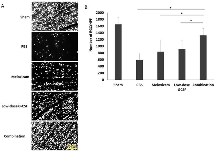
www.nature.com/scientificreports/                                                               www.nature.com/scientificreports

                            signaling pathways and is mainly dependent on phosphatidylinositol-3 kinase (PI3K)/protein kinase B (AKT)
                            activation11,12. Early intervention with G-CSF can induce M2 microglia/macrophage polarization, reduce the
                            expression of proinflammatory cytokines, and stabilize the blood–optic nerve barrier (BOB) to reduce mac-
                            rophage infiltration in the rAION model10. However, systemic treatment with G-CSF may result in leukocytosis
                            and some side effects, such as arthralgia, bone pain, headache, fatigue, nausea, fever, chills, and myalgia13. Our
                            previous study also demonstrated that subcutaneous administration of G-CSF (100 μg/kg/day) for five consec-
                            utive days could result in leukocytosis in rats11. Thus, developing a new approach to reducing the side effects of
                            G-CSF treatment and maintaining the therapeutic effects in the rAION model may provide benefits.
                                Meloxicam belongs to the enolic acid group of nonsteroidal anti-inflammatory drugs (NSAIDs) and is clin-
                            ically prescribed for treating certain types of arthritis, such as rheumatoid arthritis, for reducing inflammation
                            and pain14. The possible mechanism of meloxicam is inhibition of migration of leukocytes and blocking cycloox-
                            ygenase (COX), the enzyme responsible for the first step in the synthesis of prostaglandins15. COX can convert
                            arachidonic acid into prostaglandin H2, which is a mediator of inflammation. Meloxicam has been demon-
                            strated to primarily inhibit COX-2, the cyclooxygenase isozyme, mainly in inflamed tissues with long-lasting
                            anti-inflammatory and analgesic effects16. Preferential inhibition of COX-2 leads to high anti-inflammatory
                            potency with lower ulcerogenicity in the stomach and higher tolerability than non-selective NSAIDs. Moreover,
                            the metabolites of meloxicam are inactive16. Therefore, it has the potential to reduce inflammation in ischemic
                            optic neuropathy. However, whether meloxicam can enhance the therapeutic effects of G-CSF treatment for
                            AION remains unclear.
                                Macrophage/microglia has been shown to be polarizable and classifiable into M1 and M2 phenotype by
                            expressed cytokine/chemokine profiles, surface markers, and biological functions. M1 phenotype is responsible
                            for immunostimulation, including inflammation trigger, cell proliferation inhibition, which may cause tissue
                            damage; M2 phenotype promotes immunosuppression and inhibits inflammation, which may improve cell pro-
                            liferation and facilitate tissue repair in nervous tissues17. Activation of M2 phenotype macrophages, reduction
                            in proinflammatory cytokine expression, and stabilization of the BOB are supposed to be three vital approaches
                            to optic nerve protection in the rAION model10,18. Enhancement of anti-inflammatory actions through different
                            mechanisms may serve as a potential strategy in the treatment of ischemic optic nerve injury. The purpose of this
                            study was to investigate whether treatment with low-dose G-CSF plus meloxicam results in a synergistic effect of
                            neuroprotection and reduction of the side effect of leukocytosis in the rAION model.

                            Materials and methods
                            Experimental animals.        In this study, adult male Wistar rats weighing 150–180 g were used. The rats were
                            purchased from the breeding colony of BioLASCO Co., Taiwan. Animal care and experimental procedures were
                            performed in accordance with the Association for Research in Vision and Ophthalmology Statement for the Use
                            of Animals in Ophthalmic and Vision Research. The Institutional Animal Care and Use Committee of Tzu Chi
                            Medical Center approved all animal experiments.

                            Study design. In the drug-induced leukocytosis study, 18 rats were equally divided into six groups including
                            the sham-operated, the PBS-treated, the meloxicam-treated (0.125 mg/kg/day, Boehringer Ingelheim, Ingelheim
                            am Rhein, Germany), the low-dose G-CSF-treated (50 μg/kg/day in 0.2 mL of saline, Takasaki Pharmaceutical
                            Plant, Tokyo, Japan), high-dose G-CSF-treated (100 μg/kg/day in 0.2 mL of saline, Takasaki Pharmaceutical
                            Plant), and low-dose G-CSF (50 μg/kg/day) plus meloxicam (0.125 mg/kg/day)-treated group. The meloxicam
                            was administered via the oral route and the G-CSF via subcutaneous injection.
                                In the therapeutic evaluation, after successful induction of AION in the rats, 120 rats were equally divided
                            into four groups. Treatment with subcutaneous injection of low-dose G-CSF only once daily, meloxicam only,
                            low-dose G-CSF plus meloxicam, and phosphate-buffered saline (PBS, serving as control; 0.2 mL) alone immedi-
                            ately after the rAION procedure for a total of 5 consecutive days. Another 30 rats received sham laser treatment
                            without a photosensitizing agent to serve as normal controls. The number of rats used in this study is summarized
                            in Fig. S1. All the rats tolerated this treatment and survived until the end of the procedure.

                            Peripheral WBC count. After the drug administration, 2–3 mL of circulating blood was drawn by cardiac
                            puncture from the rats one week after rAION induction. WBC counts were obtained.

                            rAION induction.          The method of rAION induction was the same as that used in our previous report9,10.
                            Before general anesthesia, the rats were treated with Alcaine and Mydrin-P eye drops for topical anesthesia and
                            pupil dilation, respectively. The rat was administrated by intramuscular injections of a mixture of ketamine
                            (40 mg/kg body weight) and xylazine (4 mg/kg body weight; Sigma, St. Louis, MO, USA) for general anesthesia.
                            Subsequently, 2.5 mM rose bengal in PBS (1 ml/kg animal weight) was intravenously administered. After rose ben-
                            gal injection, the optic disc was immediately exposed to an argon green laser system (MC-500 Multi-color laser,
                            Nidek Co., Ltd, Tokyo, Japan, setting: 532 nm wavelength, 500 μm size and 80 mW power) for 12 1-s pulses19,20.
                            A laser fundus lens (Ocular instruments Inc.) was used to focus the laser on the optic disc. (Supplement Fig. 2)
                            Tobradex eye ointment was applied after the procedure, and the rats were monitored until complete recovery was
                            observed.

                            Retrograde labeling of RGCs with FluoroGold and morphometry of the RGCs. The detailed meth-
                            ods and protocol of FluoroGold labeling have been described in our previous reports9,18. Briefly, the retinas were
                            examined at distances of 1 mm from the center of the optic nerve for RGC counting to obtain the RGC density
                            in the central retina. We calculated eight randomly selected areas in the central retina; the total area counted was
                            about 13.5 mm2. The densities of RGC were averaged for each retina (n = 12 rats per group).

Scientific Reports |   (2020) 10:10351 | https://doi.org/10.1038/s41598-020-66977-9                                                           2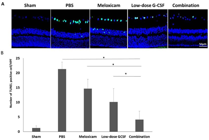
www.nature.com/scientificreports/                                                              www.nature.com/scientificreports

                            Flash visual evoked potentials (FVEP). The detailed FVEP recording method has been described in our
                            previous studies9,10. For electrode placement, the sagittal region of the skull was opened in the rats. The 4 mm
                            screw implants were pass through the skull approximate 1.5 mm and were placed at the frontal cortex and the
                            primary visual cortex region of both hemispheres using stereotaxic coordinates. A visual electrodiagnostic system
                            (Diagnosys LLC, Lowell, MA, USA) was used to measure the FVEP. The number of sweeps per average was 64 for
                            each rat. A comparison of the amplitude of the P1-N2 wave in each group was made to evaluate visual function
                            (n = 12 rats per group).

                            Optic nerve and retinal sample preparation.             The rats were euthanized after four weeks; the eyes were
                            enucleated along with the optic nerve (approximately 5 mm in length). The samples were fixed in 4% paraformal-
                            dehyde and were cryoprotected by 30% sucrose; the samples were stored at 4 °C until they settled at the bottom of
                            the tubes. Sections of 20 μm were obtained using a cryostat.

                            TUNEL assay. To ensure the use of equivalent fields for comparison, all the paraffin or frozen sections of
                            the retina were prepared with retinas at 1 to 2 mm distance from the ON head. TUNEL assay was used to detect
                            apoptotic cells in the ganglion cell layer (GCL). This assay was performed according to the manufacturer’s proce-
                            dure (DeadEnd Fluorometric TUNEL System; Promega Corporation, Madison, WI, USA). The TdT-dUTP ter-
                            minal nick-end labeling-positive cells in the GCL of each sample were calculated in 10 high-powered fields (HPF,
                            400×), and an average from three sections per retina was used for further comparison (n = 6 rats per group).

                            Immunohistochemical staining for phagocytic macrophage/microglia detection.                         ED-1 is a
                            marker for phagocytic macrophages and microglia. The longitudinal section of the optic nerve was blocked with
                            5% FBS for one hour at room temperature. The section was labeled with an ED1 primary antibody diluted in the
                            dilution buffer (2% BSA, 1 × PBS (pH 7.2), and 0.3% Triton X-100; 1: 200) overnight at 4 °C. The section was
                            incubated with Goat anti-mouse Alexa 488 (0.3% Triton X-100 and 1 × PBS (pH 7.2); 1: 500) for one hour at
                            room temperature and counterstained with DAPI (0.3% Triton X-100 and 1 × PBS (pH 7.2); 1: 500). The image
                            was acquired by using appropriate filter sets in a fluorescence microscope at × 100 magnification. ED-1+ cell
                            counting was conducted by ImageMaster 2 Platinum software.

                            Quantitative reverse transcription polymerase chain reaction. The markers of M2 macrophages
                            (Arg 1, CD206, and Fizz1) were evaluated10,18. Tissue RNA was extracted from optic nerve lysates obtained using
                            the sonication method (130 W, 30% amplitude, 5 pulses for each sample, each pulse consists of 5 seconds ON and
                            2 seconds OFF cycle) with a Qiagen RNeasy Mini Kit (Hilden, Nordrhein-Westfalen, Germany). All the RNA
                            samples were reverse transcribed at 42 °C for 30 minutes by using a high-capacity cDNA reverse transcription
                            kit (Applied Biosystems, Foster City, CA, USA). qRT-PCR was then conducted on an AB PRISM 7300 Sequence
                            Detection System (Applied Biosystems) with the QuantiTect SYBR green qRT-PCR kit (Qiagen). Expression
                            levels of CypA mRNA were used for normalization to measure the expression levels of each target gene. The cal-
                            culated data are presented as the mean relative expression levels ± standard deviation (SD). The primers used in
                            this study for target gene amplification were listed in Table S1.

                            Western blotting analysis. The optic nerve samples were collected on day seven after rAION induction
                            in each group. The protein extracts of the optic nerve were separated by using a 4–12% NuPAGE Bis-Tris gel
                            (Invitrogen, Carlsbad, CA, USA). The separated proteins were transferred onto polyvinylidene difluoride mem-
                            branes. The membranes were blocked by using 5% nonfat milk in Tris buffer saline/Tween-20 solution containing
                            20 mM Tris-HCl (pH 7.5), 0.5 M NaCl, and 0.5% Tween-20. Subsequently, the membranes were blotted with
                            mouse anti-p-Akt1 antibody (Abcam, Cambridge, MA, USA) followed by goat anti-mouse HRP. The developing
                            reaction was performed by using an enhanced chemiluminescent substrate (Perkin-Elmer Life Science, Boston,
                            MA, USA), and the relative intensities of the bands were measured by using an image analysis system (Amersham
                            Biosciences, Uppsala, Sweden).

                            Statistical analysis. All data are represented as Mean ± Standard deviation. We performed statistical anal-
                            ysis with SPSS commercial software (Chicago, IL, USA, USA). Mann–Whitney U test was used to evaluate the
                            differences between each group individually. Statistical significance was set at p < 0.05. All measurements in this
                            study were performed in a masked manner.

                            Results
                            Evaluation of leukocytosis after treatment.           After 5-day treatment with high-dose G-CSF, low-dose
                            G-CSF, meloxicam, and low-dose G-CSF plus meloxicam, the WBC counts were 6562 ± 662/μL, 6889 ± 693/μL,
                            6613 ± 557/μL, 19,633 ± 531/μL, 8897 ± 712/μL, and 9415 ± 657/μL in the sham, PBS-treated, meloxicam-treated,
                            high-dose G-CSF-treated, low-dose G-CSF-treated, and low-dose G-CSF-plus-meloxicam-treated groups,
                            respectively. The WBC counts increased by 2.99-fold in the high-dose G-CSF-treated group compared with the
                            sham group (p = 0.0011), which indicates the leukocytosis effects of high-dose G-CSF. Treatments with meloxi-
                            cam, low-dose G-CSF, and low-dose G-CSF plus meloxicam did not induce leukocytosis in the animals (Fig. 1).
                            Therefore, we used low-dose G-CSF instead of high-dose G-CSF with or without meloxicam in the following
                            experiments.

                            Combined treatment preserved more visual function than other single treatments.                       We per-
                            formed FVEP to evaluate the visual function. The FVEP of the sham, PBS-, meloxicam-, low-dose G-CSF-, and
                            the combination-treated group were recorded (Fig. 2A). The P1 latency did not exhibit a significant difference
                            among the groups in the FVEP tests. The amplitudes of the P1-N2 waves in the combination-treated group

Scientific Reports |   (2020) 10:10351 | https://doi.org/10.1038/s41598-020-66977-9                                                          3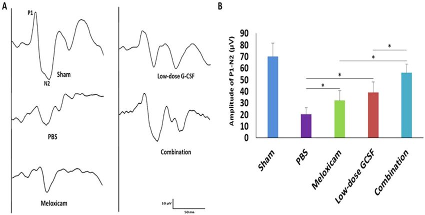
www.nature.com/scientificreports/ www.nature.com/scientificreports
Figure 1. Analysis of WBC counts in the sham-operated rats, PBS-treated rats, meloxicam-treated rats, high-
dose G-CSF-treated rats, low-dose G-CSF-treated rats, and low-dose G-CSF-plus-meloxicam-treated rats.
Treatment with meloxicam, low-dose G-CSF, and low-dose G-CSF plus meloxicam did not induce leukocytosis
in the rats, but treatment with high-dose G-CSF induced leukocytosis after a 5-day treatment. *p < 0.05.
Figure 2. Evaluation of the visual function by using FVEPs in the rAION model. (A) Representative FVEP
tracings at four weeks after rAION induction in the sham group, PBS-treated group, meloxicam-treated
group, low-dose G-CSF-treated group, and low-dose G-CSF-plus-meloxicam-treated group. (B) Bar charts
demonstrate the P1-N2 amplitude. The values of amplitude are expressed as mean ± SD in each group (n = 12
in each group). The amplitudes of the P1-N2 waves in the combination-treated group were significantly higher
than meloxicam-treated and low-dose G-CSF-treated groups, respectively. *p < 0.05.
were significantly higher than the meloxicam-treated group and low-dose G-CSF-treated group (56.3 ± 7.4 vs.
31.8 ± 8.5 and 39.2 ± 8.8 μv; p = 0.011 and 0.011, respectively; Fig. 2B). In addition, the amplitudes of the P1-N2
waves were significantly higher in the meloxicam-treated group and low-dose G-CSF-treated group compared
with PBS-treated group (31.8 ± 8.5 and 39.2 ± 8.8 vs. 20.3 ± 6.1 μv, p = 0.021 and 0.021, respectively; Fig. 2B).
The combined treatment protects RGCs from apoptosis and increases survival. In contrast to
treatment with PBS, both the low-dose G-CSF- and meloxicam-treatment groups preserved a higher density of
RGCs in the central retinas (Fig. 3A). Four weeks after rAION induction, the RGC density in the central reti-
nas in the sham, PBS-, meloxicam-, low-dose G-CSF-, and combination-treated group was 1652.3 ± 210.3/mm2,
587.8 ± 187.3/mm2, 840.1 ± 344.7/mm2, 911.6 ± 253.5/mm2, and 1321.3 ± 126.5/mm2 respectively (Fig. 3B). The
number of RGCs in the combination-treated group was 1.58- and 1.45-folds higher than the meloxicam-treated
group (p = 0.021) and low-dose G-CSF-treated group (p = 0.021), respectively. Apoptotic cells (TUNEL + cells) in
RGC layer in sham, PBS-, meloxicam-, low-dose G-CSF-, and the combination-treated group was 1.2 ± 0.8/HPF,
21.3 ± 2.4/HPF, 14.7 ± 3.2/HPF, 10.1 ± 4.6/HPF and, 4.1 ± 2.9/HPF, respectively (Fig. 4A,B). Treatment with
low-dose G-CSF plus meloxicam significantly reduced the number of apoptotic RGCs by 3.6- and 2.5-folds
(p = 0.018 and 0.021, respectively) compared with treatment with meloxicam and low-dose G-CSF.
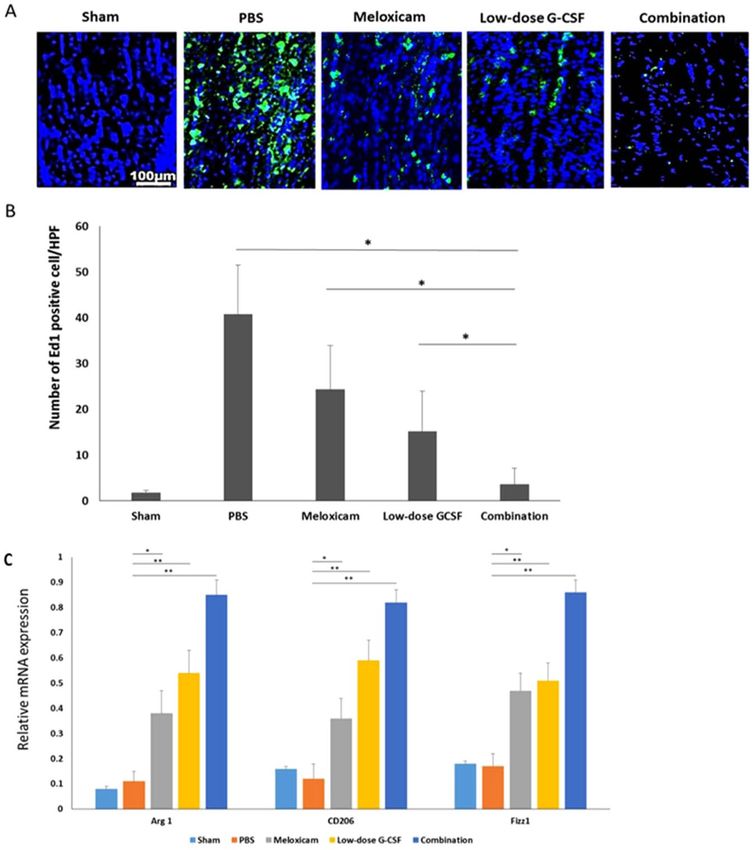
Combined treatment reduced extrinsic macrophage infiltration and increased the level of M2
phenotypic markers. Combination treatment synergistically reduced the number of ED1-positive cells
Scientific Reports | (2020) 10:10351 | https://doi.org/10.1038/s41598-020-66977-9 4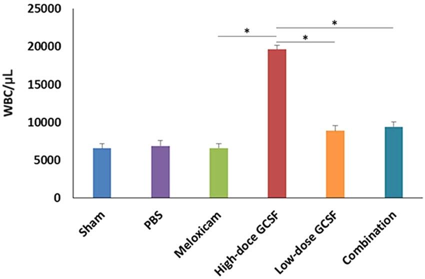
www.nature.com/scientificreports/                                                            www.nature.com/scientificreports

                            Figure 3. Survival of RGCs in rAION-induced rats with PBS treatment, meloxicam treatment, G-CSF
                            treatment, and G-CSF plus meloxicam treatment at 28 days after rAION induction. (A) A representative of flat-
                            mounted central retinas and the morphometry of RGCs in each group through FluoroGold retrograde labeling
                            at four weeks after rAION induction. (B) RGC density in the central retina in each group. Data are expressed as
                            mean ± SD for each group (n = 12). The number of RGCs in the combination-treated group was 1.58- and 1.45-
                            fold higher than in the meloxicam-treated and low-dose G-CSF-treated groups, respectively. *p < 0.05.

                            Figure 4. Analysis of RGC apoptosis in the RGC layer through TUNEL assay at four weeks after rAION
                            induction. (A) Representative images of double-stained apoptotic cells in the RGC layers in each group. The
                            apoptotic cells (TUNEL-positive cells) in green were stained with TUNEL staining, and the nuclei of the RGCs
                            in blue were labeled with DAPI staining. (B) Quantification of TUNEL-positive cells per high-power field.
                            Data are expressed as mean ± SD for each group (n = 6). Treatment with low-dose G-CSF plus meloxicam
                            significantly reduced the number of apoptotic RGC by 3.6- and 2.5-fold compared with the meloxicam-treated
                            and low-dose G-CSF-treated groups, respectively. *p < 0.05.

Scientific Reports |   (2020) 10:10351 | https://doi.org/10.1038/s41598-020-66977-9                                                        5
www.nature.com/scientificreports/ www.nature.com/scientificreports
Figure 5. Immunohistochemistry (IHC) of ED1 in the optic nerve at four weeks after rAION induction for
evaluating the inflammatory infiltration of macrophages. (A) Representative images of ED1 staining in the
longitudinal sections of the optic nerve. The ED1-positive cells in green were stained with FITC, and the
nuclei in blue were labeled with DAPI. (B) Quantification of ED1-positive cells per high-power field. Data are
expressed as mean ± SD in each group (n = 6). Macrophage recruitment was decreased by 6.75- and 4.1-fold
in the combination-treated group compared with the meloxicam-treated and low-dose G-CSF-treated groups,
respectively (C) Evaluation of M2 macrophage polarization at four weeks after rAION induction. Relative
mRNA expression levels of the markers of M2 macrophages in the optic nerve are shown as histograms. Each
value was normalized to CypA. The expression levels of Arg 1, CD206, and Fizz1 (markers of M2 macrophages)
increased after treatment with low-dose G-CSF plus meloxicam compared with treatment with PBS-treated
group, meloxicam alone, and low-dose G-CSF alone, respectively. *p < 0.05, **p < 0.01.
in the rAION model (Fig. 5A). The number of ED1-positive cells/HPF in sham, PBS-, meloxicam-, low-dose
G-CSF-, and the combination-treated group was 1.8 ± 0.5, 40.8 ± 10.7, 24.3 ± 9.6, 15.1 ± 8.9, and 3.6 ± 3.5, respec-
tively (Fig. 5B). Macrophage recruitment decreased by 6.75- and 4.1-folds in the combination-treated group
compared with the meloxicam-treated (p = 0.021) and low-dose G-CSF-treated group (p = 0.032), respectively.
Further, the qRT-PCR analysis demonstrated that the mRNA levels of Arg 1, CD206, and Fizz1 (M2 phenotypic
markers) increased after treatment with meloxicam, low-dose G-CSF, and low-dose G-CSF plus meloxicam after
rAION induction compared with PBS-treated group. In addition, the combination treatment exerted synergistic
effects on the increased expression of Arg1, CD206, Fizz; (p = 0.005) in the rAION model (Fig. 5C).
Combination treatment induced more Akt1 activation than other single treatments. To reveal
the synergetic effects of the combination treatment, the expression level of p-Akt1 was assessed at day seven after
AION induction to determine if combination treatment had an enhanced effect on p-Akt1 expression compared
to meloxicam or low dose G-CSF (Fig. 6A,B). The levels of p-Akt1 in the meloxicam-treated group (p = 0.018),
low-dose G-CSF-treated group (p = 0.021), and combination-treated group (p = 0.011) were 2.78-, 2.93-, and
4.86-fold higher than PBS-treated group, respectively. Besides, the combination treatment induced higher p-Akt1
expression than treatment with meloxicam (p = 0.021) or low-dose G-CSF (p = 0.021) in the rAION model.
Scientific Reports | (2020) 10:10351 | https://doi.org/10.1038/s41598-020-66977-9 6www.nature.com/scientificreports/ www.nature.com/scientificreports
Figure 6. Immunoblots of the optic nerve. (A) Analysis of p-Akt1 expression by using Western blotting. (B)
Quantification of the protein bands of p-Akt1. Each value was normalized to GAPDH. Data are expressed as
mean ± SD in each group (n = 6 in each group). The combination treatment induced higher p-Akt1 expression
than treatment with meloxicam or low-dose G-CSF in the rAION model. *p < 0.05.
Discussions
Consecutive high-dose G-CSF (100 μg/kg/day) injection for five days commonly triggered leukocytosis in rats,
but this undesired effect was absent with low-dose G-CSF (50 μg/kg/day) treatment. The administration of
low-dose G-CSF alone or meloxicam alone yielded preservation of visual function, better RGCs survival, reduc-
tion of macrophage infiltration, and more M2 macrophage/microglia polarization after rAION insults. Notably,
the neuroprotective effects of the combination treatment worked synergistically. In addition, the combination
treatment induced more p-Akt1 expression than meloxicam or low-dose G-CSF treatment alone in the current
model.
Based on our past work, we concluded that subcutaneous injection of high-dose G-CSF provided neuropro-
tection in both rAION and optic nerve crush (ONC) models9–12. We also found that leukocytosis was induced
by this therapy in the rat ONC model11. Moreover, this therapy has detrimental effects on patients. Commonly
encountered side effects after G-CSF use comprise arthralgia, bone pain, fatigue, nausea, fever, chills, headache,
and myalgia13. Thus, for clinical applications, reducing the undesired effects induced by G-CSF therapy is essen-
tial for ophthalmic diseases. In this study, we found that consecutively treating normal Wistar rats with high-dose
G-CSF (100 μg/kg/day) via subcutaneous injection would induce leukocytosis. By contrast, neither low-dose
G-CSF (50 μg/kg/day) treatment nor meloxicam treatment would lead to the undesired effect. As expected, the
combination treatment did not induce leukocytosis compared with normal rats. Furthermore, the addition of
meloxicam to G-CSF treatment may reduce the discomfort caused by the side effects of G-CSF because meloxi-
cam is a strong pain reliever. From the literature review, meloxicam possesses low toxicity to gastrointestinal tract
mucosa and trustworthy efficacy against inflammation and pain21–24. Therefore, treatment with a combination of
G-CSF and meloxicam is a reasonable approach for rAION.
The neuroprotective role of meloxicam on the rAION model was first disclosed in this study. Surprisingly,
meloxicam revealed comparable neuroprotective effects to low-dose G-CSF regarding antiapoptosis, inhibition
of macrophage infiltration, and macrophage M2 polarization in this model. Meloxicam breaks the inflammatory
cascade via inhibition of MAPK and p53 signaling pathway and owns antiapoptotic effects evidenced by allevi-
ating oxidative stress, avoiding mitochondrial dysfunction, and reducing endoplasmic reticulum stress response
in vitro25,26. A recent study reported that meloxicam’s neuroprotective effects maintained cell survival via the
upregulation of the PI3K/Akt pathway instead of COX-2 inhibition26. In this study, we further disclosed that
meloxicam induced Akt1 activation on day seven post-infarct. Couples of previous studies have indicated that the
upregulation of the PI3K/AKT signaling pathway benefits the survival of injured RGCs27–29. In our previous study,
we also demonstrated that the apoptosis of RGCs following optic nerve crush could be antagonized by G-CSF
via PI3K/AKT-signaling pathway12. Taken together, we consider that meloxicam can maintain Akt1 activation
to prevent RGC death after optic nerve damage. Remarkably, treatment with a combination of meloxicam plus
low-dose G-CSF induced higher Ak1 activation than treatment with meloxicam only or low-dose G-CSF only in
the rAION model. Therefore, we believe that combination treatment with meloxicam and low-dose G-CSF affords
better protection of RGCs in the rAION model via antiapoptosis of RGCs and activation of the Akt1 signaling
pathway.
After rAION induction, ED1 + phagocytes, including monocytes/macrophages of hematopoietic origin and
microglia, are recruited into the optic nerves. The hematogenous ED1 + cells found at the optic nerve following
rAION induction implicates disrupted BOB10,30. We found that ED1 + macrophage/microglial buildup at the
Scientific Reports | (2020) 10:10351 | https://doi.org/10.1038/s41598-020-66977-9 7www.nature.com/scientificreports/ www.nature.com/scientificreports
optic nerve lesion site was alleviated in the meloxicam-treated and low-dose G-CSF-treated groups. The effect of
attenuation was enhanced when the combination treatment of low-dose G-CSF plus meloxicam was used. Our
recent study, therefore, implies that the rAION-injured optic nerves may be protected by the anti-inflammatory
effects of immediate administration of meloxicam or G-CSF, and combination treatment with G-CSF plus meloxi-
cam exerts the synergistic effect of reduction in macrophage recruitment. Some studies have demonstrated that
treatment with G-CSF is able to reduce the breakdown of blood-brain barrier (BBB), brain edema, and mac-
rophage infiltration in experimental brain injury models30–33. Also, our previous report revealed that protection
from BOB disruption reduces macrophage infiltration into the optic nerve lesion site10. We believe that the possi-
ble mechanism of BOB disruption is associated with the modulation of the Akt signaling pathway in the rAION
model. Our study also noted that Akt1 was activated by treatment with meloxicam or G-CSF alone in the rAION
model. Therefore, we suggest that Akt1 activation may play a crucial role in preventing BOB breakdown after
optic nerve infarct.
Macrophage/microglia has plasticity, and one phenotype can convert into another phenotype when driven
by cytokines in different microenvironments34. We ever reported that phenotype switch of macrophages from
pro-inflammatory (M1) to anti-inflammatory (M2) prevent the cytokine-induced optic nerve injuries by reduc-
ing the pro-inflammatory cytokine expression and free radicals (e.g., IL-6, IL-1β, TNF-α, and iNOS) during the
inflammatory responses of optic nerve injury10,18. Intriguingly, our findings showed that meloxicam, a COX-2
inhibitor, alone was able to activate M2 polarization on the injured optic nerve, and the protective phenomenon
was further magnified by simultaneous G-CSF and meloxicam administration.
Hofer et al. reported that meloxicam has hematopoiesis-modulating action and can increase serum G-CSF
levels in sublethally gamma-irradiated animals35. Enhancement of neuroprotective effects and M2 polarization
from the combination treatment may be due to the increase in endogenous G-CSF after meloxicam administra-
tion. Also, we found that treatment with meloxicam alone or G-CSF alone activated Akt1 7 days after rAION
induction. Since Akt downregulation will abolish the upregulation of M2 genes, Akt activation plays an important
role in M2 macrophage differentiation36. Liu et al. demonstrated that TIPE2 activates the PI3K-AKT signaling
pathway and subsequently promotes M2 polarization37. Furthermore, recent studies have reported that Akt1 and
Akt2 kinase isoforms are critical to modulation of macrophage polarization and Akt1 ablation shifts macrophage
towards M1 status38. Taken together, we conclude that Akt1 upregulation may be crucial to promote M2 polari-
zation in the rAION model.
In conclusion, addressing anti-inflammatory therapy through different mechanisms may provide a potential
approach to the management of ischemic optic nerve injury. Combination therapy of G-CSF plus meloxicam can
reduce the leukocytosis side effects, prevent postinjury RGCs apoptosis, preserve the visual function, halt the
recruitment of macrophages to the optic nerve, and promote the M2 phenotype transition in macrophages. The
neuroprotective effects of G-CSF and meloxicam worked synergistically. However, additional works are man-
datory to more clearly elucidate the underlying mechanisms of the interaction between G-CSF and meloxicam.
Received: 14 June 2019; Accepted: 19 May 2020;
Published: xx xx xxxx
References
1. Johnson, L. N. & Arnold, A. C. Incidence of nonarteritic and arteritic anterior ischemic optic neuropathy. Population-based study
in the state of Missouri and Los Angeles County, California. J Neuroophthalmol 14, 38–44 (1994).
2. Hattenhauer, M. G., Leavitt, J. A., Hodge, D. O., Grill, R. & Gray, D. T. Incidence of nonarteritic anterior ischemic optic neuropathy.
Am J Ophthalmol 123, 103–107 (1997).
3. Lee, Y. C., Wang, J. H., Huang, T. L. & Tsai, R. K. Increased risk of stroke in patients with nonarteritic anterior ischemic optic
neuropathy: a nationwide retrospective cohort study. Am J Ophthalmol 170, 183–189, https://doi.org/10.1016/j.ajo.2016.08.006
(2016).
4. Levin, L. A. & Louhab, A. Apoptosis of retinal ganglion cells in anterior ischemic optic neuropathy. Arch Ophthalmol 114, 488–491
(1996).
5. Frampton, J. E., Lee, C. R. & Faulds, D. Filgrastim. A review of its pharmacological properties and therapeutic efficacy in
neutropenia. Drugs 48, 731–760, https://doi.org/10.2165/00003495-199448050-00007 (1994).
6. Demetri, G. D. & Griffin, J. D. Granulocyte colony-stimulating factor and its receptor. Blood 78, 2791–2808 (1991).
7. Weaver, C. H. et al. Syngeneic transplantation with peripheral blood mononuclear cells collected after the administration of
recombinant human granulocyte colony-stimulating factor. Blood 82, 1981–1984 (1993).
8. Grigg, A. P. et al. Optimizing dose and scheduling of filgrastim (granulocyte colony-stimulating factor) for mobilization and
collection of peripheral blood progenitor cells in normal volunteers. Blood 86, 4437–4445 (1995).
9. Chang, C. H., Huang, T. L., Huang, S. P. & Tsai, R. K. Neuroprotective effects of recombinant human granulocyte colony-stimulating
factor (G-CSF) in a rat model of anterior ischemic optic neuropathy (rAION). Exp Eye Res 118, 109–116, https://doi.org/10.1016/j.
exer.2013.11.012 (2014).
10. Wen, Y. T., Huang, T. L., Huang, S. P., Chang, C. H. & Tsai, R. K. Early applications of granulocyte colony-stimulating factor (G-CSF)
can stabilize the blood-optic-nerve barrier and ameliorate inflammation in a rat model of anterior ischemic optic neuropathy
(rAION). Dis Model Mech 9, 1193–1202, https://doi.org/10.1242/dmm.025999 (2016).
11. Tsai, R. K., Chang, C. H. & Wang, H. Z. Neuroprotective effects of recombinant human granulocyte colony-stimulating factor
(G-CSF) in neurodegeneration after optic nerve crush in rats. Exp Eye Res 87, 242–250, https://doi.org/10.1016/j.exer.2008.06.004
(2008).
12. Tsai, R. K., Chang, C. H., Sheu, M. M. & Huang, Z. L. Anti-apoptotic effects of human granulocyte colony-stimulating factor
(G-CSF) on retinal ganglion cells after optic nerve crush are PI3K/AKT-dependent. Exp Eye Res 90, 537–545, https://doi.
org/10.1016/j.exer.2010.01.004 (2010).
13. McCullough, J., Clay, M., Herr, G., Smith, J. & Stroncek, D. Effects of granulocyte-colony-stimulating factor on potential normal
granulocyte donors. Transfusion 39, 1136–1140 (1999).
14. Barner, A. Review of clinical trials and benefit/risk ratio of meloxicam. Scand J Rheumatol Suppl 102, 29–37 (1996).
15. Noble, S. & Balfour, J. A. Meloxicam. Drugs 51, 424–430; discussion 431–432 (1996).
16. Engelhardt, G. Pharmacology of meloxicam, a new non-steroidal anti-inflammatory drug with an improved safety profile through
preferential inhibition of COX-2. Br J Rheumatol 35(Suppl 1), 4–12 (1996).
Scientific Reports | (2020) 10:10351 | https://doi.org/10.1038/s41598-020-66977-9 8www.nature.com/scientificreports/ www.nature.com/scientificreports
17. Mills, C. D. M1 and M2 Macrophages: Oracles of Health and Disease. Crit Rev Immunol 32, 463–488 (2012).
18. Georgiou, T. et al. Neuroprotective Effects of Omega-3 Polyunsaturated Fatty Acids in a Rat Model of Anterior Ischemic Optic
Neuropathy. Invest Ophthalmol Vis Sci 58, 1603–1611, https://doi.org/10.1167/iovs.16-20979 (2017).
19. Bernstein, S. L., Guo, Y., Kelman, S. E., Flower, R. W. & Johnson, M. A. Functional and cellular responses in a novel rodent model of
anterior ischemic optic neuropathy. Investigative ophthalmology & visual science 44, 4153–4162, https://doi.org/10.1167/iovs.03-
0274 (2003).
20. Slater, B. J., Mehrabian, Z., Guo, Y., Hunter, A. & Bernstein, S. L. Rodent anterior ischemic optic neuropathy (rAION) induces
regional retinal ganglion cell apoptosis with a unique temporal pattern. Investigative ophthalmology & visual science 49, 3671–3676,
https://doi.org/10.1167/iovs.07-0504 (2008).
21. Furst, D. E. Meloxicam: selective COX-2 inhibition in clinical practice. Semin Arthritis Rheum 26, 21–27 (1997).
22. Monteiro, B. P. et al. Analgesic efficacy of an oral transmucosal spray formulation of meloxicam alone or in combination with
tramadol in cats with naturally occurring osteoarthritis. Veterinary anaesthesia and analgesia 43, 643–651, https://doi.org/10.1111/
vaa.12360 (2016).
23. Tiurin, V. P. & Rakov, A. L. [Efficacy and intolerance of meloxicam in injection and tablet form]. Voen Med Zh 324, 54–58 (2003).
24. Tsvetkova, E. S. [Meloxicam: intramuscular administration in rheumatology]. Ter Arkh 75, 96–97 (2003).
25. Park, J. H. et al. Meloxicam inhibits fipronil-induced apoptosis via modulation of the oxidative stress and inflammatory response in
SH-SY5Y cells. J Appl Toxicol 36, 10–23, https://doi.org/10.1002/jat.3136 (2016).
26. Tasaki, Y. et al. Meloxicam protects cell damage from 1-methyl-4-phenyl pyridinium toxicity via the phosphatidylinositol 3-kinase/
Akt pathway in human dopaminergic neuroblastoma SH-SY5Y cells. Brain Res 1344, 25–33, https://doi.org/10.1016/j.
brainres.2010.04.085 (2010).
27. Levkovitch-Verbin, H. Retinal ganglion cell apoptotic pathway in glaucoma: Initiating and downstream mechanisms. Prog Brain Res
220, 37–57, https://doi.org/10.1016/bs.pbr.2015.05.005 (2015).
28. Li, H. B. et al. Long Non-Coding RNA-MALAT1 Mediates Retinal Ganglion Cell Apoptosis Through the PI3K/Akt Signaling
Pathway in Rats with Glaucoma. Cell Physiol Biochem 43, 2117–2132, https://doi.org/10.1159/000484231 (2017).
29. Nie, X. G. et al. Downregulation of microRNA-149 in retinal ganglion cells suppresses apoptosis through activation of the PI3K/Akt
signaling pathway in mice with glaucoma. American journal of physiology. Cell physiology 315, C839–c849, https://doi.org/10.1152/
ajpcell.00324.2017 (2018).
30. Bernstein, S. L., Johnson, M. A. & Miller, N. R. Nonarteritic anterior ischemic optic neuropathy (NAION) and its experimental
models. Prog Retin Eye Res 30, 167–187, https://doi.org/10.1016/j.preteyeres.2011.02.003 (2011).
31. Li, L. et al. G-CSF attenuates neuroinflammation and stabilizes the blood-brain barrier via the PI3K/Akt/GSK-3beta signaling
pathway following neonatal hypoxia-ischemia in rats. Exp Neurol 272, 135–144, https://doi.org/10.1016/j.expneurol.2014.12.020
(2015).
32. Ghorbani, M. et al. G-CSF administration attenuates brain injury in rats following carbon monoxide poisoning via different
mechanisms. Environ Toxicol 32, 37–47, https://doi.org/10.1002/tox.22210 (2017).
33. Wei, X. et al. Granulocyte Colony-Stimulating Factor Attenuates Blood-Brain Barrier Damage and Improves Cognitive Function in
Spontaneously Hypertensive Rats. CNS Neurol Disord Drug Targets 16, 781–788, https://doi.org/10.2174/187152731666617020715
5730 (2017).
34. Hesketh, M., Sahin, K. B., West, Z. E. & Murray, R. Z. Macrophage Phenotypes Regulate Scar Formation and Chronic Wound
Healing. Int J Mol Sci 18, https://doi.org/10.3390/ijms18071545 (2017).
35. Hofer, M. et al. Meloxicam, an inhibitor of cyclooxygenase-2, increases the level of serum G-CSF and might be usable as an auxiliary
means in G-CSF therapy. Physiol Res 57, 307–310 (2008).
36. Vergadi, E., Ieronymaki, E., Lyroni, K., Vaporidi, K. & Tsatsanis, C. Akt Signaling Pathway in Macrophage Activation and M1/M2
Polarization. J Immunol 198, 1006–1014, https://doi.org/10.4049/jimmunol.1601515 (2017).
37. Liu, R. et al. Negative Immune Regulator TIPE2 Promotes M2 Macrophage Differentiation through the Activation of PI3K-AKT
Signaling Pathway. PloS one 12, e0170666, https://doi.org/10.1371/journal.pone.0170666 (2017).
38. Arranz, A. et al. Akt1 and Akt2 protein kinases differentially contribute to macrophage polarization. Proc Natl Acad Sci USA 109,
9517–9522, https://doi.org/10.1073/pnas.1119038109 (2012).
Acknowledgements
The authors thank Mr. Yu-Chieh Ho and Ms. I-Ping Tsai for their assistance in animal handling and figure
preparation. This study was supported by a grant from the Ministry of Science and Technology, Taiwan (MOST
103-2314-B-303-007-MY3).
Author contributions
Y.T.W. and R.K.T. designed the experiment. Y.T.W., P.K.L. and W.L. performed the experiments. Y.T.W., P.K.L.
analyzed the data. K.K., Y.T.W. and P.K.L. made the figures. K.K., Y.T.W., P.K.L., M.H.T. and R.K.T. wrote and
edited the manuscript. M.H.T. and R.K.T. provided resources and financial support. All authors reviewed and
approved the manuscript.
Competing interests
The authors declare no competing interests.
Additional information
Supplementary information is available for this paper at https://doi.org/10.1038/s41598-020-66977-9.
Correspondence and requests for materials should be addressed to M.T. or R.-K.T.
Reprints and permissions information is available at www.nature.com/reprints.
Publisher’s note Springer Nature remains neutral with regard to jurisdictional claims in published maps and
institutional affiliations.
Scientific Reports | (2020) 10:10351 | https://doi.org/10.1038/s41598-020-66977-9 9www.nature.com/scientificreports/ www.nature.com/scientificreports
Open Access This article is licensed under a Creative Commons Attribution 4.0 International
License, which permits use, sharing, adaptation, distribution and reproduction in any medium or
format, as long as you give appropriate credit to the original author(s) and the source, provide a link to the Cre-
ative Commons license, and indicate if changes were made. The images or other third party material in this
article are included in the article’s Creative Commons license, unless indicated otherwise in a credit line to the
material. If material is not included in the article’s Creative Commons license and your intended use is not per-
mitted by statutory regulation or exceeds the permitted use, you will need to obtain permission directly from the
copyright holder. To view a copy of this license, visit http://creativecommons.org/licenses/by/4.0/.
© The Author(s) 2020
Scientific Reports | (2020) 10:10351 | https://doi.org/10.1038/s41598-020-66977-9 10You can also read