Sorafenib for the Treatment of Unresectable Hepatocellular Carcinoma: Preliminary Toxicity and Activity Data in Dogs - MDPI
←
→
Page content transcription
If your browser does not render page correctly, please read the page content below
cancers
Article
Sorafenib for the Treatment of Unresectable
Hepatocellular Carcinoma: Preliminary Toxicity and
Activity Data in Dogs
Laura Marconato 1, *, Silvia Sabattini 1 , Giorgia Marisi 2 , Federica Rossi 3 ,
Vito Ferdinando Leone 3 and Andrea Casadei-Gardini 4
 1   Department of Veterinary Medical Sciences, University of Bologna, via Tolara di Sopra 50,
     Ozzano dell’Emilia, 40064 Bologna, Italy; silvia.sabattini@unibo.it
 2   Biosciences Laboratory, Istituto Scientifico Romagnolo per Lo Studio e La Cura Dei Tumori (IRST) IRCCS,
     Via Piero Maroncelli 40, Meldola, 47014 Forlì-Cesena, Italy; giorgia.marisi@irst.emr.it
 3   Centro Oncologico Veterinario, via San Lorenzo 1-4, Sasso Marconi, 40037 Bologna, Italy;
     rossi@centroncologicovet.it (F.R.); leone@centroncologicovet.it (V.F.L.)
 4   Division of Oncology, Department of Oncology and Hematology, University Hospital Modena,
     41124 Modena, Italy; casadeigardini@gmail.com
 *   Correspondence: laura.marconato@unibo.it

 Received: 3 April 2020; Accepted: 15 May 2020; Published: 18 May 2020

 Abstract: Unresectable nodular and diffuse hepatocellular carcinoma (HCC) have a poor prognosis
 with limited treatment options. Systemic traditional chemotherapy has been only rarely reported,
 with unsatisfactory results. The aim of this prospective, non-randomized, non-blinded, single center
 clinical trial was to investigate safety profile, objective response rate, time to progression and overall
 survival of sorafenib in comparison with metronomic chemotherapy (MC) consisting of thalidomide,
 piroxicam and cyclophosphamide in dogs with advanced, unresectable HCC. Between December
 2011 and June 2017, 13 dogs were enrolled: seven received sorafenib, and six were treated with MC.
 Median time to progression was 363 days (95% CI, 191–535) in dogs treated with sorafenib versus
 27 days (95% CI, 0–68) in dogs treated with MC (p = 0.044). Median overall survival was 361 days
 (95% CI, 0–909) in dogs receiving sorafenib, while 32 days (95% CI, 0–235) in those receiving MC
 (p = 0.079). Sorafenib seems to be a good candidate for the treatment of dogs with advanced HCC,
 due to a benefit in disease control and an acceptable safety profile, offering a good basis on which new
 randomized prospective clinical trials should be undertaken to compare the efficacy and drawback of
 sorafenib versus MC or traditional chemotherapy.

 Keywords: dog; hepatocellular carcinoma; metronomic therapy; outcome; sorafenib; toxicity;
 spontaneous model

1. Introduction
     In dogs, hepatocellular carcinoma (HCC) is the most common primary liver tumor [1,2], and its
prognosis depends on the morphological type. The massive form is typically confined to one liver
lobe and may be amenable to surgical resection [3]. Also, the metastatic potential is generally low to
moderate. Conversely, the nodular and diffuse forms of HCC typically involve multiple liver lobes and
are not amenable to surgical resection [1–3]. Also, the metastatic rate exceeds 90% for both forms [1–3].
Thus, unresectable nodular or diffuse HCC harbors a poor prognosis, as there are limited treatment
options for non-surgical cases.
     Interdisciplinary approaches, including local tumor ablation, transarterial embolization,
and radiotherapy, remain uninvestigated in veterinary medicine. Systemic traditional chemotherapy

Cancers 2020, 12, 1272; doi:10.3390/cancers12051272                              www.mdpi.com/journal/cancers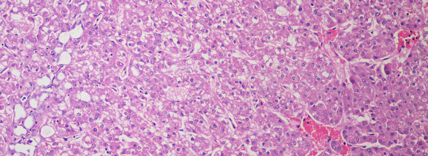
Cancers 2020, 12, 1272                                                                               2 of 14

has only been rarely reported, with unsatisfactory results. Resistance of tumor cells to chemotherapeutic
drugs is an important component of clinical treatment failure and recurrence for patients with HCC [4].
In one study, seven dogs with unresectable HCC were treated with intravenous gemcitabine [5].
Median progression-free interval and median survival time were 150 days and 197 days, respectively [5].
In another study, two dogs with unresectable and metastatic HCC did not respond to gemcitabine and
carboplatin [6]. Thus, new treatment approaches are urgently needed.
      Metronomic chemotherapy (MC) has gained traction recently as an attractive treatment modality
due to its favorable toxicity profile and ease of administration in comparison to traditional chemotherapy.
MC refers to the practice of administering cytotoxic drugs without prolonged drug-free breaks and
at doses significantly lower than dose-intense chemotherapy, with the therapeutic target of both
anti-angiogenic and immune-modulatory effects [7]. In veterinary oncology, MC is mainly used in
a palliative setting. Drugs that are commonly used in metronomic regimens include, among others,
cyclophosphamide and piroxicam [8–10]. There are fewer reports of thalidomide use in dogs, either used
as a single agent or as part of a metronomic regimen in combination with cyclophosphamide and
piroxicam. These reports have shown a favorable toxicity profile and some antitumor activity [11–15].
      In people with advanced or intermediate stage HCC, in those with refractory cancer, or in patients no
longer amenable to locoregional therapies and preserved liver function (child pugh A), the multi-kinase
inhibitor sorafenib remains the only FDA-approved first-line treatment option for systemic therapy [16].
Sorafenib is a multi-kinase inhibitor that targets Raf kinase, vascular endothelial growth factor receptors
1, 2, and 3, and platelet-derived growth factor receptor β with anti-proliferative and anti-angiogenic
activities. Unfortunately, in a substantial percentage of human patients, acquired sorafenib resistance
remains a major clinical obstacle, leading to treatment failure. Several mechanisms are implicated in the
reduction of tumor cell sensitivity to sorafenib, including metabolic rewiring [17–22].
      The most common drug-related side effects are fatigue, hypertension, anorexia, diarrhea,
rash/desquamation, and hand–foot skin reaction [16,23].
      In dogs, the drug sorafenib showed potent antitumor activity against canine osteosarcoma and
hemangiosarcoma cells in vitro [24,25]. According to preclinical toxicology studies conducted in
healthy female beagle dogs, sorafenib administered orally at dosages of 30 mg/kg/day was associated
with significant gastrointestinal, cutaneous, renal, adrenal, bone and hematologic toxicity after long
term administration (3–12 months), whereas at a 10 mg/kg/day dosing there were no significant side
effects observed [26,27]. To date sorafenib was also evaluated clinically in an early phase tolerability
study on dogs with various cancers, including one animal with HCC [28]. Single oral doses of sorafenib
were tolerable up to 3 mg/kg when given on a once-weekly basis [28]. Thus, it seems plausible that the
dose can be further escalated.
      The aim of this prospective, non-randomized, non-blinded, single center clinical trial was to
investigate safety profile, objective response rate (RR), time to progression (TTP) and overall survival
(OS) of sorafenib in comparison with MC consisting of thalidomide, piroxicam and cyclophosphamide
in dogs with advanced, unresectable HCC. It was hypothesized that sorafenib would lead to a better
outcome than MC.

2. Results

2.1. Dogs and Tumor Characteristics
     Between December 2011 and June 2017, 13 client-owned dogs were enrolled at one single Centre
(Centro Oncologico Veterinario): seven received sorafenib, and six were treated with MC.
     Represented breeds are listed in Table 1. There were 10 females (seven spayed) and three intact males.
Median age was 11 years (range, 5 to 13 years), and median weight was 10.6 kg (range, 2.7 to 41.1 kg).
     Complete staging diagnostic tests were performed in each case on the day of initial presentation:
nine dogs underwent TBCT, whereas four dogs were staged by means of abdominal ultrasound and
thoracic radiographs.
Cancers 2020, 12, 1272 3 of 14
Table 1. Demographic and clinical details of 13 dogs with hepatocellular carcinoma.
Clinical Altered Liver Surgical Treatment Treatment Antitumor OS (Cause of
Breed Sex Age Weight
Stage Enzymes Debulking Protocol Toxicity Response, TTP Death)
G1 erythema and 784
Golden retriever M 11 41.1 T2N0M1 Yes No Sorafenib PR, 363
alopecia (cancer)
G1
CR, 1579 1706
Golden retriever SF 5 27.8 T1N2M0 No Yes Sorafenib hyperpigmentation;
(no progression) (alive)
G1 diarrhea
PR, 1250 1398
Yorkshire terrier SF 10 3.6 T2N1M0 Yes Yes Sorafenib G2 alopecia
(no progression) (alive)
PR, 228 255
Bloodhound F 8 20.5 T3N1M1 No No Sorafenib None
(no progression) (leishmaniasis)
361
Cross-breed M 11 10 T3N0M0 Yes No Sorafenib None SD, 168
(cancer)
Sorafenib,
327
Pomeranian SF 13 10.1 T2N1M0 Yes No Then MC None SD, 230
(cancer)
crossover
Sorafenib,
279
Poodle F 13 2.7 T2N0M1 No No Then MC None SD, 205
(cancer)
crossover
27
Shih-tzu SF 11 6.7 T3N1M1 Yes Yes MC None PD, 27
(cancer)
1390
Beagle SF 11 13.1 T3N1M0 Yes No MC None SD, 703
(cancer)
G1
196
Scottish terrier SF 7 10.6 T2N0M1 Yes No MC gastrointestinal; SD, 35
(cancer)
G1 renal
French G1 hemorrhagic 390
SF 6 11.7 T3N0M0 No No MC SD, 219
bouledogue cystitis (cancer)
Jack Russell 19
M 12 8.1 T3N0M0 Yes No MC None PD, 1
Terrier (cancer)
G2 32
German Shepherd F 11 29.6 T3N1M0 No No MC PD, 1
gastrointestinal (cancer)
M = male; F = female; SF = spayed female; MC = metronomic chemotherapy; G = grade; TTP = time to progression; OS = overall survival; CR = complete remission; PR = partial remission;
SD = stable disease; PD = progressive disease.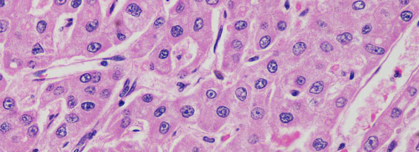
Cancers 2020, 12, x 4 of 13
Complete staging diagnostic tests were performed in each case on the day of initial presentation:
Cancersdogs
nine 2020,underwent
12, 1272 TBCT, whereas four dogs were staged by means of abdominal ultrasound4 of and
14
thoracic radiographs.
Imaging workup revealed a hepatic round mass with a mean maximum diameter of 6.4 cm
Imaging
(range, 3 to 10workup
cm). Forrevealed a hepatic TBCT,
dogs undergoing round HCCmass hadwitha aheterogeneous
mean maximum diameter ofwhereas
enhancement; 6.4 cm
(range, 3 to 10 cm). For dogs undergoing TBCT, HCC had a heterogeneous
for those undergoing abdominal ultrasound, the tumor appeared sonographically heterogeneous enhancement; whereas for
those undergoing abdominal ultrasound, the tumor appeared sonographically heterogeneous solid.
solid.
In nine
In nine dogs,
dogs,HCCsHCCsappeared
appeared as as a single
a single largelarge
massmass
locatedlocated
in theinquadrate
the quadrate
lobe (nlobe the=left
= 3),(n 3),
the left lateral and medial (n = 4), the right lateral (n = 1) and right medial lobe
lateral and medial (n = 4), the right lateral (n = 1) and right medial lobe (n = 1). In three of these nine (n = 1). In three of
these nine
cases, cases, nodules
secondary secondary werenodules
observedwere observed
adjacent adjacent
to the primaryto the primary
lesion, lesion,with
compatible compatible with
intrahepatic
intrahepatic
metastases. metastases.
In the
In the other
other four
four cases,
cases, multiple
multiple hepatic
hepatic nodules
nodules of of similar
similar size
size disseminated
disseminated in in all
all liver
liver lobes
lobes
were
were found,
found, ranging
ranging from
from 0.4
0.4 and
and 9 9
cm cmin in diameter.
diameter. Additional
Additional findings
findings were were regional
regional lymph lymph
node
node enlargement (n = 5), peritoneal nodules and ascites (n = 2), sternal lymphadenopathy
enlargement (n = 5), peritoneal nodules and ascites (n = 2), sternal lymphadenopathy (n = 1), thoracic (n = 1),
thoracic nodules
nodules (n = 1) and (n =a 21)cm
and a 2 cm
right rightnodule
adrenal = 1). (n = 1).
adrenal(nnodule
A histopathological diagnosis of HCC was obtained in in
A histopathological diagnosis of HCC was obtained all all
dogsdogs by means
by means of imaging-guided
of imaging-guided tru-
tru-cut biopsy (n = 10) or surgical biopsy (n = 3). Histologically, all
cut biopsy (n = 10) or surgical biopsy (n = 3). Histologically, all tumors were characterizedtumors were characterized by by aa
predominant
predominant trabecular pattern, with
trabecular pattern, with neoplastic
neoplastic cells
cells arranged
arranged in in irregular
irregular trabeculae
trabeculae up up to
to 20
20 cells
cells
thick often separated by dilated sinusoids, occasionally forming cavernous
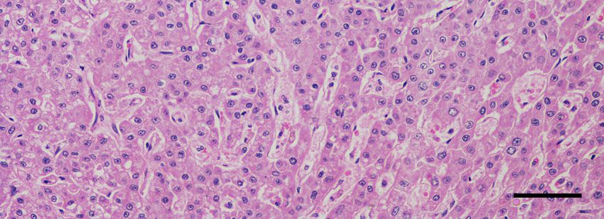
thick often separated by dilated sinusoids, occasionally forming cavernous spaces (Figures 1 and 2). spaces (Figures 1 and 2).
Colliquative necrosis
Colliquative necrosis waswas aa common
common finding
finding (Figure
(Figure 3). Less frequently,
3). Less frequently, areas
areas of pseudoglandular or
of pseudoglandular or
solid differentiation
differentiation were were observed
observed in in aa subset
subset ofof tumors.
tumors.
Figure 1. Dog,
Dog, liver.
liver. Histological
Histological sample
sample of
of hepatocellular
hepatocellular carcinoma.
carcinoma. The
The tumor
tumor is
iswell-differentiated
well-differentiated
and composed by variably thick trabeculae formed by neoplastic hepatocytes
hepatocytes (trabecular pattern).
(trabecular pattern).
Hematoxylin and eosin. Bar, 100 µm.
Figure
Figure 2.2.Dog,
Dog,liver.
liver. Histological
Histological sample
sample of hepatocellular
of hepatocellular carcinoma.
carcinoma. At high magnification,
At high magnification, neoplastic
neoplastic cells show moderate anisocytosis and anisokaryosis, prominent nucleoli and mitotic
cells show moderate anisocytosis and anisokaryosis, prominent nucleoli and mitotic figures. Hematoxylin
figures. Hematoxylin
and eosin. Bar, 50 µm. and eosin. Bar, 50 µm.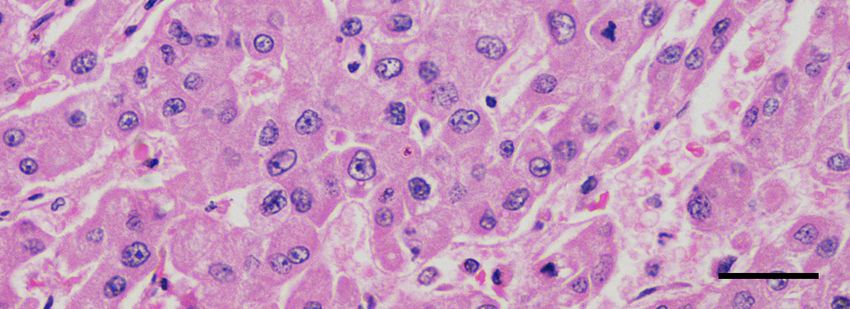
Cancers 2020, 12, 1272                                                                                    5 of 14

     At enrollment, all dogs had measurable HCC: 3 dogs had T2N0M1 disease (stage IV), 3 dogs
had T3N0M0 disease (stage III), 2 had T2N1M0 disease (stage IV), 2 had T3N1M1 disease (stage IV),
2 dogs had T3N1M0 disease (stage IV), and one dog had T1N2M0 disease (stage IV). Enlarged regional
lymph nodes and other suspected metastatic lesions were sampled by ultrasound-guided fine-needle
aspiration in all N1 or M1 cases, confirming metastatic disease.
     Five dogs had a normal CBC, renal and hepatic function, as documented by pre-therapeutic
bloodwork. Eight dogs had an increased ALP (4–53 times the upper reference limit), three dogs had an
increased GGT (2.2–569 times the upper reference limit), two dogs had an increased bilirubin (2 and
3 times the upper reference limit, respectively), and one dog had an increased AST (2.5 times the upper
reference limit).
     Of the seven dogs receiving sorafenib, two underwent prior surgical debulking of the largest
hepatic lesion, 38 and 42 days prior to enrollment, respectively. Both dogs had incomplete margins
based on histopathological evaluation and measurable regional metastatic lymph nodes at enrollment.
These dogs had the longest TTP (1250 and 1579 days, respectively) and were both still alive at data
analysis closure, after 1398 and 1706 days.
     Of the six dogs receiving MC, one underwent prior surgical debulking of the largest hepatic lesion
31 days before initiation of MC; two hepatic lesions and metastasis to the regional lymph nodes and
lungs were present at enrollment. This dog survived 27 days only after initiation of MC.
     Demographic features and potential prognostic variables were homogeneously distributed
between the two treatment groups (Table 2).

      Table 2. Demographic information and distributions of variables potentially associated with prognosis
      of 13 dogs with hepatocellular carcinoma treated with sorafenib or metronomic chemotherapy.

                                         Sorafenib           Metronomic Chemotherapy
                    Variable                                                                   p
                                          (n = 7)                    (n = 6)
                       Sex
                      Male                      2                             1              >0.999
                     Female                     5                             5
                    Age a
                   ≤11 Years                    5                             5              >0.999
                   >11 Years                    2                             1
                    Weight a
                    ≤10.6 kg                    4                             2               0.592
                    >10.6 kg                    3                             4
              Surgical Debulking
                      No                        5                             5              >0.999
                      Yes                       2                             1
                 Clinical Stage
                       III                      2                             1              >0.999
                       IV                       5                             5
             Hepatic Enzymes
            Within Normal Limits                3                             2              >0.999
                   Altered                      4                             4
              Treatment Toxicity
                     No                         4                             3              >0.099
                     Yes                        3                             3
                                          a   Median used as cut-off value.
Cancers 2020, 12, 1272 6 of 14
Cancers 2020, 12, x 5 of 13
Figure 3.
Figure 3. Dog,
Dog, liver.
liver.Histological
Histologicalsample
sampleofofhepatocellular
hepatocellular carcinoma.
carcinoma. TheThe trabeculae
trabeculae of hepatocytes
of hepatocytes are
are separated by multiple irregular vascular channels and cavernous spaces. The cytoplasm
separated by multiple irregular vascular channels and cavernous spaces. The cytoplasm of neoplastic of
neoplastic cell is often pale staining or vacuolated due to glycogen or lipid filling. Multifocally,
cell is often pale staining or vacuolated due to glycogen or lipid filling. Multifocally, the neoplastic the
neoplastic
tissue tissue
is loss and is loss andby
replaced replaced
granularbyeosinophilic
granular eosinophilic and karyorrhectic
and karyorrhectic debris (colliquative
debris (colliquative necrosis).
necrosis). Hematoxylin
Hematoxylin and eosin.andBar,eosin. Bar, 100 µm.
100 µm.
2.2. Treatment and Toxicity
At enrollment, all dogs had measurable HCC: 3 dogs had T2N0M1 disease (stage IV), 3 dogs had
T3N0M0Seven disease (stage III),
dogs received 2 had
oral T2N1M0
sorafenib. diseasetreatment
Median (stage IV), 2 had T3N1M1
duration was 228 disease (stage168
days (range, IV),to2
dogs had T3N1M0 disease (stage IV), and one dog had T1N2M0 disease (stage
321 days). At data analysis closure, five dogs had discontinued treatment due to drug unavailability,IV). Enlarged regional
lymph
and onenodes andto
dog due other
PD suspected metastatic
after 168 days. One doglesions
had were
died sampled by ultrasound-guided
for tumor-unrelated fine-needle
causes (leishmaniasis)
aspiration
while in all N1
still under or M1 cases,
treatment. The dogconfirming metastatic
experiencing disease.one trans-arterial chemoembolization
PD received
Five dogs had a normal CBC, renal and
after having interrupted sorafenib. Two of the five dogs hepatic function, as documented
that discontinued by pre-therapeutic
treatment due to drug
bloodwork. Eight dogs had an increased ALP (4–53 times the upper
unavailability were crossed over to MC, whereas the remaining three received no further reference limit), threetreatment
dogs had
an increased
based on owners’GGTpreference.
(2.2–569 times the uppertwo
Interestingly, reference limit),
of the latter twostill
were dogs hadinan
alive CRincreased bilirubin (2
and PR, respectively,
and 3 times the upper reference limit, respectively), and one dog had an increased
at data analysis closure, whereas the other 2 switching to MC progressed and died due to tumor-related AST (2.5 times the
upper reference
causes after 28 andlimit).
34 days from crossover.
Of the seven dogs
Six dogs were treated receiving
with MC. sorafenib,
Mediantwo underwent
treatment priorwas
duration surgical
21 daysdebulking
(range, 4oftothe
703largest
days).
hepatic lesion, 38 and 42 days prior to enrollment, respectively. Both
Only one dog interrupted treatment due to PD after 703 days and was treated with toceranib dogs had incomplete margins
in a
based on
rescue setting.histopathological evaluation and measurable regional metastatic lymph nodes at
enrollment. These dogs had the longest TTP (1250 and 1579 days, respectively)
Concerning toxicity, in the sorafenib group three dogs developed cutaneous toxicity, consisting of and were both still
aliveeach
one at data analysis
of the following:closure,
grade after 1398 and
2 alopecia, 17061 days.
grade hyperpigmentation, and grade 1 localized erythema
Of the six dogs receiving MC, one
and alopecia. One dog also developed grade 1 diarrheaunderwent prior
thatsurgical
resolveddebulking
uneventfully of the
withlargest hepatic
symptomatic
lesion 31 days before initiation of MC; two hepatic lesions and metastasis
treatment. Routine liver tests were monitored during sorafenib treatment. Hyperbilirubinemia to the regional lymph nodes
and
and lungs
serum were present
transaminase at enrollment.
elevations did not This dog survived 27 days only after initiation of MC.
occur.
Demographic
In the MC group, features
three and
dogspotential prognostic
experienced variablestoxicity
gastrointestinal were homogeneously
(2 of grade 1, 1 of distributed
grade 2),
between the two treatment groups (Table 2).
one dog experienced grade 1 hemorrhagic cystitis, and one dog experienced grade 1 renal toxicity.
In the two groups, treatment-related mortality was not observed.
Table 2. Demographic information and distributions of variables potentially associated with
Based on the questionnaire results, QoL was improved in 6 of the 7 dogs receiving sorafenib,
prognosis of 13 dogs with hepatocellular carcinoma treated with sorafenib or metronomic
whereas in one dog QoL was maintained. Only 3 of the 6 dogs receiving MC lived long enough to
chemotherapy.
assess QoL. In two of them, QoL was improved, whereas in the last dog QoL was maintained.
Sorafenib Metronomic Chemotherapy
Variable p
2.3. Response Rate and Outcome (n = 7) (n = 6)
Sex
Of the seven dogs treated with sorafenib, 1, 3, and 3 dogs had CR, PR, and SD, respectively. CR was
Male 2 1 > 0.999
documented in one dog that underwent hepatic debulking and where hepatic, splenic, and sternal
Female 5 5
lymphadenopathy was Age evident
a at presentation, resolving at the first imaging follow-up and until
1706 days. PR was observed
≤ 11 Yearsin three dogs and
5 consisted of a decrease
5 of the liver lesions size (n = 3),
> 0.999
resolution of malignant
> 11 Years (n = 2) and2 reduction of lung nodules
effusion 1 (n = 1). The response rate was
57.1%, and the disease-control
Weighta rate was 100%.
≤ 10.6 kg 4 2 0.592
> 10.6 kg 3 4Cancers 2020, 12, 1272 7 of 14
Of the six dogs receiving MC, three dogs had SD and three dogs had PD. Among the three dogs
with SD, the hepatic mass size was stable respectively for 35, 219 and 703 days and subsequently
increased in size with lesion rupture and secondary haemoabdomen. The response rate was 0% and
the disease-control
Cancers
Cancers 2020, 12,
2020, 12, xx rate was 50%. 77 of
of 13
13
At the end of the study, five dogs receiving sorafenib had died (4 due to PD and 1 due to
convulsive crisis
leishmaniasis).
convulsive crisisTumor-related
(n == 2),
(n 2), most
most likely
likely attributable
deaths attributable
were due toto torupture
hepaticofencephalopathy.
hepatic encephalopathy.
HCC with hemoabdomen Because necropsy
Because necropsy was
(n = 2) and
was
not permitted,
permitted,
convulsive
not crisiscerebral metastases
(n = 2),
cerebral most likely
metastases could not be
be completely
attributable
could not completely ruled
to hepaticruled out in
in these
encephalopathy.
out these cases.
cases.
Because Median TTP was
necropsy
Median TTP was
363
not days (95%
363 permitted,
days (95% CI, CI, 191–535);
cerebral median
metastases
191–535); median OS
could was
OS was 361
not 361 days (95%
be completely CI,
days (95% ruled 0–909).
out in these cases. Median TTP was
CI, 0–909).
All dogs
363 days
All dogs
(95% in in the
CI,the MC group
191–535);
MC group
median hadOS
had died
was
died due
due todays
361to PD.(95%
PD. Tumor-related deaths were
CI, 0–909). deaths
Tumor-related were duedue to
to rupture
rupture of of
HCCAll
HCC with
dogs
with hemoabdomen
in the MC group
hemoabdomen (n had
(n == 4),
4),died
peritoneal
due to PD.
peritoneal andTumor-related
and adrenal metastases
adrenal metastases
deaths were(n == due
(n 1), and
1), and
to disseminated
rupture of HCC
disseminated
intravascular
with hemoabdomen
intravascular coagulopathy (n = 1).
(n = 4), peritoneal
coagulopathy Median
(n = 1). Median TTP
and adrenal
TTP waswas 27 days
metastases (95% CI,
(n = 1),
27 days (95% CI,and0–68); median
disseminated
0–68); OS was 32 days
median OSintravascular
was 32 days
(95% CI,
CI, 0–235).
0–235).
coagulopathy
(95% (n = 1). Median TTP was 27 days (95% CI, 0–68); median OS was 32 days (95% CI, 0–235).
TTP was
TTP was significantly
significantly longer
longer among
among dogsdogs receiving
receiving sorafenib
receiving sorafenib(p
sorafenib (p =
(p == 0.044),
0.044), whereas
whereas nono significant
significant
difference
difference
difference waswas observed
was observed
observed for for OS
for OS
OS (p (p
(p == 0.079) (Figures 4 and
= 0.079) (Figures 4 and 5). 5).
Figure 4. Time toto progression
progression for
for 13
13 dogs
dogs with
with hepatocellular
hepatocellular carcinoma
carcinoma treated
treated with
with sorafenib
sorafenib (solid
(solid
Figure 4. Time to progression for 13 dogs with hepatocellular carcinoma treated with sorafenib (solid line)
line)
line)
or or metronomic
or metronomic
metronomic chemotherapy
chemotherapy
chemotherapy (dashed
(dashed(dashed line).
line). In line). In the sorafenib
In the sorafenib
the sorafenib group,
group,
group, dogs dogs
had adogs had
had
longer a longer
a longer
time time
time to
to progressionto
progression
progression
(363 (363
versus 27(363 versus 27 days;
days; pp == 0.044).
p = 0.044).
versus
days; 27 0.044).
Figure
Figure 5.
Figure 5. Overall
5. Overallsurvival
Overall survivalfor
survival for1313
for 13dogs
dogs
dogs
with hepatocellular
with
with
carcinoma
hepatocellular
hepatocellular
treated
carcinoma
carcinoma
withwith
treated
treated
sorafenib (solid(solid
sorafenib
with sorafenib
line)
(solid
or metronomic
line) chemotherapy
or metronomic
metronomic (dashed
chemotherapy line). Median
(dashed overall survival
line). Median
Median was 361 days
overall survival
survival for the
was 361
361 sorafenib
days for the
the
line) or chemotherapy (dashed line). overall was days for
group and group
sorafenib 32 daysand
for the
32 metronomic
days for the chemotherapy
metronomic group; however,
chemotherapy the difference
group; however, was
the not statistically
difference was
sorafenib group and 32 days for the metronomic chemotherapy group; however, the difference was
significant (p = 0.079).
not statistically
statistically significant (p
(p == 0.079).
0.079).
not significant
None of
None of the
the other
other examined
examined variables
variables were
were significantly
significantly associated
associated with
with TTP
TTP or
or OS
OS (Table
(Table 3).
3).Cancers 2020, 12, 1272 8 of 14
None of the other examined variables were significantly associated with TTP or OS (Table 3).
Table 3. Log-rank test for the variables potentially associated with increased risk of tumour progression
and overall survival in 13 dogs with hepatocellular carcinoma.
Time to Progression Overall Survival
Variable
Median Median
p p
(95% CI) (95% CI)
Sex
Male 168 (0–435) 0.301 327 (172–481) 0.474
Female 219 (184–253) 361 (0–908)
Age a
≤11 Years 219 (0–488) 0.235 390 (0–979) 0.059
>11 Years 205 (0–531) 212 (0–695)
Weight a
≤10.6 kg 168 (0–509) 0.237 279 (66–492) 0.194
>10.6 kg 363 (81–645) 784 (0–1606)
Surgical Debulking
No 205 (126–284) 0.107 327 (213–441) 0.104
Yes not reached not reached
Clinical Stage
III 168 (0–435) 0.102 361 (0–908) 0.330
IV 230 (18–442) 327 (0–1030)
Hepatic Enzymes
Within Normal Limits 219 (189–249) 0.643 390 (93–687) 0.675
Altered 168 (0–438) 327 (98–556)
Treatment
Metronomic Chemotherapy 27 (0–68) 0.044 * 32 (0–235) 0.079
Sorafenib 363 (191–535) 361 (0–909)
Treatment Toxicity
No 205 (110–300) 0.357 327 (1–653) 0.194
Yes 219 (0–613) 390 (0–1096)
a Median used as cut-off value. * Significant.
3. Discussion
The aim of this study was to evaluate the safety and efficacy of sorafenib in dogs with unresectable
HCC compared with those that received MC.
MC is based on the principle of administering low doses of chemotherapeutic drug continuatively
for long periods of time, resulting in an increased antitumor activity through the inhibition of
neo-angiogenesis and at the same time in a low toxicity profile due to the reduced dose of drug at
each administration. On the wave of the good results reported by our center in treating dogs with
various advanced cancer [11,14,15], we tested MC in canine HCC, confirming a favorable safety profile.
In people with HCC, MC with capecitabine showed a good safety profile and antitumor efficacy [29,30].
While the toxicity profile of MC is well documented, little is known about sorafenib. When the
current clinical trial was started, there were no data concerning treatment toxicity in dogs, thus the
dose that was considered to be safe in healthy beagles was adopted [26,27].
The schedule and dosage of sorafenib in the treatment of HCC used in the present study was found
to be safe and tolerable. Three (42.9%) cases of cutaneous toxicity and 1 (14.3%) case of diarrhea occurred
as adverse events, but none of them were dose-limiting toxicities. However, within the realm of this
small non dose-escalating trial, our results need confirmation by other studies. Notably, the analysis of
QoL revealed an improvement in 6/7 dogs receiving sorafenib.Cancers 2020, 12, 1272 9 of 14
Conversely, sorafenib has significant toxicity in humans, and the most common treatment-related
adverse events include hand-foot syndrome, hypertension, and diarrhea [16]. Nevertheless, according to
several studies, the development of one or more adverse events was associated with longer TTP,
disease control rate and OS [16].
It was reported that in dogs with unresectable HCC there are no efficacious non-surgical treatment
options [1–3]. Currently, there is no standard systemic chemotherapy with cytotoxic agents for dogs
with diffuse or nodular HCC. Traditional chemotherapy with gemcitabine as single agent or alternated
to carboplatin was tested in dogs, with no evidence of survival benefit [5,6].
Sorafenib is the first molecular targeted drug approved for the treatment of human advanced
HCC and is a potent small molecule inhibitor of multiple kinases with anti-proliferative and
anti-angiogenic activities.
After 10 years of research into sorafenib in human advanced HCC, there are still no validated
prognostic or predictive factors of response. Moreover, the exact molecular mechanisms of resistance
have not been fully elucidated [31,32].
Our population treated with sorafenib had an improved TTP in comparison with the dogs
treated with MC. Similarly, although not formally compared, the disease response rate, TTP and OS
observed in this study were higher than the outcomes reported by other studies focusing on cytotoxic
chemotherapy [5,6].
The small sample size did not allow determining if this observation translated into a better
survival outcome, although dogs receiving sorafenib showed a median OS (361 days) 10 times higher
than those receiving MC (32 days). It must be noted that five out of the seven dogs receiving sorafenib
discontinued treatment due to drug unavailability. Thus, it may be possible that continuous therapy
with sorafenib would have translated into a prolonged survival as well.
On the other hand, although unlikely, the trend to live longer in the sorafenib group may be due to
the lack of effective treatments for dogs with unresectable HCC that failed MC. Indeed, dogs receiving
sorafenib were allowed to crossover to MC or other treatments if they progressed or if the drug was
no longer available, whereas no post-progression therapy was recommended for those dogs on MC.
Also, owners willing to treat their dogs with an investigational drug may be willing to continue
treatment once the disease progresses, especially in the face of an improved QoL. This may have led to
a better outcome in the group of dogs receiving sorafenib. It must be acknowledged that in veterinary
oncology, quality (rather than quantity) of life is the primary goal of any antitumoral treatment. As a
consequence, survival time is intimately linked to QoL, possibly biasing outcome results. However, it is
important to note that dogs crossing over received MC for a short time (28 and 34 days, respectively),
thereby reducing the likelihood of a significant survival benefit attributable to the therapeutic switch.
In the current study, two dogs in the sorafenib treatment arm were pretreated with debulking
surgery prior to the beginning of systemic therapy and were exceptional responders despite the presence
of residual local and metastatic disease. This finding was not unexpected, as bulky disease is challenging
to treat with current systemic therapies for non-hematologic cancers. According to the Norton-Simon
hypothesis, the highest degree of chemosensitivity occurs when the tumor is not bulky. As such,
a debulking surgery may result in a residual tumor burden that is more chemosensitive, implying that
the ability to receive local therapy could have contributed to the better outcome in these dogs.
Beside the small population size, the present study has additional limitations. First, there was no
randomization, and the treatments were selected at the clinician and owners’ discretion. This may
have resulted in a selection bias for dogs treated with sorafenib, although there were no significant
differences in the dogs’ characteristics between the two groups Second, some dogs in both groups
received prior debulking surgery. Lastly, low sorafenib unavailability precluded continued use of
sorafenib in five out of seven dogs.
With these limitations in mind, sorafenib seems to be a good candidate for the treatment of dogs
with advanced HCC, due to a benefit in disease control and an acceptable safety profile.Cancers 2020, 12, 1272 10 of 14
4. Material and Methods
4.1. Inclusion and Exclusion Criteria
Dogs with histologically confirmed HCC of any clinical stage were qualified for recruitment
(Table 4). Dogs were eligible for enrollment if they were ineligible for surgical excision or if nodal or
distant metastatic disease was confirmed at admission, regardless of prior surgical debulking of the
primary tumor.
Table 4. TNM staging system for canine hepatocellular carcinoma.
Primary tumor (T)
T0: no evidence of primary tumor
T1: solitary tumor of any size involving one lobe
T2: multiple tumors of any size involving multiple lobes
T3: tumor(s) with direct invasion of adjacent organs
Regional lymph nodes (N)
N0: no regional lymph nodes metastasis
N1: regional lymph node metastasis
N2: distant lymph node metastasis
Distant metastasis (M)
M0: no distant metastasis
M1: distant metastasis
Inclusion criteria were as follows: (1) at least one unidimensional lesion measurable by ultrasound
or computed tomography (CT) according to the canine Response Evaluation Criteria In Solid Tumors
version 1.0 (cRECIST v1.0; 23); (2) not being a candidate for surgical resection if clinical stage evaluations
indicated stage T3 or N1 or M1, or if imaging revealed insufficient apparently normal hepatic tissue
for liver lobectomy to be safely performed; (3) no prior systemic antitumoral treatments; (4) an
adequate bone marrow, cardiac, renal, and hepatic function, as documented by a normal CBC,
renal and hepatic serum chemistry values. Specifically, dogs were required to have an absolute
neutrophil count ≥ 1500 cells/µL, hematocrit ≥ 25%, platelet count ≥ 100,000/µL, serum creatinine
concentration and alanine transaminase activity ≤ 3 times the upper limit of normal, lethargy/fatigue
status (VCOG-CTCAE version 1.0) of either 0 or 1 [24].
Exclusion criteria included hepatic encephalopathy, second malignancies, concurrent serious
systemic diseases, or previous systemic chemotherapy or molecular target therapy.
Enrollment began in December 2011, and the study closed in June 2017.
Written, informed consent to participate in this study was obtained from each owner. All owners
were informed of the advantages and disadvantages of the treatment options, including unknown
treatment outcomes and treatment-related morbidities. The final treatment decision was made jointly
by each owner and attending clinician, with full respect for the option to decline participation.
4.2. Treatment Protocol
Sorafenib treatment was offered to all dogs. If owners rejected sorafenib, dogs received MC.
Sorafenib was administered orally at a dose of 5 mg/kg twice daily on an empty stomach; the dose
was administered to the nearest 50 mg.
The drug was discontinued in the event of disease progression, protocol-defined unacceptable
toxicity, a dose interruption of more than 30 days, owner choice, the recommendation of the attendingCancers 2020, 12, 1272 11 of 14
clinician, or drug unavailability. In case of sorafenib discontinuation, regardless of the reason, dogs were
allowed to crossover to alternative treatments, including MC.
MC was administered orally and consisted of low-dose cyclophosphamide (10 mg/m2 q24 h),
piroxicam (0.3 mg/kg q24 h) and thalidomide (2 mg/kg q24 h). Owners administering thalidomide
were comprehensively informed of its known teratogenic effect.
4.3. Safety Monitoring and Efficacy of Treatment
Safety evaluation was performed 14 days after the beginning of treatment, and included medical
history obtained from the owners, physical examination, CBC with differential and platelet count,
serum biochemical analysis, and urinalysis with microscopic examination of urine sediment. The dog’s
vital signs, temperature and systolic blood pressure were recorded at each visit.
During follow-up, the levels of ALT, AST, γGTP, serum albumin, total bilirubin, urea, and creatinine
were determined every 4–6 weeks to evaluate liver function. Toxic effects were graded in accordance
with VCOG-CTCAE guidelines [33]. Unacceptable toxicity was defined as side effects of grade 3 or
more in any tissue or organ.
Quality of life (QOL) was assessed using a questionnaire designed for the study based on
investigators’ clinical expertise and this was assessed at baseline (before starting treatment) and at
every recheck during treatment (Questionnaire S1).
Tumor measurements, based on RECIST [34], were performed at baseline (within 14 days before
initiation of treatment) by means of abdominal ultrasound or total body computed tomography (TBCT).
Thoracic radiographs were also included in the initial work-up and repeated in the follow-up if
clinically indicated. The first follow-up imaging was done after 4 weeks in both treatment arms and
every 4–8 weeks thereafter.
The therapeutic effect was defined by the RECIST as follows: complete response (CR), disappearance
of all measurable lesions for >4 weeks; partial response (PR), >30% decrease in the sum of the largest
target lesion diameters and no development of a new lesion for >4 weeks; progressive disease (PD),
>20% increase in the sum of the largest target lesion diameters or appearance of a new lesion; and stable
disease (SD), neither PR nor PD seen for >4 weeks.
Dogs that died before their first radiographic assessment were classified as having PD. The number
of dogs achieving CR, PR, SD, and PD was analyzed. The response rate was defined as the percentage
of dogs whose best response RECIST rating of CR or PR was maintained for at least four weeks after
the first demonstration of such a rating. The disease control rate was defined as the percentage of dogs
whose best response RECIST rating of CR, PR, or SD was maintained for at least four weeks after the
first demonstration of such a rating. For dogs with SD or an objective response, follow-up assessments
for survival were performed every 4 weeks until PD and/or death.
4.4. Statistical Analysis
Differences in the demographic and clinical features between dogs receiving the two protocols
were evaluated with Mann–Whitney U-test and Fisher’s exact test.
Time to progression (TTP) was defined as the time from the initiation of treatment to the date of
disease progression. Overall survival (OS) was defined as the time from the initiation of treatment
to the date of death or the dog’s last follow-up. For survival analysis, dogs were censored if they
were alive at the time of study closure or died of tumor-unrelated causes, whereas for TTP dogs were
censored if, by the last examination, disease had not progressed.
Survival curves were generated according to the Kaplan-Meier product-limit method.
Survival estimates are presented as medians with the corresponding 95% confidence intervals (95% CI).
The influence of potential prognostic variables (sex, age, weight, multiplicity of lesions, clinical stage,
altered liver enzymes, surgical debulking, treatment protocol, treatment toxicity) on TTP and OS was
investigated with the log-rank test.Cancers 2020, 12, 1272 12 of 14
Data were analyzed by use of commercial software programs (SPSS Statistics v. 24, IBM, Somers,
NY, and Prism v. 5.0, GraphPad, San Diego, CA, USA). p values ≤ 0.05 were considered significant.
5. Conclusions
The results of this study offer a good basis on which new randomized prospective clinical trials
should be undertaken to compare the efficacy and drawback of sorafenib versus MC or traditional
chemotherapy in dogs with advanced HCC.
Supplementary Materials: The following are available online at http://www.mdpi.com/2072-6694/12/5/1272/s1,
Questionnaire S1 Questionnaire for evaluating health-related quality-of-life in 13 dogs with advanced hepatocellular
carcinoma receiving metronomic chemotherapy or sorafenib.
Author Contributions: Conceptualization, L.M. and A.C.-G.; Data curation, L.M. and S.S.; Formal analysis, S.S.;
Investigation, F.R. and V.F.L.; Methodology, S.S., G.M. and A.C.-G.; Project administration, L.M.; Writing—Original
draft, L.M. and S.S.; Writing—Review and editing, A.C.-G. All authors have read and agreed to the published
version of the manuscript.
Funding: This research received no external funding
Conflicts of Interest: The authors declare no conflict of interest.
References
1. Patnaik, A.K.; Hurvitz, A.I.; Lieberman, P.H.; Johnson, G.F. Canine hepatocellular carcinoma. Vet. Pathol.
1981, 18, 427–438. [CrossRef] [PubMed]
2. Liptak, J.M.; Dernell, W.S.; Monnet, E.; Powers, B.E.; Bachand, A.M.; Kenney, J.G.; Withrow, S.J. Massive
hepatocellular carcinoma in dogs: 48 cases, (1992–2002). Am. Vet. Med. Assoc. 2004, 225, 1225–1230.
[CrossRef] [PubMed]
3. Kosovsky, J.E.; Matthiesen, D.T. Results of partial hepatectomy in 18 dogs with hepatocellular, carcinoma.
J. Am. Anim. Hosp. Assoc. 1989, 25, 203–206.
4. Marin, J.J.; Romero, M.R.; Briz, O. Molecular bases of liver cancer refractoriness to pharmacological, treatment.
Curr. Med. Chem. 2010, 17, 709–740. [CrossRef] [PubMed]
5. Elpiner, A.K.; Brodsky, E.M.; Hazzah, T.N.; Post, G.S. Single-agent gemcitabine chemotherapy in dogs with
hepatocellular carcinomas. Vet. Comp. Oncol. 2011, 9, 260–268. [CrossRef] [PubMed]
6. Dominguez, P.A.; Dervisis, N.G.; Cadile, C.D.; Sarbu, L.; Kitchell, B.E. Combined gemcitabine carboplatin
therapy for carcinomas in dogs. J. Vet. Intern. Med. 2009, 23, 130–137. [CrossRef] [PubMed]
7. Kareva, I.; Waxman, D.L.; Klement, G.L. Metronomic chemotherapy: An attractive alternative to maximum
tolerated dose therapy that can activate anti-tumor immunity minimize therapeutic resistance. Cancer Lett.
2015, 358, 100–106. [CrossRef]
8. Lana, S.; U’ren, L.; Plaza, S.; Elmslie, R.; Gustafson, D.; Morley, P.; Dow, S. Continuous low-dose oral
chemotherapy for adjuvant therapy of splenic hemangiosarcoma in, dogs. Vet. Intern. Med. 2007, 21, 764–769.
[CrossRef]
9. Elmslie, R.E.; Glawe, P.; Dow, S.W. Metronomic therapy with cyclophosphamide piroxicam effectively
delays tumor recurrence in dogs with incompletely resected soft tissue sarcomas. J. Vet. Intern. Med.
2008, 22, 1373–1379. [CrossRef]
10. London, C.A.; Gardner, H.L.; Mathie, T.; Stingle, N.; Portela, R.; Pennell, M.L.; Clifford, C.A.; Rosenberg, M.P.;
Vail, D.M.; Williams, L.E.; et al. Impact of toceranib/piroxicam/cyclophosphamide maintenance therapy
on outcome of dogs with appendicular osteosarcoma following amputation carboplatin chemotherapy:
A multi-institutional, study. PLoS ONE 2015, 10, e0124889. [CrossRef]
11. Finotello, R.; Henriques, J.; Sabattini, S.; Stefanello, D.; Felisberto, R.; Pizzoni, S.; Ferrari, R.; Marconato, L.
A retrospective analysis of chemotherapy switch suggests improved outcome in surgically removed
biologically aggressive canine haemangiosarcoma. Vet. Comp. Oncol. 2017, 15, 493–503. [CrossRef] [PubMed]
12. Bray, J.P.; Orbell, G.; Cave, N.; Munday, J.S. Does thalidomide prolong survival in dogs with splenic
haemangiosarcoma? Small Anim. Pract. 2018, 59, 85–91. [CrossRef] [PubMed]Cancers 2020, 12, 1272 13 of 14
13. de Campos, C.B.; Lavalle, G.E.; Fialho Ligório, S.; Camargo Nunes, F.; Carneiro, R.A.; Amorim, R.L.;
Cassali, G.D. Absence of significant adverse events following thalidomide administration in bitches
diagnosed with mammary gland carcinomas. Vet. Rec. 2016, 179, 514. [CrossRef] [PubMed]
14. Cancedda, S.; Marconato, L.; Meier, V.; Laganga, P.; Roos, M.; Leone, V.F.; Rossi, F.; Bley, C.R. Hypofractionated
radiotherapy for macroscopic canine soft tissue sarcoma: A retrospective study of 50 cases treated with a 5x6
Gy protocol with or without metronomic chemotherapy. Vet. Radiol. Ultrasound 2016, 57, 75–83. [CrossRef]
[PubMed]
15. Polton, G.; Finotello, R.; Sabattini, S.; Rossi FLaganga, P.; Vasconi, M.E.; Barbanera, A.; Stiborova, K.;
Rohrer Bley, C.; Marconato, L. Survival analysis of dogs with advanced primary lung carcinoma treated by
metronomic cyclophosphamide piroxicam thalidomide. Vet. Comp. Oncol. 2018, 16, 399–408. [CrossRef]
[PubMed]
16. Llovet, J.M.; Ricci, S.; Mazzaferro, V.; Hilgard, P.; Gane, E.; Blanc, J.F.; de Oliveira, A.C.; Santoro, A.; Raoul, J.L.;
Forner, A.; et al. Sorafenib in advanced hepatocellular carcinoma. N. Engl. J. Med. 2008, 359, 378–390.
[CrossRef]
17. De Matteis, S.; Ragusa, A.; Marisi, G.; De Domenico, S.; Gardini, A.C.; Bonafè, M.; Giudetti, A.M. Abberant
metabolism in hepatocellular carcinoma provides diagnostic therapeutic, opportunities. Oxid. Med. Cell
Longev. 2018, 2018, 7512159. [CrossRef]
18. Casadei-Gardini, A.; Del Coco, L.; Marisi, G.; Conti, F.; Rovesti, G.; Ulivi, P.; Canale, M.; Frassineti, G.L.;
Foschi, F.G.; Longo, S.; et al. 1H-NMR based serum metabolomics highlights different specific biomarkers
between early advanced hepatocellular carcinoma, stages. Cancers (Basel) 2020, 12, 241. [CrossRef]
19. De Matteis, S.; Scarpi, E.; Granato, A.M.; Vespasiani-Gentilucci, U.; La Barba, G.; Foschi, F.G.; Bandini, E.;
Ghetti, M.; Marisi, G.; Cravero, P.; et al. Role of SIRT-3, p-mTOR and HIF-1α in Hepatocellular carcinoma
patients affected by metabolic dysfunctions and in chronic treatment with metformin. Int. J. Mol. Sci.
2019, 20, 1503. [CrossRef]
20. Fiume, L.; Manerba, M.; Vettraino, M.; Di Stefano, G. Effect of sorafenib on the energy metabolism of
hepatocellular carcinoma cells. Eur. J. Pharmacol. 2011, 670, 39–43. [CrossRef]
21. Kim, M.J.; Choi, Y.K.; Park, S.Y.; Jang, S.Y.; Lee, J.Y.; Ham, H.J.; Kim, B.G.; Jeon, H.J.; Kim, J.H.; Kim, J.G.; et al.
PPARδ reprograms glutamine metabolism in sorafenib-resistant HCC. Mol. Cancer Res. 2017, 15, 1230–1242.
[CrossRef] [PubMed]
22. Cassim, S.; Raymond, V.A.; Lacoste, B.; Lapierre, P.; Bilodeau, M. Metabolite profiling identifies a signature
of tumorigenicity in hepatocellular carcinoma. Oncotarget 2018, 9, 26868–26883. [CrossRef] [PubMed]
23. Strumberg, D.; Clark, J.W.; Awada, A.; Moore, M.J.; Richly, H.; Hendlisz, A.; Hirte, H.W.; Eder, J.P.; Lenz, H.J.;
Schwartz, B. Safety, pharmacokinetics, and preliminary antitumor activity of sorafenib: A review of four
phase I trials in patients with advanced refractory solid tumors. Oncologist 2007, 12, 426–437. [CrossRef]
24. Wolfesberger, B.; Tonar, Z.; Gerner, W.; Skalicky, M.; Heiduschka, G.; Egerbacher, M.; Thalhammer, J.G.;
Walter, I. Pharmacologic inhibition of MEK signaling prevents growth of canine hemangiosarcoma. Mol. Cancer
Ther. 2013, 12, 1701–1714.
25. Wolfesberger, B.; Tonar, Z.; Gerner, W.; Skalicky, M.; Heiduschka, G.; Egerbacher, M.; Thalhammer, J.G.;
Walter, I. The tyrosine kinase inhibitor sorafenib decreases cell number and induces apoptosis in a canine
osteosarcoma cell line. Res. Vet. Sci 2010, 88, 94–100. [CrossRef] [PubMed]
26. Wilhelm, S.; Chien, D.S. BAY 43-9006: Preclinical data. Curr. Pharm. Des. 2002, 8, 2255–2257. [CrossRef]
[PubMed]
27. European Medical Agency [Webpage on the Internet]. Initial Marketing Authorization Study of Nexavar.
2007. Available online: http://www.ema.europa.eu/ema/index.jsp?curl=pages/medicines/human/medicines/
000690/human_med_000929.jsp&mid=WC0b01ac058001d124 (accessed on 4 March 2017).
28. Foskett, A.; Manley, C.; Naramore, R.; Gordon, I.K.; Stewart, B.M.; Khanna, C. Tolerability of oral sorafenib
in pet dogs with a diagnosis of cancer. Vet. Med. (Auckl.) 2017, 8, 97–102. [CrossRef]
29. Casadei Gardini, A.; Foca, F.; Scartozzi, M.; Silvestris, N.; Tamburini, E.; Faloppi, L.; Brunetti, O.; Rudnas, B.;
Pisconti, S.; Valgiusti, M.; et al. Metronomic capecitabine versus best supportive care as second-line treatment
in hepatocellular carcinoma: A retrospective study. Sci. Rep. 2017, 7, 42499. [CrossRef]
30. Trevisani, F.; Brandi, G.; Garuti, F.; Barbera, M.A.; Tortora, R.; Casadei Gardini, A.; Granito, A.; Tovoli, F.;
De Lorenzo, S.; Inghilesi, A.L.; et al. Metronomic capecitabine as second-line treatment for hepatocellular
carcinoma after sorafenib discontinuation. J. Cancer Res. Clin. Oncol. 2018, 144, 403–414. [CrossRef]Cancers 2020, 12, 1272 14 of 14
31. Marisi, G.; Cucchetti, A.; Ulivi, P.; Canale, M.; Cabibbo, G.; Solaini, L.; Foschi, F.G.; De Matteis, S.; Ercolani, G.;
Valgiusti, M.; et al. Ten years of sorafenib in hepatocellular carcinoma: Are there any predictive and/or
prognostic markers? World J. Gastroenterol. 2018, 24, 4152–4163. [CrossRef]
32. Di Costanzo, G.G.; Casadei Gardini, A.; Marisi, G.; Foschi, F.G.; Scartozzi, M.; Granata, R.; Faloppi, L.;
Cascinu, S.; Silvestris, N.; Brunetti, O.; et al. Validation of a simple scoring system to predict sorafenib
effectiveness in patients with hepatocellular carcinoma. Target. Oncol. 2017, 12, 795–803. [CrossRef]
[PubMed]
33. Veterinary Co-operative Oncology Group. Veterinary Co-operative oncology group- common terminology
criteria for adverse events (VCOG-CTCAE) following chemotherapy or biological antineoplastic therapy in
dogs and cats v1.0. Vet. Comp. Oncol. 2004, 2, 194–213.
34. Nguyen, S.M.; Thamm, D.H.; Vail, D.M.; London, C.A. Response evaluation criteria for solid tumours in
dogs (v1.0): A veterinary cooperative oncology group (VCOG) consensus document. Vet. Comp. Oncol.
2015, 13, 176–183. [CrossRef] [PubMed]
© 2020 by the authors. Licensee MDPI, Basel, Switzerland. This article is an open access
article distributed under the terms and conditions of the Creative Commons Attribution
(CC BY) license (http://creativecommons.org/licenses/by/4.0/).You can also read