Vivid 7 Dimension Real-time 4D imaging and Real-time 4D color imaging - GE Healthcare
←
→
Page content transcription
If your browser does not render page correctly, please read the page content below
GE Healthcare

Vivid 7 Dimension
Real-time 4D imaging and
Real-time 4D color imaging
The Vivid™ 7 Dimension is a premier cardiovascular ultrasound         Technical description:
system that builds on the strength of GE’s powerful imaging
                                                                      The foundation for 4D imaging is the unique 3V transducer.
platform, acquiring more clinical information – and in fewer steps.
                                                                      This transducer has a true, non-sparse 2D array with
GE’s innovative transducer technology allows you to use               thousands of transducer elements. With such a high number
only one probe to acquire multiple planes of images at the            of transducer elements, part of the beam forming is made
same time, and from the same heartbeat, without changing              within the transducer itself. Cutting-edge material and
probe positions.                                                      electronic technology has made it possible to have a very high
                                                                      dynamic range beam forming in the transducer. This results
This single, unique transducer gives you multiple imaging
                                                                      in a probe capable of scanning all modes, including the
capabilities, such as Coded Octave Harmonics, color Doppler,
                                                                      challenging CW Doppler mode. This technology makes it
Tissue Velocity Imaging, PW and CW Doppler – plus multi-
                                                                      possible to steer the ultrasound beam in any direction, so
dimensional and 4D imaging.
                                                                      that real-time 4D images can be acquired. For color imaging,
GE’s real-time 4D imaging and real-time, full-volume imaging          this translates into unprecedented sensitivity in 4D color flow
enable the user to acquire artifact-free depictions of the            imaging. Even with such probe complexity, the design has
entire heart, to increase diagnostic confidence.                      been made with an ergonomically shaped transducer that
Real-time 4D color Doppler, and real-time, full-volume color          allows easy access to imaging windows.
Doppler imaging, offer instant displays of 4D color for
improved visualization, including the ability to quantify
regurgitant lesions.
So, one transducer can be used to perform a complete
exam, including stress and contrast imaging, which can
significantly reduce exam time and streamline workflow.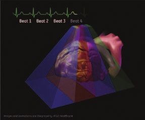
Real-time, non-gated 4D imaging
The 3V probe and the Vivid 7 Dimension are capable of
acquiring real-time, non-gated 4D tissue and color imaging.
The volume data is displayed in real time with volume
rendering techniques for visualization of valves and
structures. The size and shape of the volume can be
easily adjusted.
This system can even perform real-time, non-gated 4D color
imaging. The volume color data is simultaneously rendered
with the volume tissue data. Tools like Volume Size enable
the user to optimize the size and frame rate for the
respective region of interest.
Raw data capabilities offer all the same post-processing
functionality for 4D images that are available during the
routine examination. For example, controls like color baseline
or color maps can be adjusted on the EchoPAC™ workstation
during analysis.

Real-time, full-volume 4D imaging
The Vivid 7 Dimension system enables the user to acquire
large tissue volumes with gated acquisition. The powerful
processing capabilities allow for real-time display of the
data while the rendering continuously updates, so all
throughout the scanning process the user can see when
the correct data is acquired.
The highly efficient acquisition of color Doppler gives high-
volume rates in real-time, 4D full-volume color imaging.
The user can easily change both the size of the color region
of interest and the acquired frame rates using the Volume
Size control.
The volume rendering technology for real-time, 4D full-volume
color Doppler allows for effortless jet flow detection.
The turbulent flow pattern can easily be rendered within
the color-volume data.
The scan setup for real-time, 4D full-volume color imaging
also includes the flexibility to set the number of required
cardiac cycles for acquisition. In addition, a number of
navigation and cropping tools are available that allow
for optimization of the 4D data.
Tips:
• Four-color render maps are available.
• Different color maps may improve depth perception
for a particular image or for an individual viewer
• The gray/red map may assist those that have
difficulties differentiating colors
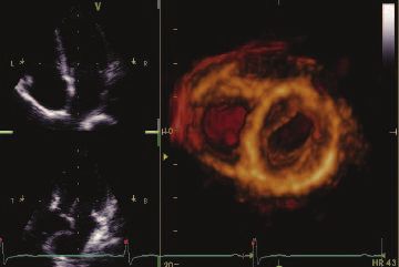
Figure 1. Red/Bronze Depth Color Render map
Enhanced 4D Visualization 4D Stereo Vision
4D Ultra-Definition Clarity 4D Stereo Vision is a technique where two different volume
Ultra-Definition Clarity Control is a one button setting to optimize renderings are displayed with two different colors and overlaid
4D volumes for streamlined 4D workflow. Combines multiple at slightly different viewing angles. For the user to see the
post-processing controls into one, to provide personalization stereoscopic effect, they must wear special Stereo Vision
of image quality from smooth to crisp resolution. glasses, which are provided with your 4D system or upgrade
purchase. 4D Stereo Vision gives the user a new perspective
How to activate on the Vivid 7 of depth and volume for the rendered image.
• Display a 4D image
Workflow Tip: Adjust Volume Optimization and Ultra-
• Bottom row of LED display, press more Definition Clarity to improve the effect of both Depth Color
• Press “4D colorize” then choose the Depth Render maps and Stereo Vision
Color Render map of your choice
Tip: Stereo angle rotary increases or decreases the angle
of the image to the left and right eye. Adjust to optimize
How to activate on the EchoPAC
the effect on an individual basis.
• Display a 4D image
How to activate on the Vivid 7
• Press 4D colorize on the control panel
• Wear the provided Stereo Vision glasses
• Scroll down to Depth Color Render map of your choice
• Display a 4D image
• Bottom row of LED display, press more
Depth Color Render maps
• Press 4D colorize, then Stereo
Other visual enhancements such as Depth Color Render maps
have been integrated to improve the perception of depth in
How to activate on the EchoPAC
volume-rendered images. Every pixel along each scan line, in the
volume rendering is assigned a color according to its depth. • Put on GE viewing glasses
This enables the user to better distinguish the perception • Display 4D rendering
of depth in the volume rendered image. (Figure 1)
• Press 4D colorize
• Select StereoCase study one
The following are images from a clinical case study evaluating a patient with a history
of pacemaker insertion.
Optimization tip:
Increase 2D gain to visualize valves
and increase 4D gain to visualize
the back wall of the chamber.
Real-time 4D imaging enables the user to see the The patient’s left ventricular function was
4D rendering in real time, as the patient’s image assessed with real-time, 4D full-volume imaging.
is continuously updated. This patient was treated Following the full-volume acquisition, 9-Slice
with a single chamber pacemaker. This patient mode can be used to further assess regional
was followed with transthoracic echocardiography wall motion of the left ventricle.
including real-time 4D imaging.
Navigation and reference plane:
Use the navigation image located
in the middle of the screen for
orientation of the 4D dataset.
The patient’s right ventricular function was also To assess the interaction of the pacemaker wire
assessed with real-time 4D imaging. During the and the tricuspid valve, real-time, 4D full-volume
routine echo examination, mild to moderate color imaging was utilized. Here, the system
tricuspid regurgitation was visualized. demonstrates the extent of the tricuspid
regurgitation as it extends into the base of the
right atrium and is forced into the atrial septum
by the pacemaker wire.Case study two
The following are images from a clinical case study evaluating mitral regurgitation.
These images are all acquired from
the same transducer, improving
clinical workflow.
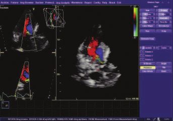
This patient presented with “moderate” mitral Multi-dimensional imaging, including tri-plane
regurgitation. This patient’s mitral regurgitation imaging, offers additional clinical confidence by
was assessed by combining tools of continuous evaluating color Doppler from three orthogonal
wave (CW) and acquiring multiple-imaging views views simultaneously, from the same heart cycle
with color Doppler. as seen. This patient’s mitral regurgitation, PISA,
can be seen here from all three planes.
Optimization tip:
Use tissue transparency to suppress
the appearance of the tissue to
evaluate the interaction of this tissue
structure with the color-volume.
Vena contracta width ranges3:
.7cm Severe has been quantitatively evaluated from two evaluate the vena contracta. Following the
dimensions can now be evaluated from a color- acquisition of a full-volume dataset, the user
volume acquisition. To assess this patient’s can use the 6-Slice mode to quantify the color-
mitral regurgitation, we acquired a real-time volume acquisition in 6 short-axis slices, giving
full-volume image. The advantage of GE’s real- the user the ability to isolate the vena contracta
time 4D color acquisition is that the clinician to perform a planimetry area of the narrowest
views the information as the 4D image continu- part of the regurgitation.
ously updates in real time.Acquiring and rendering 4D datasets are easy to learn
and adopt into clinical practice.
Real-time 4D acquisition Real-time 4D color acquisition
1. Select 3V probe 1. Press 2D and enter Color mode
2. Optimize IQ settings (gain, depth, TGC, etc.) (Optimize IQ – depth, gain, TGC and ROI)
3. Press 4D button 2. Press 4D (RT Color)
4. Adjust 4D (Active) Gain as necessary 3. Press 4D CF Prepare Mode
a. Increase 2D gain to see valves 4. Press Full Volume – (RT 4D Color FV)
5. Press Image Store or Freeze
b. Increase 4D (Active) Gain to see back
of the chamber
4D color rendering
5. Select Full Volume
6. Press Angle
6. Press Image store or Freeze
7. Use Trackball (Translate/Rotate)
to optimize dataset for region of interest
Tissue rendering
8. Increase Tissue Transparency
7. Press Angle
Evaluate Flow Transparency
8. Trackball (Translate/Rotate) for
9. Press Store
image orientation
9. Use Volume Optimize or Clarity if necessary
10. Press Image Store
Not all features may be available in your current software package.
Please consult your sales representative to inquire about additional
features for your Vivid 7 Dimension or your EchoPAC.References:
1. Fang L, Hsiung MC, Miller AP, Nanda NC, Yin WH, Young MS, Velayudhan DE. Assessment of aortic
regurgitation by live three-dimensional transthoracic echocardiographic measurements of vena
contracta area: usefulness and validation. Echocardiography. 2005 Oct;22(9):775-81.
2. Khanna D, Vengala S, Miller AP, Nanda NC, Lloyd SG, Ahmed S, Sinha A, Mehmood F, Bodiwala K,
Upendram S, Gownder M, Dod HS, Nunez A, Pacifico AD, McGiffin DC, Kirklin JK, Misra VK.
Quantification of mitral regurgitation by live three-dimensional transthoracic echocardiographic
measurements of vena contracta area. Echocardiography. 2004 Nov;21(8):737-43.
3. Zoghbi WA, Enriquez-Sarano M, Foster E, Grayburn PA, Kraft CD, Levine RA, Nihoyannopoulos P,
Otto CM, Quinones MA, Rakowski H, Stewart WJ, Waggoner A, Weissman NJ. Recommendations
for evaluation of the severity of native valvular regurgitation with two-dimensional and Doppler
echocardiography. J Am Soc Echocardiogr. 2003 Jul;16(7):777-802.
4. A report from the American Society of Echocardiography’s Nomenclature and Standards
Committee and The Task Force on Valvular Regurgitation, developed in conjunction with the
American College of Cardiology Echocardiography Committee, The Cardiac Imaging Committee
Council on Clinical Cardiology, the American Heart Association, and the European Society of
Cardiology Working Group on Echocardiography, represented by: William A. Zoghbi, MD, Maurice
Enriquez-Sarano, MD, Elyse Foster, MD, Paul A. Grayburn, MD, Carol D. Kraft, RDMS, Robert A. Levine,
MD, Petros Nihoyannopoulos, MD, Catherine M. Otto, MD, Miguel A. Quinones, MD, Harry Rakowski,
MD, William J. Stewart, MD, Alan Waggoner, MHS, RDMS, and Neil J. Weissman, MD.
©2007 General Electric Company – All rights reserved.
GE Healthcare
GE Medical Systems Ultrasound & Primary Care Diagnostics, LLC,
9900 Innovation Drive a General Electric company, doing business as GE Healthcare.
Wauwatosa, WI 53226 General Electric Company reserves the right to make changes in
specifications and features shown herein, or discontinue the product
U.S.A. described at any time without notice or obligation. Contact your
GE representative for the most current information.
www.gehealthcare.com GE, GE Monogram, Vivid and EchoPAC are trademarks
of General Electric Company.
imagination at work
ULTC-0190-11.07-EN-USYou can also read