Rare Presentation of Chorioadenoma Destruens as Acute Haemoperitoneum Mimicking Ruptured Ectopic Pregnancy
←
→
Page content transcription
If your browser does not render page correctly, please read the page content below
KATHMANDU UNIVERSITY MEDICAL JOURNAL

Rare Presentation of Chorioadenoma Destruens as Acute
Haemoperitoneum Mimicking Ruptured Ectopic Pregnancy
Sinha M,1 Kaur R,1 Gupta R,2 Rani R,1 Aggarwal A1

Department of Obstetrics & Gynaecology
1

Kasturba Hospital, Delhi, India
                                                     ABSTRACT
West Virginia University- Charleston Division
2
                                                     Gestational trophoblastic neoplasms (GTN) are proliferative degenerative disorders
Charleston Area Medical Center                       of placental elements and include complete or partial mole (90%), invasivemole
                                                     (5-8%), choriocarcinoma (1-2%) and placental site tumor (1-2%). Chorioadenoma
Charleston, West Virginia, USA
                                                     destruens is a trophoblastic tumor, characterized by myometrial invasion through
                                                     direct extension or via venous channels. We present a case of invasive mole eroding
                                                     uterus and uterine vasculature, causing sudden rupture of uterus with massive
Corresponding Author
                                                     haemoperitoneum mimicking ectopic pregnancy. A 20 year old G1P0 at 6 weeks
Ramandeep Kaur                                       gestation presented in Casualty of Kasturba Hospital complaining of severe acute
                                                     onset lower abdominal pain for one hour. Clinical examination revealed shock.
Department of Obstetrics & Gynaecology
                                                     Sonography suggested ectopic pregnancy and immediate exploratory laparotomy
Kasturba Hospital, Delhi, India                      was decided. On laparotomy, 2000cc of haemoperitoneum was noted. Grape like
                                                     vesicles protruding through fundal perforation with profuse active bleeding was
E-mail: ramandeepkh@yahoo.in
                                                     seen. Bleeding persisted despite evacuation. Step wise uterine devascularisation
                                                     failed to achieve haemostasis. Total abdominal hysterectomy was performed as a
                                                     life saving measure.
Citation

Sinha M, Kaur R, Gupta R, Rani R, Aggarwal A. Rare
Presentation of Chorioadenoma Destruens as Acute     KEY WORDS
Haemoperitoneum Mimicking Ruptured Ectopic
Pregnancy. Kathmandu Univ Med J 2014;48(4):288-91.   Ectopic pregnancy, haemoperitonem, invasive mole

INTRODUCTION
Invasive mole or chorioadenoma destruens comprises 15                  chorioadenoma destruens may present rarely as ruptured
% of all gestational trophoblastic neoplasia (GTN).1 GTN               ectopic pregnancy in shock, incomplete abortion,
is characterized by histological abnormalities of chorionic            menorraghia or carcinoma endometrium.6,7
villi with oedema of villous stroma and varying degrees of
                                                                       We present a case of invasive mole which gradually eroded
trophoblastic proliferation. Absence or presence of fetal or
                                                                       in the uterus and the uterine vasculature, leading to
embryonic elements classifies complete or partial mole.
                                                                       sudden rupture of uterus, massive hemoperitoneum and
Molar pregnancies which fail to regress result in invasive
                                                                       shock, mimicking ectopic pregnancy.
mole, choriocarcinoma, persistent trophoblastic tumor or
placental site trophoblastic tumor.2
Molar pregnancies become Invasive moles in 20%                         CASE REPORT
and are commoner in complete molar pregnancies.3,4                     A 20 year old G1P0 at 6 weeks gestational age presented
Chorioadenoma destruens is a trophoblastic tumor,                      to the emergency room of Kasturba Hospital complaining
characterized by myometrial invasion through direct                    of severe lower abdominal pain, acute in onset and for the
extension or via venous channels with persistence of                   past one hour. Her pain was severe, colicky in nature and
edematous chorionic villi and trophoblastic proliferation              radiated to the back and thighs. She denied any nausea,
invading myometrium.5 The presence of villi in the                     vomiting or fever. Her past medical and surgical histories
trophoblastic tissue differentiates an invasive mole                   were unremarkable. She denied any known drug allergies
from choriocarcinoma Instead of vaginal bleeding (97%)                 and was not on any medications. She attained menarche at

Page 288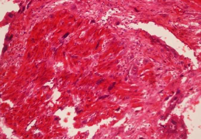
Case Note VOL. 12 | NO. 4 | ISSUE 48 | OCT- DEC 2014
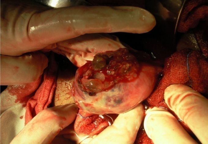
Figure 1. Ultra Sonography prior to surgery Figure 2. Uterine fundal perforation showing Figure 3. Uterine fundal perforation
grape like vesicles protruding through the showing grape like vesicles protruding
perforation through the perforation& profuse bleeding
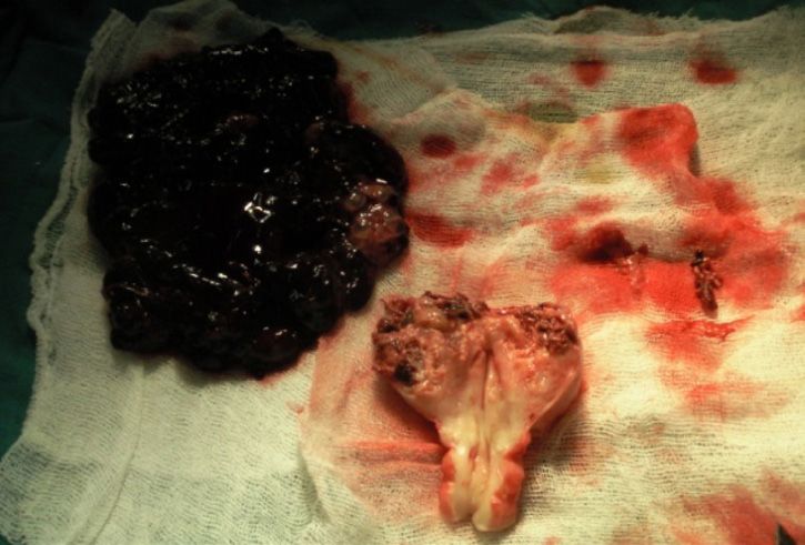
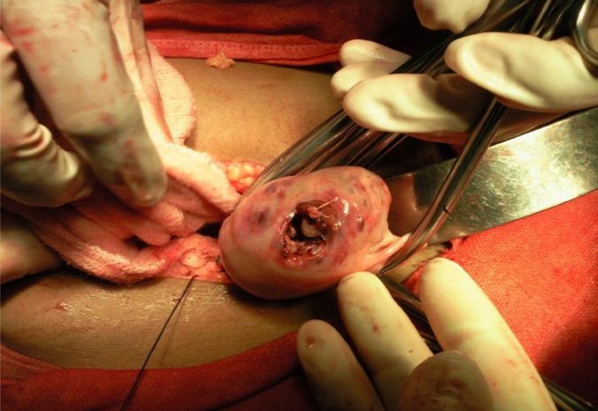
Figure 4. During hysterectomy- uterus showing Figure 5. Cut section of the uterus following hysterectomy
large perforation showing chorioadenoma destruens with tissue & Blood clot
the age of 12 years and her menstrual cycles were regular. On, exploratory laparotomy, 2000cc of hemoperitoneum
She denied any pregnancies in the past and had a marriage was noted, with peritoneal cavity full of blood and clots.
history of one year. Uterus was perforated at the fundus and profuse active
bleeding was noted at the site of perforation. Grape like
On examination patient was awake, alert and oriented but
vesicles were protruding through the uterine perforation
was in apparent distress. She was hypotensive with BP of
which measure approximately 3 cm x 3 cm (Fig 2, Fig 3). The
90/60 mm of Hg, tachycardic with pulse of 112 b/min. She
uterus was eight weeks in size, soft and enlarged, bilateral
was pale clinically, afebrile and her respiratory rate was
adenxae were normal. On further abdominal exploration,
16/min. Her cardiac, respiratory and neurological system
no ectopic tissue or metastatic tissue was identified.
evaluation was unremarkable
Profuse ongoing bleeding was seen from site of perforation
On abdominal examination, she was severely tender in
following complete evacuation of the uterus and patient
bilateral lower quadrants, with presence of guarding,
continued to remain in a state of shock despite adequate
rigidity and rebound tenderness. On bimanual examination,
fluid replacement, 4 units of blood transfusion and 20 units
cervical os was closed, the uterine size could not be assessed
of continuous oxytocin infusion. To achieve haemostasis,
due to presence of guarding and tenderness. These
step wise systematic uterine devascularization was
findings were highly suggestive of ectopic pregnancy and
attempted but complete haemostasis could be not be
emergency transvaginal ultrasound (TVS) was performed,
achieved and patient continued to bleed profusely from
which demonstrated anteverted uterus, measuring
the perforated site. Decision was made to proceed with
approximately – 46 x 60 x 76 mm with irregular echogenic
total abdominal hysterectomy as a life saving measure.
products seen in uterine cavity. Pouch of Douglas showed
Post surgery patient became haemodynamically stable.
moderate amount of free fluid. However, a definitive
Hysterectomized uterus was then sent for histopathological
diagnosis of ectopic pregnancy could not be established.
examination (Fig 4, Fig 5).
During TVS, patient became drowsy and slipped into a semi
Her postoperative course was uncomplicated.
comatose condition. Her vitals deteriorated with pulse
of 140/min and systolic blood pressure of 60 mm of Hg. On postoperative day one, her vitals improved and her
Patient was immediately rushed to operation theatre for hemoglobin was 8 gm/dl. She received additional two units
Emergency laparotomy with a definitive suspicion of a of blood transfusion and made an uneventful recovery.
ruptured ectopic pregnancy. On laboratory investigation,
The postoperative metastatic work-up included
her haemoglobin and hematocrit was 6 gram and 20 mg
serum biochemistry, chest x-ray and upper abdominal
percent, random blood sugar values of 130 mg/dl, Serum
Ultrasonography and no evidence of metastasis was
Bilirubin 1.2 mg/dl, and Serum creatinine 0.9 mg/dl. Her
detected. In view of low levels of serum ß-hCG, patient
beta HCG could not be sent as emergency laboratory
was kept under surveillance with serial quantitative ß-hCG
facilities of Kasturba Hospital does not include this
estimation to consider chemotherapy if ß-hCG levels
investigation.
Page 289
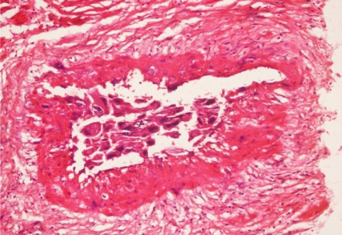
KATHMANDU UNIVERSITY MEDICAL JOURNAL

Figure 6. Section from uterus showing a Figure 7. The tumour cells are showing     Figure   8. Syncytiotrophoblasts and
tumour arising from endometrial surface and vascular invasion. (H&E, X 20)         intermediate trophoblasts are infiltrating
composed of intermediate trophoblasts and                                          the myometrium as single cell infiltrate.
afew multinucleated syncytiotrophoblasts,                                          (H&E, X 20)
arranged in sheets. (H&E, X40)
remains at a plateau or rises. Postoperatively ß-hCG level      of gestational trophoblastic neoplasia with uterine rupture
was 110 mIU/ml. Patient was discharged home in stable           are often catastrophic owing to profuse bleeding, which
condition on postoperative day 3. Her ß-hCG level was           could potentially be lethal.
Case Note                                                                                          VOL. 12 | NO. 4 | ISSUE 48 | OCT- DEC 2014

is either unavailable or not affordable and chances of losing             Use of chemotherapy in the management of invasive mole is
the woman to follow-up are high. The recommendations                      still debatable, with the evidence of spontaneous regression
for post-molar follow-up include serum βhCG levels every                  of metastatic mole in the literature. Chemoprophylaxis may
1–2 weeks after evacuation until normal, hCG levels 2–4                   be particularly useful in patients with a high-risk complete
weeks after the first normal level, and hCG surveillance                  mole when hormonal follow-up is either unavailable or not
every 1–2 months for 6 months after the first normal hCG                  affordable and chances of losing the woman to follow-up
level. Serial hCG monitoring of women with disease in                     are high. We did not consider chemotherapy in our case in
prolonged remission (greater than 1 year) often provides                  view of no evidence of metastasis and low ß-HCG levels on
early evidence of recurrence, though GTT may rarely be                    subsequent follow up. The potential risk of patient being
associated with undetectable levels of hCG.8                              lost to follow up was very much there but did not happen
                                                                          in our case.
In our patient, we were unable to achieve adequate
haemostasis by step wise uterine devasculaization
attempted with uterine and ovarian vessel ligation.                       CONCLUSION
Unfortunately the surgical emergency team was not well
versed with the technique of Internal Iliac Ligation, hence it            Chorioadenoma destruen has a potential for myometrial
was not attempted and total abdominal hysterectomy was                    and vascular invasion, leading to uterine perforation
                                                                          and massive hemorrhage. Therefore, to avoid adverse
then performed. Although the development of effective
                                                                          consequences it is necessary to identify such cases by early
chemotherapy has resulted in improved survival of patients                first trimester ultrasound. Continued reporting of these
with gestational trophoblastic tumor, hysterectomy                        cases are important so that the obstetricians are aware
remains an important adjuncts in the treatment of a                       about the possibility of ruptured invasive mole and it should
selected subset of patients.                                              be kept as a differential diagnosis in all the young pregnant
                                                                          women presents with acute onset lower abdominal pain.

REFERENCES
1. Kumar S, Vimla N, Mittal S. Invasive mole presenting as acute          8. Miller FM, Laing FC. Gestational trophoblatic disease http://
    hemoperitoneum. J K science. 2004;6: 159-160.                             brighamrad. havard.edu/cases/bwh/hcache/34/full.html

2. Berkowitz RS, Goldstein DP, In: Berck JS. Gestational trophoblastic    9. Mackenzie F, Mathers A, Kennedy, J Invasive hydatidiform mole
    neoplasm. PhiladelphiaLipincott, Williams and Wilkins, 2002.1353-         presenting as an acute primary haemoperitoneum. Br J Obstet
    74.                                                                       Gynecol. 1993; 100: 953-54.

3. D. Berek & Novak’s Gynecology, 14th ed, Philadelphia, Lippincott       10. Wilson RB, Hunter IS, Dockerty MB. Chorioadenoma Destruens. Am J
    Williams & Wilkins, 2007. p1581-1604.                                     Obstet Gynecol. 1961; 81: 546-59.

4. Kittur S, Venktesh, Ramlingappa A. A rare case of invasive mole with   11. Hol K, Junnare K, Shekhawat GS, Damle H. Case of Invasive Mole with
    silent uterine perforation. Int J. Reprod Contracept Obstet Gynecol       Uterine Rupture Presenting as a Hemoperitoneum. Global Research
    2013;2:109-10.                                                            Analysis, 2013 Dec; 2(12): 195-96.

5. Atala C, Riedemann R, Biotti M, Ramírez F, Paublo M. Invasive mole     12. Branka N, Jelena L. Invasive mole case report of massive uterine
    with uterine rupture. Rev Chil Obstet Ginecol. 1992;57(5):356-8.          destruction- Med Arh. 2008;62:242–243.

6. Singh A and, Ratnani RJ. Heterogenous presentation of Chorioadenoma    13. Mitani Y, Jimi S, Takao N. Partial resection of the uterus for
    destruens. J Obstet Gynaecol India. Dec 2012; 62(Suppl 1): 71–74.         chorioadenoma destruens. J Jpn Obstet Gynecol Soc. 1968
                                                                              Jul;15(3):163-7.
7. Mandal D, Nandi N, Dey RP, Biswas RR, Bhattacharya AK, Biswas SC.
    Partial Invasive Molar Pregnancy – Two Case Reports. Al Ameen J Med   14. Goldstein DP and Berkowitz RS. Gestational Trophoblastic Neoplasia.
    Sci. 2010; 3(1): 91-93                                                    WB Saunders, Philadelphia; 1982. 98-121.

                                                                                                                                       Page 291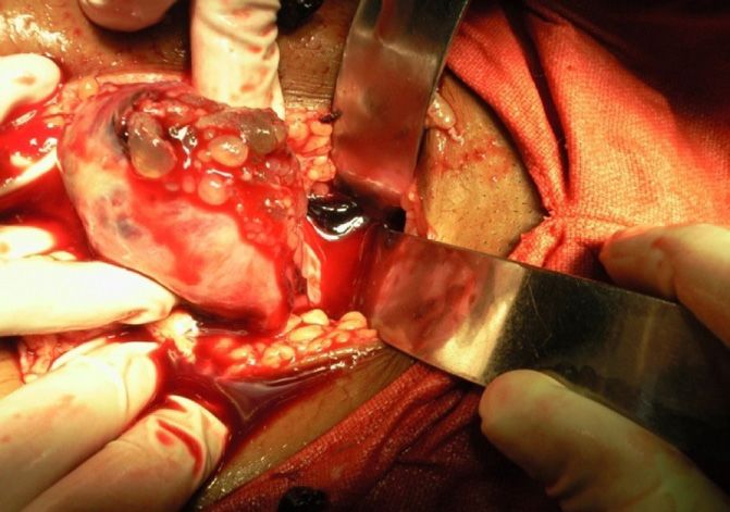

You can also read